Nerve Blocks App
 Pain Medicine Assistant App
 POCUS App
 IV Access App
 MSK Knee App
 VetRA App
 Nerve Block Manual
 Regional Anesthesia Updates
 Anesthesiology Manual
 Anesthesiology Review
 Anesthesia Updates 2025
 Anesthesia Updates 2026
 Pediatric Anesthesia Updates
 Airway Management Updates
 US Interventional Pain Manual
 Pain Medicine Updates
 Mastering Difficult IV Access
 PACU Nursing Manual
 RA Veterinary Manual
 About
AboutGeneration of ultrasound
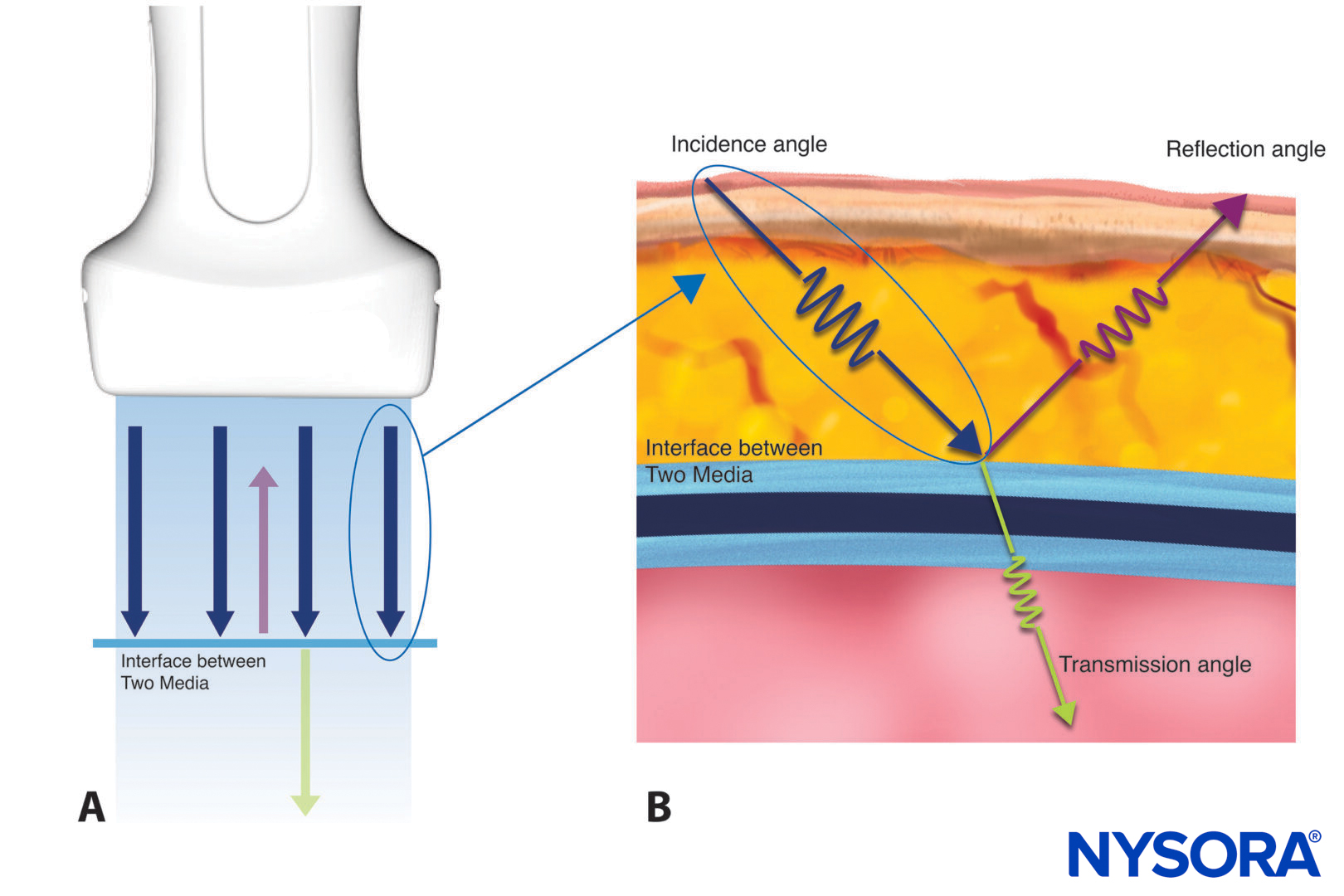
Ultrasound transducers can generate mechanical or ultrasound waves that are generated by a material with a piezoelectric effect. Application of an alternating electric field to such material causes mechanical vibrations, which translate into ultrasound. The material can also convert mechanical vibrations (the received echoes) into an electric charge, which is then translated into an ultrasound image on a computer screen. The brightness of each dot corresponds to the echo strength, producing what is known as a grayscale image.
Types of ultrasound transducers
Different types of ultrasound transducers: A. Curved array transducer, B. Linear array transducer, C. Phased array transducer.
Curved array transducer (A)
A curved transducer yields a curvilinear scan and an arc-shaped image at lower frequencies.
- Advantages: Deep penetration, wide depth of field.
- Disadvantages: Lower near-field resolution
- Use: Curved array transducers are generally used for visualizing deeper structures, such as intra-abdominal structures.
Linear array transducer (B)
A linear transducer produces parallel scan lines and a rectangular display, called a linear scan. They usually produce higher frequencies.
- Advantages: High-resolution images.
- Disadvantages: Low penetration power.
- Use: Linear array transducers are generally used for visualizing superficial structures, such as blood vessels and superficial musculoskeletal structures.
Phased array transducer (C)
Phased array transducers produce an arc-shaped image at lower frequencies by systematically directing ultrasound waves in different directions.
- Advantages: Small footprint, narrow beam
- Disadvantages: Lower near-field resolution
Use: The smaller footprint facilitates imaging between the ribs or other anatomical structures that interfere with insonating.
Interesting facts
- Transducers have a marker on the side to indicate their orientation relative to the image on the screen.
- The index mark typically corresponds to the right side of the patient, except in cardiac ultrasound imaging, where it corresponds to the left.
- As per convention, when the transducer is positioned longitudinally, the marker orientation should indicate cranial.
Tip
- Always rest the palmar side of the wrist on the patient to stabilize and improve the image.
Transducer maneuvers
- Sliding: Transducer alignments to find a window.
- Tilting: To tilt the transducer to planes perpendicular to the index mark.
- Rotation: Clockwise or counter-clockwise rotation
- Rocking (heeling): Pivoting the transducer to planes parallel to the index mark.
![]()