Indications
Possible causes of hydronephrosis and obstructive uropathy:
- Kidney stones or bladder stones
- Benign prostatic hyperplasia (BPH)
- Cancer (bladder, renal, uterine, colon, cervical)
- Dysfunctional bladder (urinary retention)
Tip
It is important to understand that abnormal findings in the kidney require a sonographic assessment of the bladder.
Functional anatomy
Anatomy of the abdomen
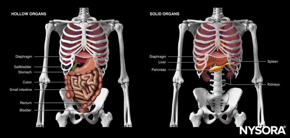
Hollow and solid organs of the abdomen.
Anatomy of the kidney

Anatomy of the kidney.
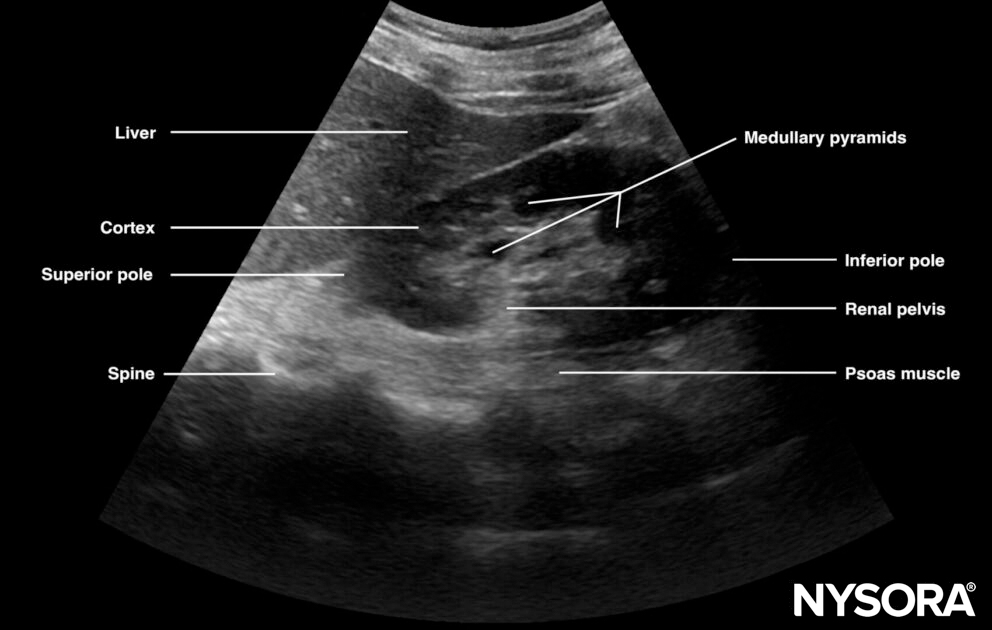
Ultrasound anatomy of the kidney.
- Outer kidney: cortex
- Inner kidney: medulla: renal pyramids (loops of Henlé)
- Size of a kidney: longitudinal: 10-12cm, width: 5-6cm
- Anatomy: renal cortex, parenchyma (pyramids), sinus and hilum
- Hypoechoic tissue: cortex, parenchyma, pyramids
- Hyperechoic tissue: fatty sinus
Tip
Always try to visualize both poles of the kidney.
Ultrasound machine setup
- Transducer: curvilinear (or phased array)
- Ultrasound preset: abdominal
- Orientation: index mark towards the head of the patient
- Depth: 10 – 15 cm
Patient position
Position the patient supine and completely flat, with the arms abducted.

Patient position for a kidney ultrasound.
Landmarks
- Costal margin: ribs in the upper abdomen protect the upper abdominal organs but may limit the acoustic window to the liver, spleen, and kidneys
- Xiphoid process: the upper border of the abdomen
- Linea alba: midline of the abdomen that separates the rectus abdominis muscles and connects the xiphoid process with the pubic bone.
- Umbilicus: virtually separates the abdomen into four quadrants
- Pubic bone: a bony structure and the lower margin of the abdomen. The pelvis starts at the level of the pubic bone.
- Anterior superior iliac spine: a bony structure that forms the lateral border of the pelvis
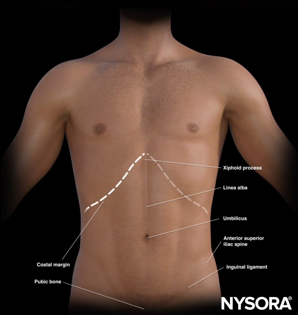
External landmarks of the abdomen.
The umbilicus divides the abdomen into different quadrants that allocate different organs.
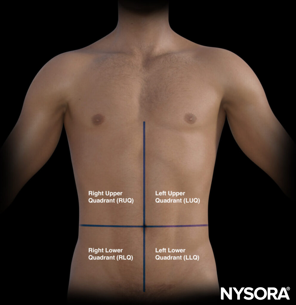
The quadrants of the abdomen.
- Right upper quadrant (RUQ): Liver, gallbladder, kidney
- Left upper quadrant (LUQ): Stomach, spleen, kidney
- Pelvic/paracolic region (lower quadrants): Colon, small intestines, rectum, bladder, male or female reproductive organs
Transducer position
A scan of the kidneys is performed in two areas:
1: The right upper quadrant (RUQ): place the transducer between the mid- and the posterior axillary line at the level of the xiphoid process and start scanning caudally until you visualize the first the liver and then both poles of the right kidney
2: The Left upper quadrant (LUQ): place the transducer between the mid- and the posterior axillary line at the level of the xiphoid process and start scanning caudally until you visualize both poles of the left kidney
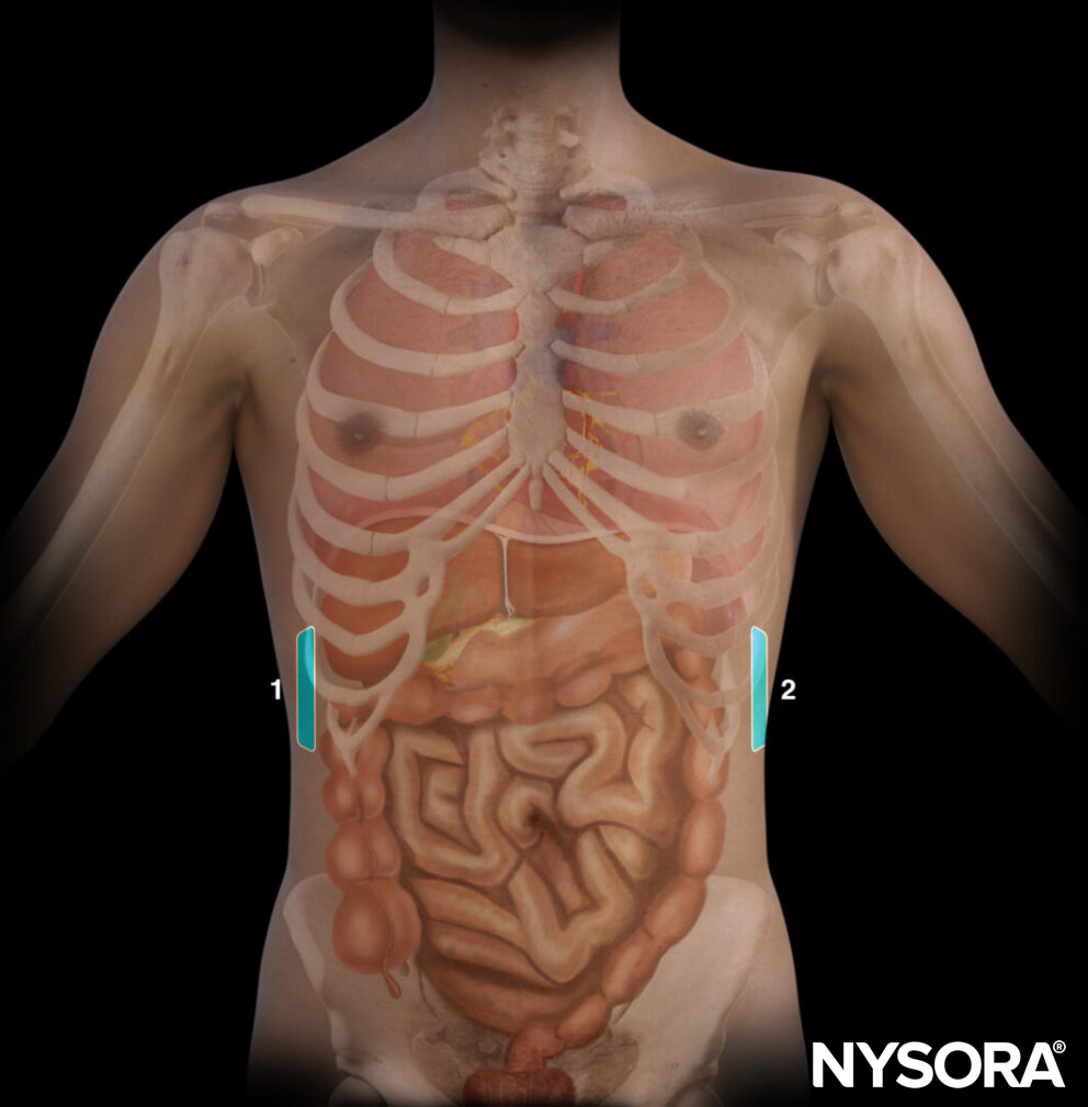
Different areas for transducer positioning for a renal ultrasound: 1, RUQ; 2, LUQ.
Tips
- Respiratory variation might compromise ultrasound imaging during inspiration or expiration
- Small rotational movements of the probe can help to find an acoustic window between the ribs.
Scanning
Right kidney (RUQ)
Position the transducer on the midaxillary line in the RUQ. The kidney can be found adjacent to and inferior to the liver.
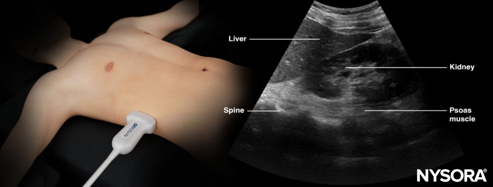
Transducer position and ultrasound anatomy of the right kidney.
Tips
- For difficult ultrasound imaging, try to scan more posteriorly or ask the patient to inhale.
- Try to scan the kidneys dynamically by fanning or tilting to get a full understanding of the kidney.
Left kidney (LUQ)
Position the transducer on the mid/posterior axillary line in the LUQ. The kidney can be found adjacent to and inferior to the spleen.
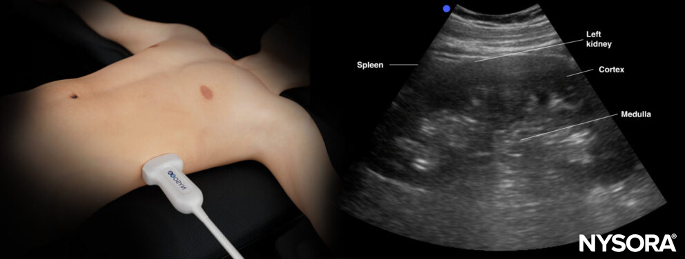
Transducer position and ultrasound anatomy of the right kidney.
Bladder ultrasound
Refer to the bladder ultrasound chapter.
Interpretation
In the absence of obstruction, the central sinus fatty tissue looks homogenous and collapsed with dark, small pockets of urine within the sinus.

Sonoanatomy of a normal kidney.
In the presence of hydronephrosis, a central collection of hypoechoic fluid or urine will be present in the center of the kidney. The size of the collection will determine the degree of hydronephrosis.
Degrees of hydronephrosis:
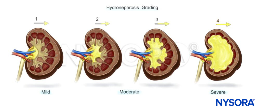
Overview of the degrees of hydronephrosis.
The degrees of hydronephrosis are based on anatomical landmarks
- Mild: enlargement of calyces with preservation of papilla (and renal anatomy).
- Moderate: rounding of calyces with obliteration of papilla and blunting of the pyramids (bear claw).
- Severe: calyceal ballooning with cortical filling and grossly dilated renal pelvis.
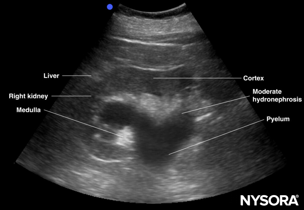
Sonoanatomy of a kidney with moderate hydronephrosis.
Tips
- The ureter is usually obscured by bowel gas
- Cortical cysts (connection pelvis), polycystic kidney disease (bilateral), extrarenal pelvis or parapelvic cysts (round shape & well-defined capsule), vascular malformation, and renal masses can interfere with the diagnosis of hydronephrosis.
- Acoustic shadows in the kidney pelvis may be due to kidney stones. These shadows can be visualized with color Doppler as a comet tail artifact, also called the ‘twinkling artifact’.
- Hydronephrosis always requires a bladder ultrasound
- Other, more specific hydronephrosis grading systems exist, but they are considered more advanced.
Clinical updates
Moses and Fernandez (POCUS Journal, 2022) review the expanding role of kidney POCUS in AKI, emphasizing that cortical thickness (normal 8–10 mm) correlates more closely with eGFR than kidney length and that combined findings of decreased length plus increased echogenicity are specific for advanced CKD, while early ATN shows increased corticomedullary differentiation with renal enlargement. They highlight integration of vascular Doppler, including resistive index (>0.7 associated with persistent AKI and mortality) and the VExUS grading system, to distinguish pre-renal azotemia from parenchymal injury and venous congestion, proposing a stepwise ultrasound algorithm (Figure 5) to systematically evaluate pre-renal, intrinsic, and post-renal causes at the bedside.
- Moses AA, Fernandez HE. Ultrasonography in Acute Kidney Injury. POCUS J. 2022;7(Kidney):35-44.