Nerve Blocks App
 Pain Medicine Assistant App
 POCUS App
 IV Access App
 MSK Knee App
 VetRA App
 Nerve Block Manual
 Regional Anesthesia Updates
 Anesthesiology Manual
 Anesthesiology Review
 Anesthesia Updates 2025
 Anesthesia Updates 2026
 Pediatric Anesthesia Updates
 Airway Management Updates
 US Interventional Pain Manual
 Pain Medicine Updates
 Mastering Difficult IV Access
 PACU Nursing Manual
 RA Veterinary Manual
 About
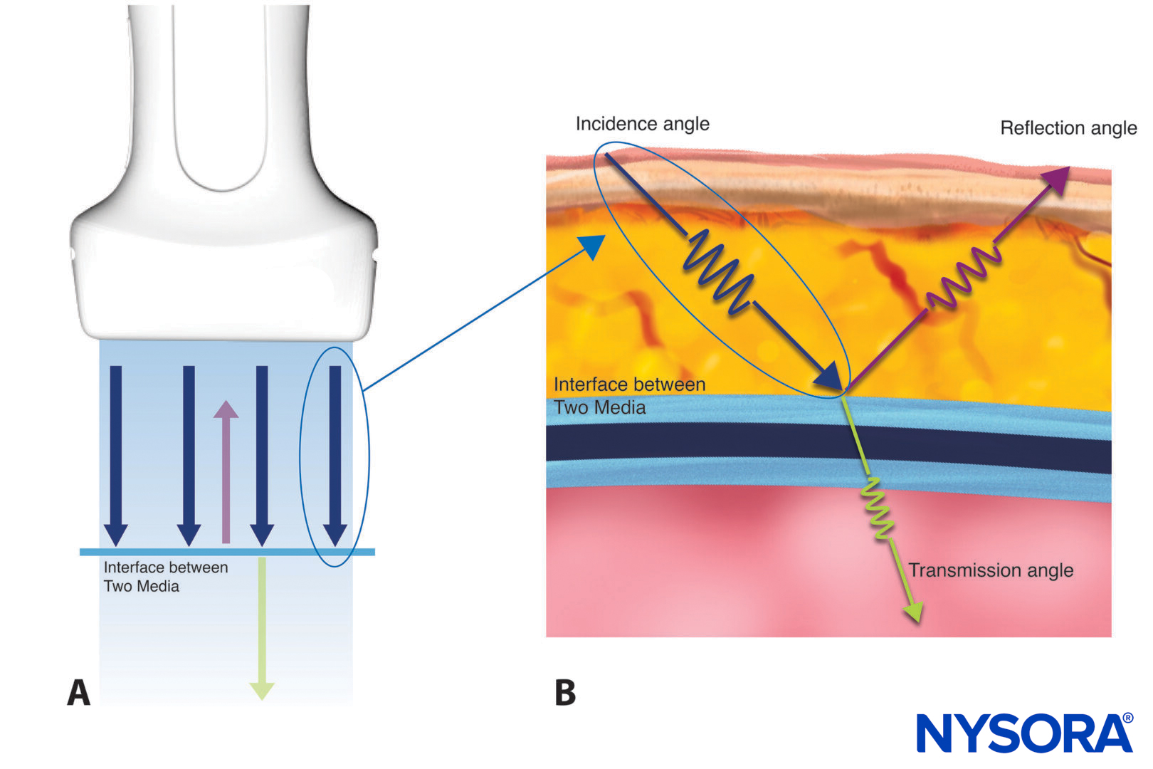
AboutImaging artifacts will arise due to false interpretations by ultrasound machines. It is important to understand that image artifacts can aid or impede ultrasound imaging interpretation. POCUS often relies on the understanding of artifacts to obtain qualitative information on the structure of interest.
Types of artifacts:
- Acoustic shadowing
- Refraction
- Reverberation
- Enhancement
- Mirror imaging
Acoustic shadowing
Acoustic shadowing is caused by a structure that is more or less dense than soft tissue (e.g., bone or lung). It will result in the loss of the ultrasound signal due to scattering and reflection of the ultrasound beam.
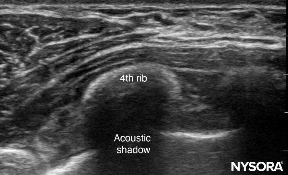
Acoustic shadowing.
Refraction
Refraction is the appearance of the sound wave being bent when the ultrasound beam obliquely crosses an interface with tissues with different densities and, thus, different propagation speeds. It may result in an acoustic shadow at the interface boundary or a duplication artifact.
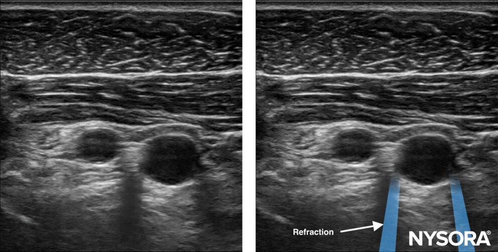
Refraction artifacts occur at the edges of vessels.
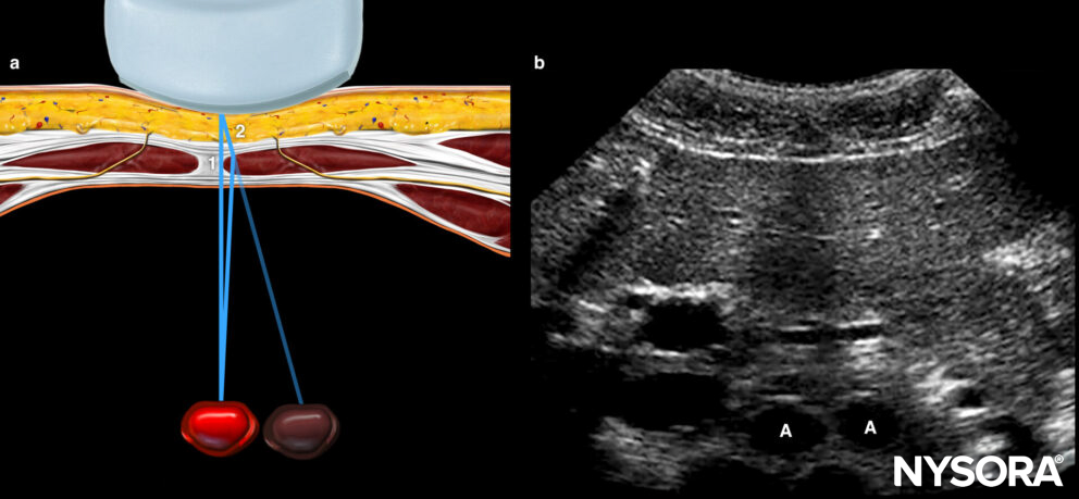
Illustration (a) shows how sound beam refraction results in a duplication artifact. (b) is a transverse midline view of the upper abdomen showing duplication of the aorta (A) secondary to rectus muscle refraction. The ultrasound figure was published in Atlas of Ultrasound-guided Procedures in Interventional Pain Management, Copyright Elsevier (2004).
Reverberation
Reverberation is the repetitive reflection of the sound wave between two reflective layers. It will result in horizontal lines with the same interspace.
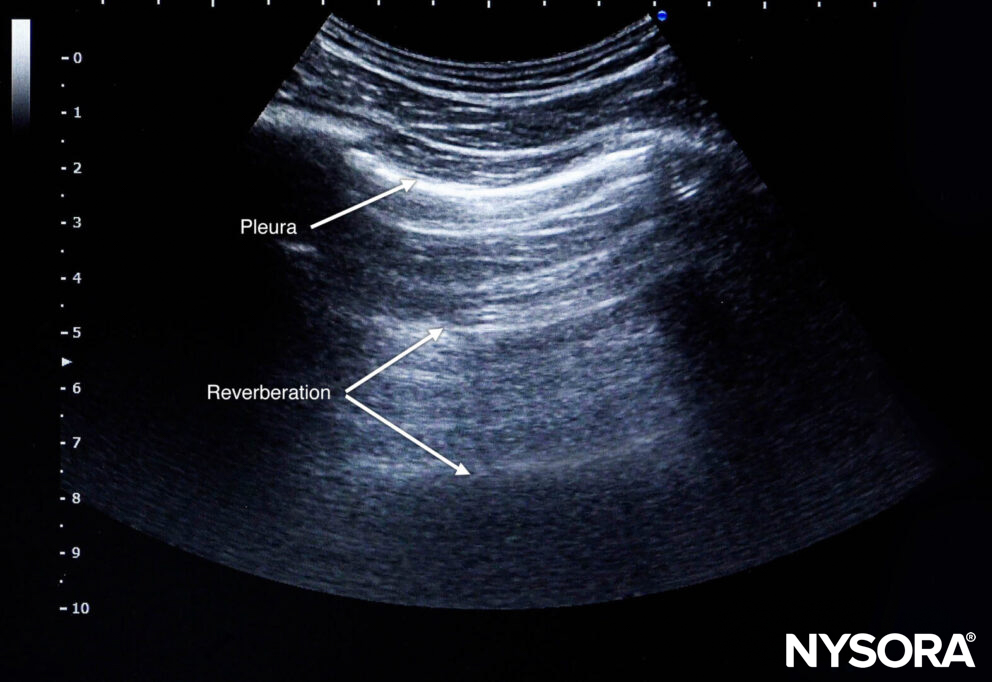
Reverberation.
Enhancement
Sound waves travel faster in structures with low acoustic impedance, resulting in brighter areas directly below these structures (e.g., bladder, vessels). Enhancement can make it harder to visualize underlying structures.
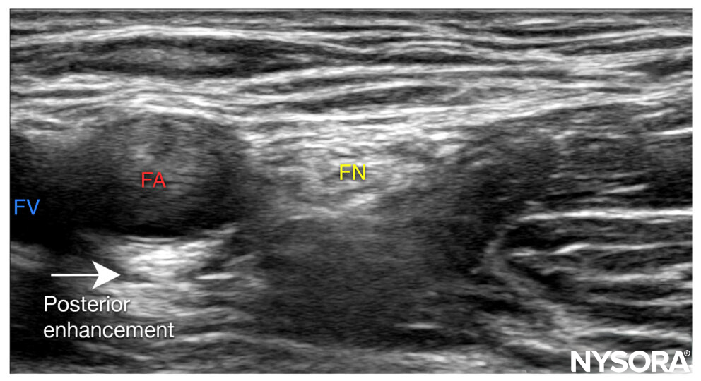
Posterior enhancement.
Mirror imaging
Reflective layers can result in ‘ghost’ images by the reflection of ultrasound waves from different directions. Mirrored structures can be found on the ultrasound image but not in the original structure.
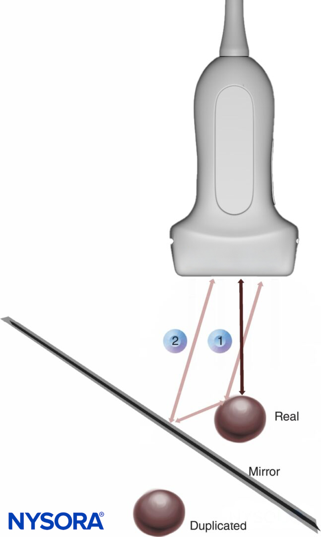
Mirror image: 1. Real or direct reflection of the ultrasound wave, 2. False or indirect reflection of the ultrasound wave.
Interesting fact
- By moving the transducer, the mirrored structure will disappear, but the original structure will remain.
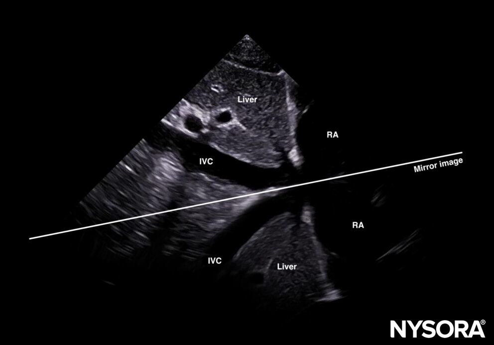
Mirror imaging of the inferior caval vein (IVC), liver, and right atrium (RA).