Learning objectives
- Identify patients at risk for cardiac contusion
- Diagnose cardiac contusion
- Manage patients with (suspected) cardiac contusion
Definition & mechanisms
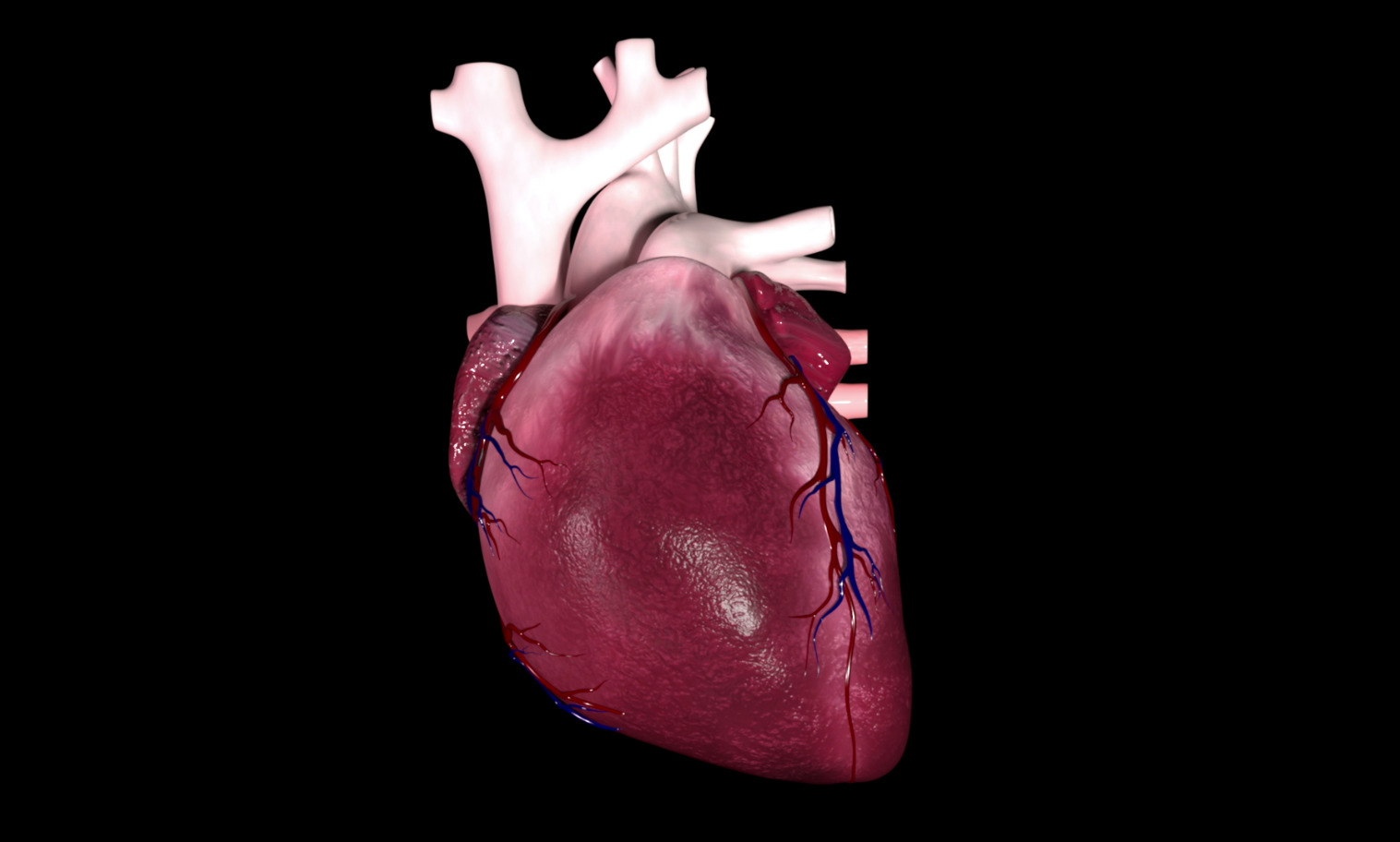
- Bruising or (microscopically small) hemorrhaging of the heart muscle
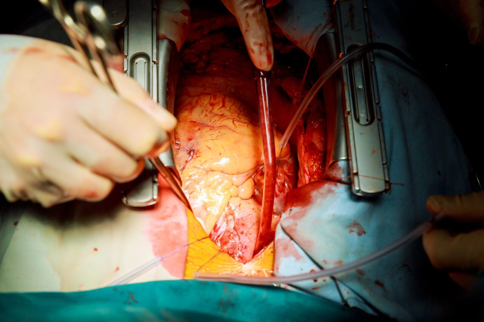
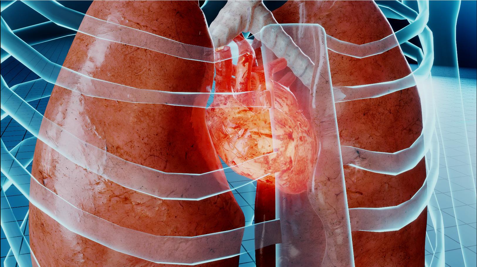
- Generally caused by blunt thoracic trauma: A decelerating force on the anterior side of the thorax
Signs and symptoms
- Mechanical injuries (e.g., rupture of atria or chordae)
- Arrhythmias (premature ventricular complexes, atrial fibrillation, ventricular fibrillation), most commonly within 24 h after trauma
- In the emergency department, many patients do not show symptoms of cardiac contusion after trauma, but severe arrhythmia or even cardiac arrest can occur within 72 h
- Patients with hemodynamic changes but without clear bleeding or cardiac tamponade are suspected of cardiac contusion
Diagnosis
Diagnosis for cardiac contusion remains controversial, diagnostic tools include:
- ECG
- Echocardiography
- Measurement of cardiac biomarkers (troponin T, troponin I, CK-MB)
Management
- Determine the extent of trauma (ECG, ultrasound, cardiac biomarkers)
- Assess cardiac function and intracardiac volume
- ICU admission for monitoring
- Cardiogenic shock management:
- Invasive angiography
- Revascularization
- Inotropes/vasopressors
- Fluid resuscitation
- Ventilation
- Mechanical support
Suggested reading
- Van Lieshout EMM, Verhofstad MHJ, Van Silfhout DJT, Dubois EA. Diagnostic approach for myocardial contusion: a retrospective evaluation of patient data and review of the literature. Eur J Trauma Emerg Surg. 2021;47(4):1259-1272.
- Thiele H, Ohman EM, Desch S, Eitel I, de Waha S. Management of cardiogenic shock. European Heart Journal. 2015;36(20):1223-30.