Back to News

 1 min read



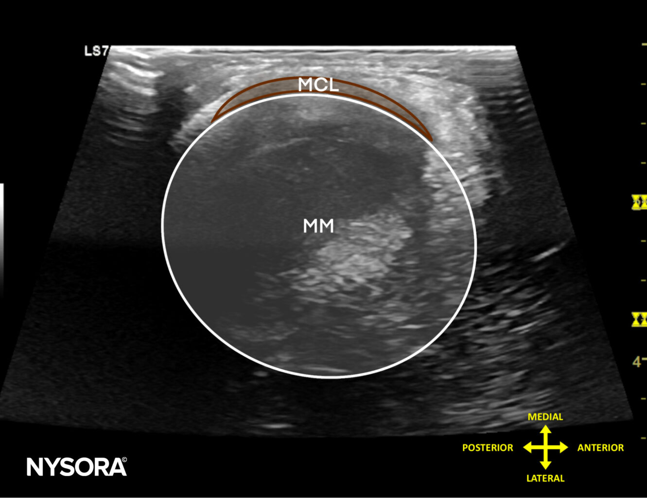
An excessively common sight of injury in the knee is the medial meniscus. The medial meniscus (internal semilunar fibrocartilage) is a fibrocartilage semi-circular band that spans the knee joint medially, located between the medial condyle of the femur and the medial condyle of the tibia. We’re going to dive into three pro tips for scanning the medial meniscus properly.
During scanning, the recognizable points of interest are:
- The clefts in the medial meniscus.
- Extrusion of the rim of the medial meniscus.
- Hyperechoic debris in the center of the medial meniscus, which indicates mucoid degeneration of the medial meniscus.
Here are our 3 tips for scanning the medial meniscus (transverse scan)
- Place the patient in a supine position with the knee flexed 90°.
- Position the transducer transverse over the medial joint line.
- Identify the medial meniscus and medial collateral ligament superficial to the meniscus.
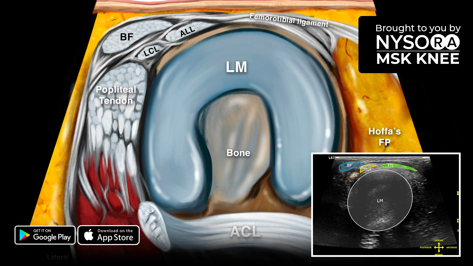
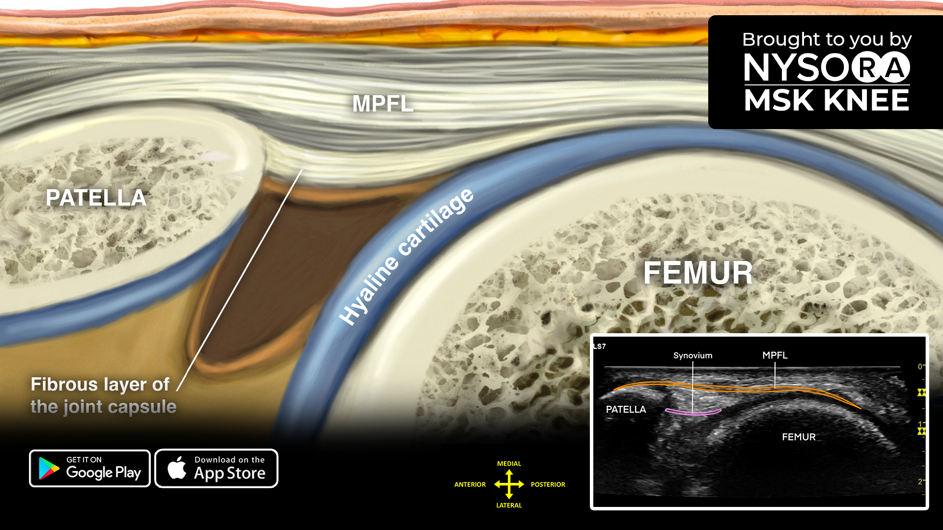
Sonoanatomy
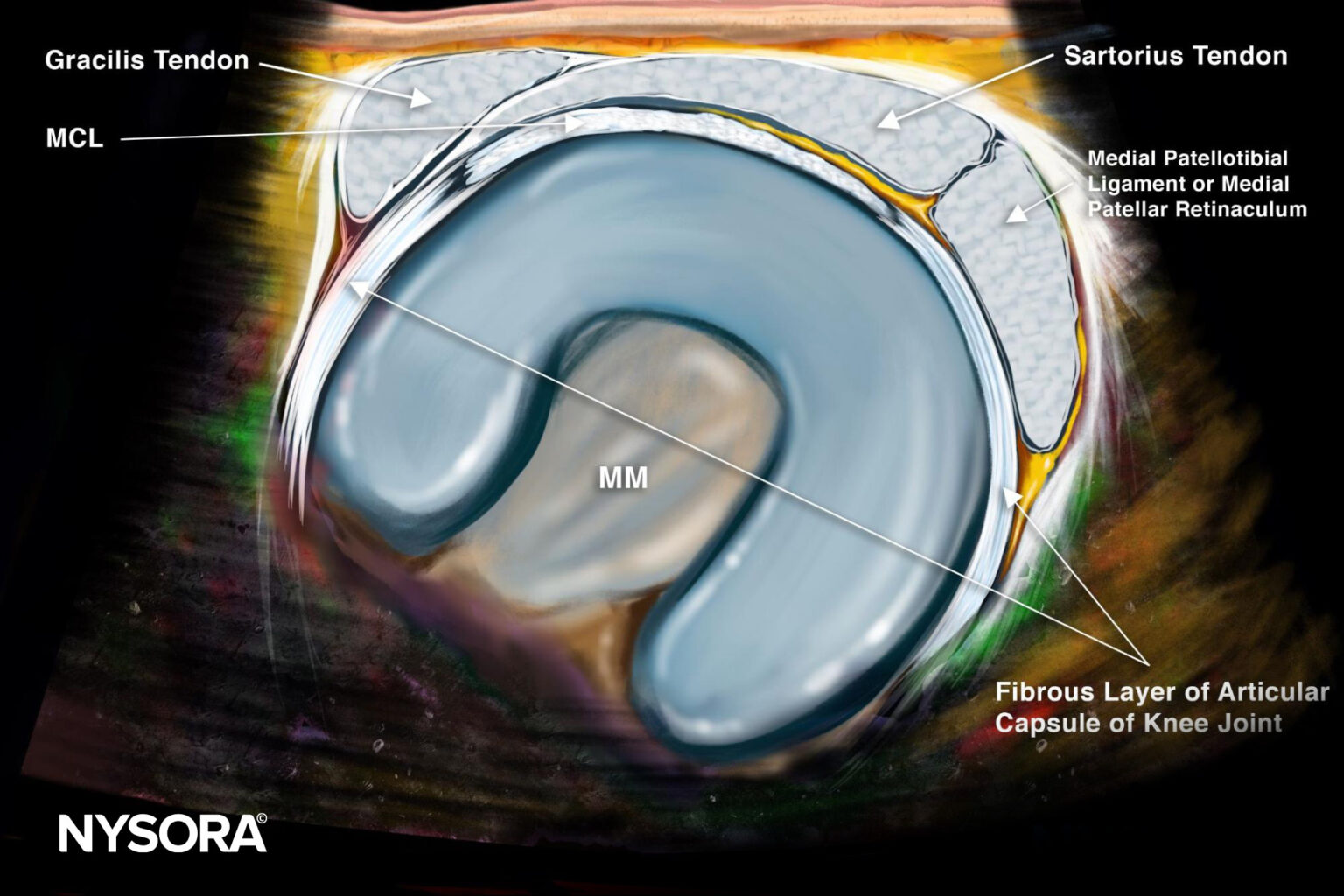
Reverse Ultrasound Anatomy
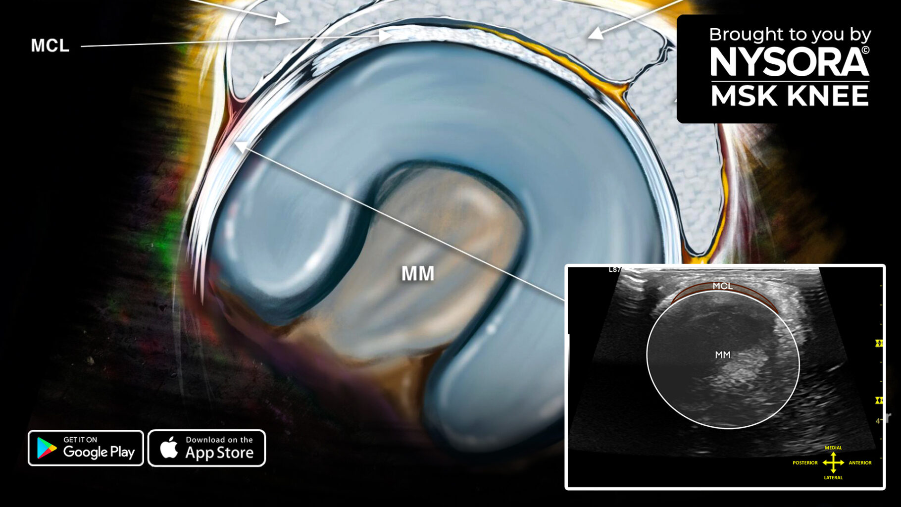
Comparison of sonoanatomy and reverse ultrasound anatomy of the medial meniscus (transverse scan).
Download the MSK App for more tips and the most practical and applicable techniques in musculoskeletal ultrasound anatomy and regenerative therapy of the knee.