Nerve Blocks App
 Pain Medicine Assistant App
 POCUS App
 IV Access App
 MSK Knee App
 VetRA App
 Nerve Block Manual
 Regional Anesthesia Updates
 Anesthesiology Manual
 Anesthesiology Review
 Anesthesia Updates 2025
 Anesthesia Updates 2026
 Pediatric Anesthesia Updates
 Airway Management Updates
 US Interventional Pain Manual
 Pain Medicine Updates
 Mastering Difficult IV Access
 PACU Nursing Manual
 RA Veterinary Manual
 About
About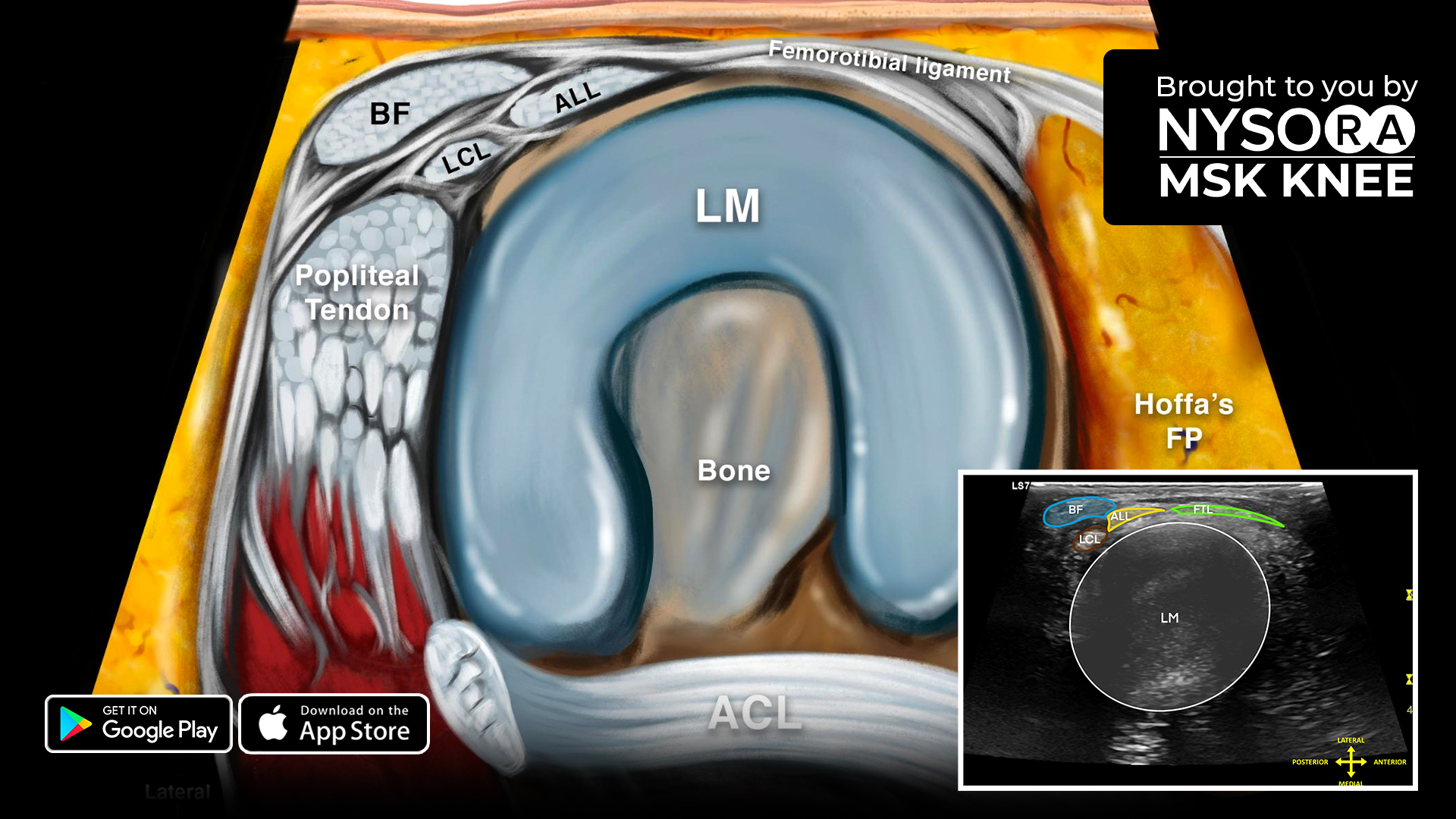


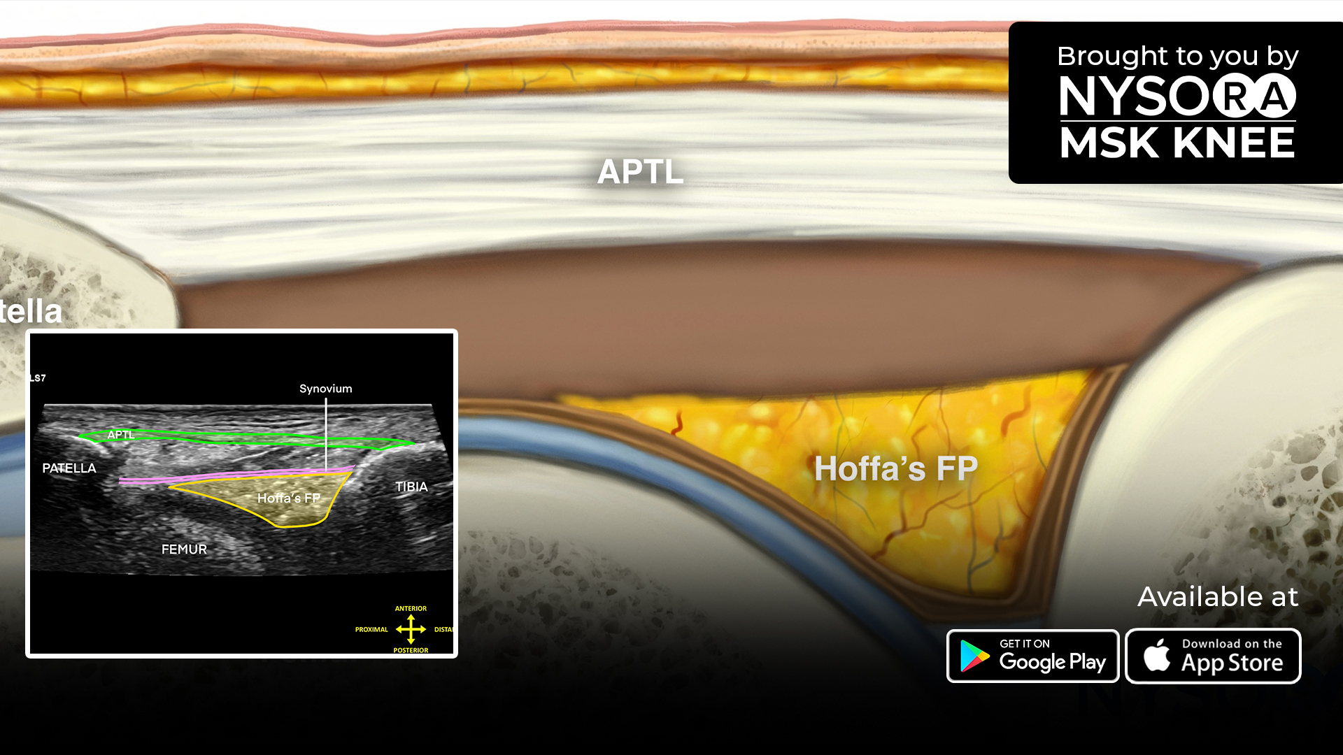
When scanning the lateral meniscus, look for any protrusion during vulgar stress, clefts, or disruptions between the lateral meniscus and tibia, and significant increases i hyperechogenicity over the center and white zone of the lateral meniscus.
Here are 4 easy-to-apply tips for scanning the lateral meniscus (transverse scan):
- Position the patient supine with the knee flexed 90°
- Place the transducer across the lateral femorotibial joint line
- Identify the lateral meniscus in the middle of the ultrasound image
- The other visualized structures are the: Biceps femoris, lateral collateral ligament, anterolateral ligament, and femorotibial ligament
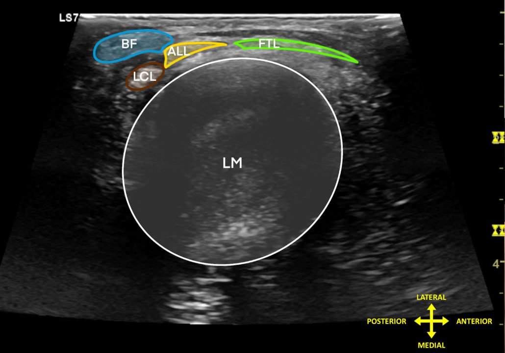
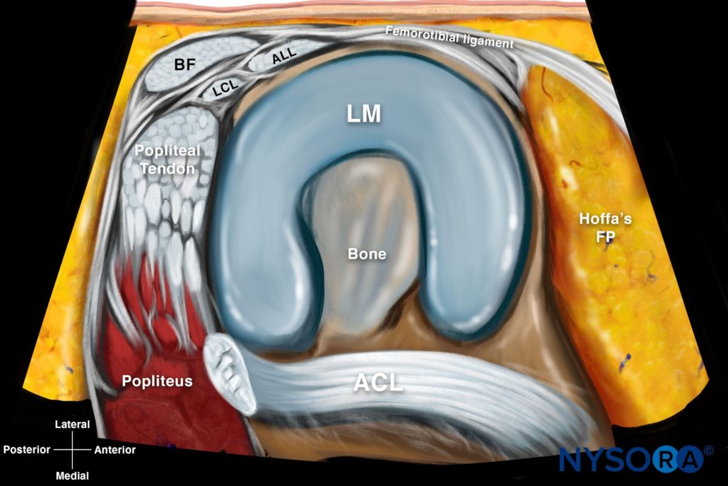
Comparison of sonoanatomy and reverse ultrasound anatomy of the lateral meniscus (transverse scan).
Check out the all-new reverse ultrasound anatomy illustration and slider image added in “Sonoanatomy of the Lateral Knee > Lateral Meniscus”. Download the MSK App for more tips and the most practical and applicable techniques in musculoskeletal ultrasound anatomy and regenerative therapy of the knee.