Nerve Blocks App
 Pain Medicine Assistant App
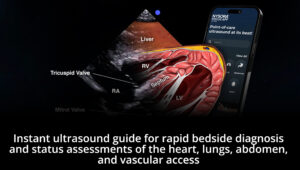
 POCUS App
 IV Access App
 MSK Knee App
 VetRA App
 Nerve Block Manual
 Regional Anesthesia Updates
 Anesthesiology Manual
 Anesthesiology Review
 Anesthesia Updates 2025
 Anesthesia Updates 2026
 Pediatric Anesthesia Updates
 Airway Management Updates
 US Interventional Pain Manual
 Pain Medicine Updates
 Mastering Difficult IV Access
 PACU Nursing Manual
 RA Veterinary Manual
 About
About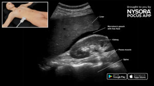
The exam for free intraperitoneal fluid is a “rule-in” exam and is specifically focused on the detection of intra-abdominal fluid.
Abdominal free fluid is often present with:
- Hepatic, renal, or cardiac failure (ascites)
- Inflammation or abdominal sepsis
- Hemorrhage
- Malignancy
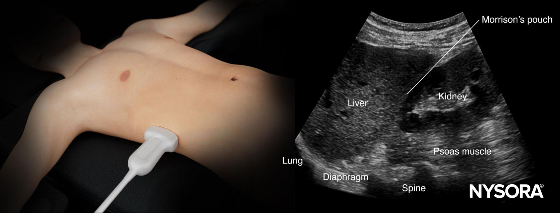
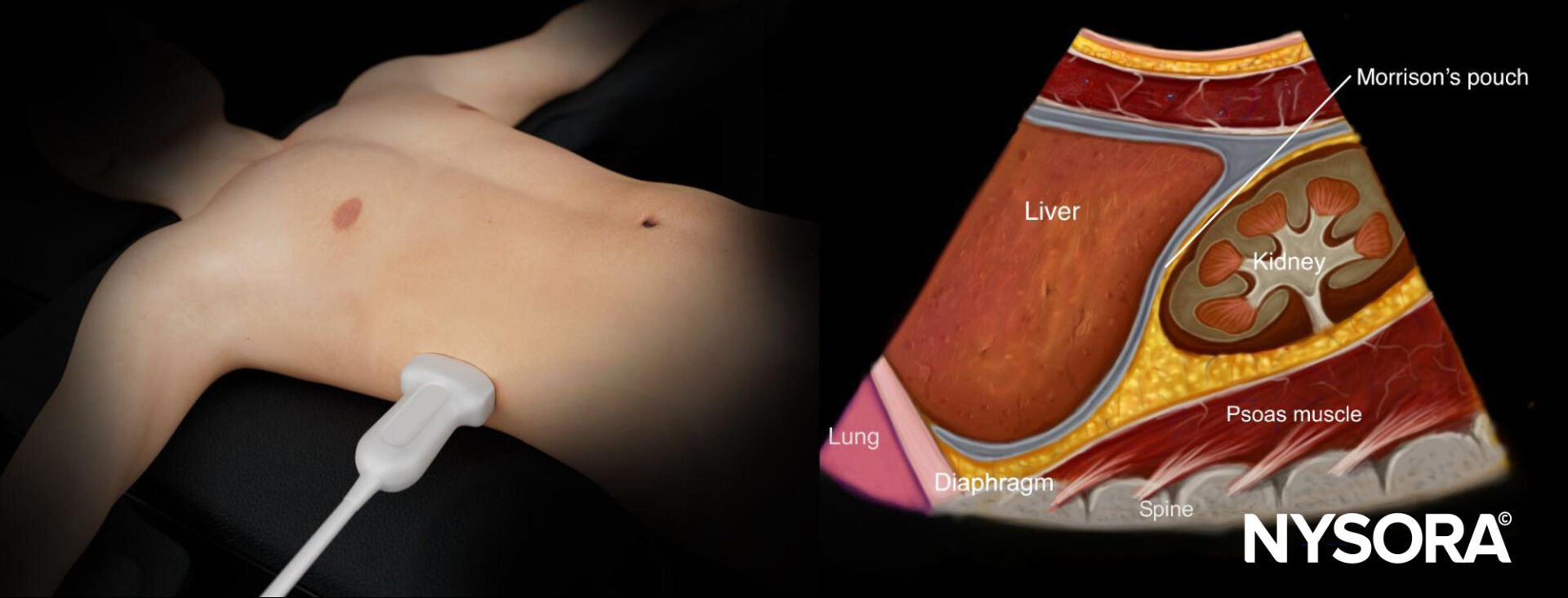
Assess this view to detect free fluid in the right thorax and abdomen.
- Position the transducer between the mid- and posterior axillary line at the level of the xiphoid process with the orientation marker toward the head of the patient.
- Scan caudally until you visualize the liver and kidney.
- Structures of interest: Lung, diaphragm, liver, kidney, Morrison’s pouch (virtual space between liver and kidney).
Normal sonoanatomy
Ultrasound anatomy of the right upper quadrant and relevant anatomical structures
Reverse Ultrasound Anatomy of the right upper quadrant and relevant anatomical structures
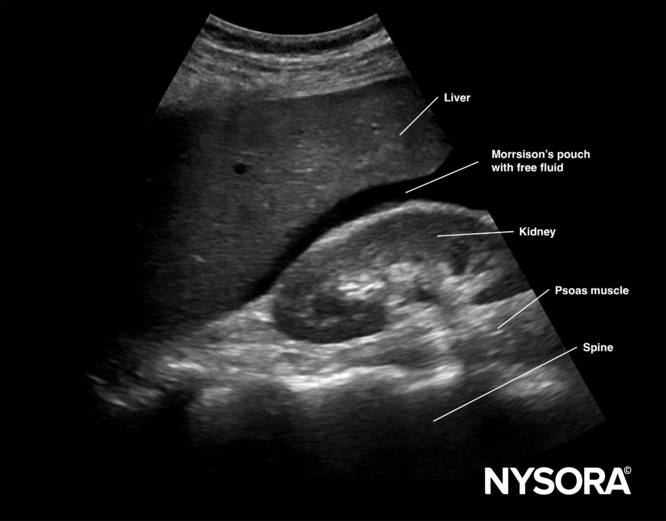
Free intraperitoneal fluid
Free right intraperitoneal fluid collects between the liver and kidney (Morrison’s pouch). If the intrathoracic free fluid is present, it can be identified above the diaphragm.
Unleash the potential of POCUS with NYSORA’s POCUS App and elevate your practice, expand your capabilities, and deliver exceptional patient care. Download HERE.