
Explore NYSORA knowledge base for free:
Explore NYSORA knowledge base for free:
Rapid bedside diagnosis app for the heart, lungs, abdomen, vascular access, and more. Master your emergency diagnostics skills on the go!


Vascular, lung, abdominal, and more.
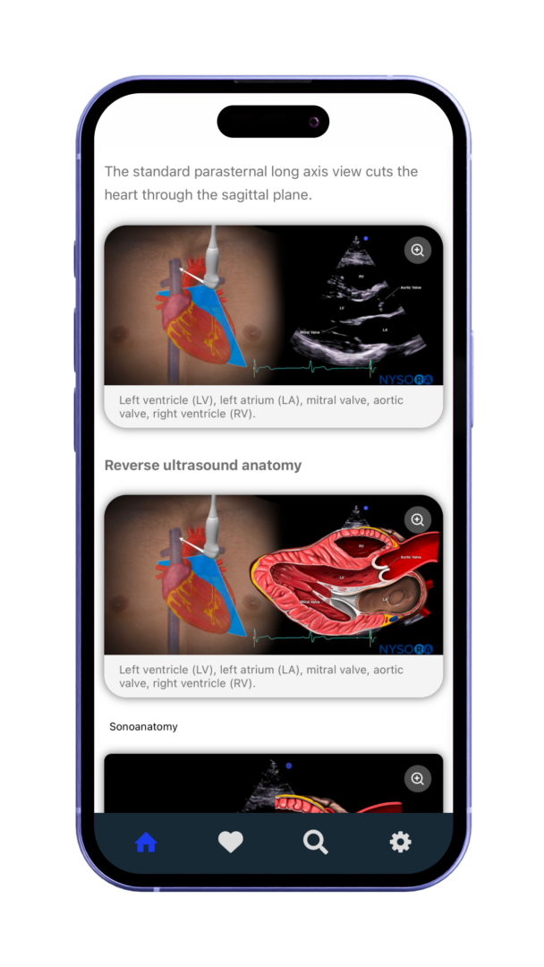
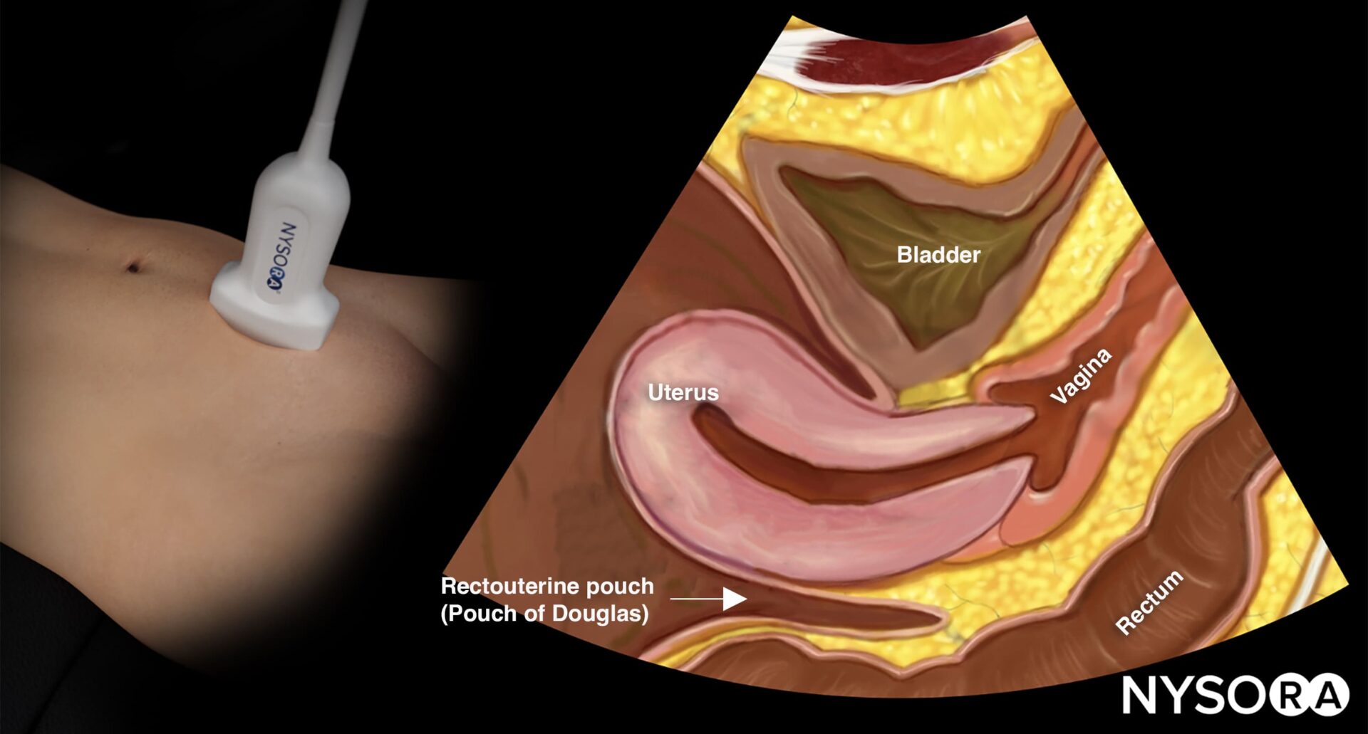
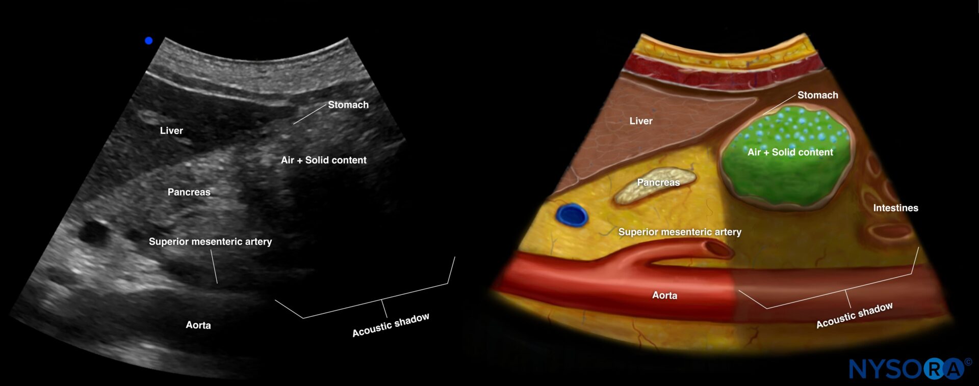
Reverse Ultrasound Anatomy illustrations make sonoanatomy easier to understand and apply.
Optimized for fast, easy access on mobile and tablet devices in clinical practice.
Vascular, lung, abdominal, and more.
Reverse Ultrasound Anatomy illustrations make sonoanatomy easier to understand and apply.
Optimized for fast, easy access on mobile and tablet devices in clinical practice.
The best mobile guide for point-of-care-ultrasound. Trusted by physicians worldwide.


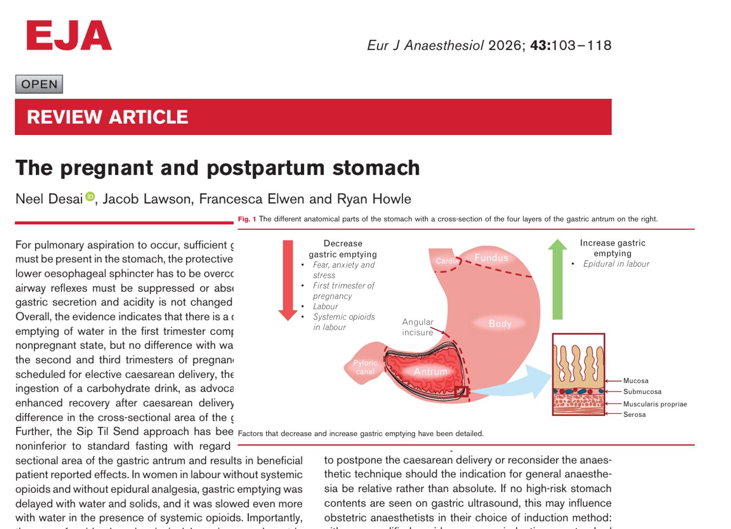
Introduction Pulmonary aspiration remains one of the most feared complications in obstetric anesthesia. Although rare, it carries significant maternal and neonatal morbidity. A comprehensive 2026 review published in the European Journal of Anaesthesiology provides updated insights into gastric physiology during pregnancy, labor, and the postpartum period, along with the evolving role of gastric ultrasound. Anatomy and physiology of the stomach The stomach is a distensible muscular organ divided into four main regions: Cardia Fundus Body Pylorus Its primary physiological roles include: Temporary storage of food and fluids Mechanical and chemical digestion Regulation of gastric emptying into the duodenum Secretion of intrinsic factor Modulation of appetite and satiety Key physiological insight Basal gastric acid secretion and acidity do not change during pregnancy What causes pulmonary aspiration? For aspiration to occur, three conditions must be met: Presence of sufficient gastric contents Reduced lower oesophageal sphincter (LOS) tone Suppressed airway reflexes Pregnancy-specific changes LOS pressure decreases progressively, reaching a nadir at ~36 weeks Intragastric pressure increases due to the gravid uterus Risk increases significantly under general anesthesia Gastric emptying during pregnancy First trimester Gastric emptying of liquids is delayed Likely influenced by hormonal changes and early pregnancy physiology Second and third trimesters No significant difference in gastric emptying for: Liquids Solids Clinical takeaway Early pregnancy poses a higher risk of delayed gastric emptying than later stages Gastric emptying during labor Labor significantly alters gastric physiology. Without analgesia or opioids Gastric emptying is delayed for both liquids and solids With systemic opioids Further slowing of gastric emptying With epidural analgesia Gastric emptying improves However, it does not return to nonpregnant levels Postpartum gastric physiology Gastric emptying returns to nonpregnant baseline levels No significant differences observed within the first 5 days postpartum Gastric ultrasound: a game-changing tool Gastric ultrasound has become a critical bedside tool […]
We are thrilled to announce the launch of our Airway Assessment course, now available in the POCUS App! This comprehensive course is designed to equip healthcare professionals with the knowledge and skills to perform advanced airway management using ultrasound techniques. Learning objectives You will gain expertise in: Understanding the anatomy and physiology of the upper airway. Identifying key upper airway structures. mastering the scanning techniques to assess the upper airway Why airway ultrasound? Safe and non-invasive: Quick and painless for patients. Highly accurate: Provides real-time assessment of airway structures and conditions. Improved outcomes: Reduces complications in difficult airway scenarios, a leading cause of anesthesia-related mortality. Evidence-based: Confirming ETT placement via ultrasound shows 98% sensitivity and specificity. What you’ll learn This course covers essential topics in airway ultrasound, including: Upper airway anatomy and physiology Gain a solid understanding of the functional anatomy. Identification of upper airway structures with ultrasound Detailed descriptions help you identify structures clearly, for instance: Hyoid bone – recognizable on ultrasound as a linear hyperechoic inverted U-shaped structure with two greater horns (cornua). Thyroid cartilage – ultrasound appearance as a hypoechoic inverted V-shaped or triangular structure. Cricothyroid membrane – appears as a hyperechoic horizontal line. Ultrasound imaging techniques Suprahyoid area Place the transducer longitudinally below the mandible and above the hyoid bone. Ultrasound image shows a fan-shaped tongue with a typical striated appearance. Underneath the mylohyoid and geniohyoid muscles, the tongue appears fan-shaped, with intrinsic muscles providing a striated appearance. Infrahyoid area: Sagittal ‘string of pearls’ technique Start from the trachea, sliding the probe cranially. The tracheal rings appear as dark hypoechoic structures, resembling a pearl necklace. Infrahyoid transverse approach Position the transducer transversely on the midline of the neck. Scan cranially to caudally, visualizing key landmarks: Hyoid bone Thyrohyoid membrane Thyroid cartilage Cricothyroid membrane Cricoid cartilage Trachea […]


The best mobile guide for point-of-care-ultrasound. Trusted by physicians worldwide.
The app includes:
The NYSORA POCUS App provides quick access to expertly-curated content, helping you:
The app covers:
The app can be used in multiple ways:
It’s a mobile-friendly reference tool offering:
Get practical anesthesia insights & focused literature updates you can apply in your next case.