Nerve Blocks App
 Pain Medicine Assistant App
 POCUS App
 IV Access App
 MSK Knee App
 VetRA App
 Nerve Block Manual
 Regional Anesthesia Updates
 Anesthesiology Manual
 Anesthesiology Review
 Anesthesia Updates 2025
 Anesthesia Updates 2026
 Pediatric Anesthesia Updates
 Airway Management Updates
 US Interventional Pain Manual
 Pain Medicine Updates
 Mastering Difficult IV Access
 PACU Nursing Manual
 RA Veterinary Manual
 About
About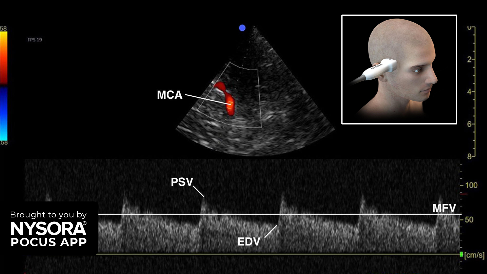
Transcranial Doppler (TCD) ultrasound is a non-invasive tool utilized in point-of-care ultrasound (POCUS) for assessing cerebral blood flow dynamics. This case study explores the application of TCD in detecting intracranial hypertension in a clinical setting.
Case presentation:
- A 45-year-old male presented to the emergency department with severe headache, nausea, and blurred vision.
- Medical history included hypertension and a recent head trauma from a minor car accident.
Physical Examination:
- The patient was conscious but exhibited signs of increased intracranial pressure (ICP), such as papilledema and bradycardia.
- Initial neurological assessment showed no focal deficits.
Clinical Decision:
- Given the suspicion of intracranial hypertension, a TCD ultrasound was performed at the bedside to quickly assess cerebral blood flow dynamics and evaluate for raised ICP.
Indications for TCD
- Intracranial hypertension
- Suspected diagnosis of cerebral circulatory arrest
- Vasospasm detection
- Identification of midline shift
Essential Information on TCD
- TCD offers real-time information and can be performed at the bedside.
- It is not a replacement for CT scans but provides trending capabilities and immediate data.
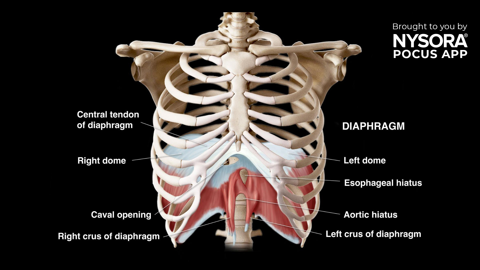
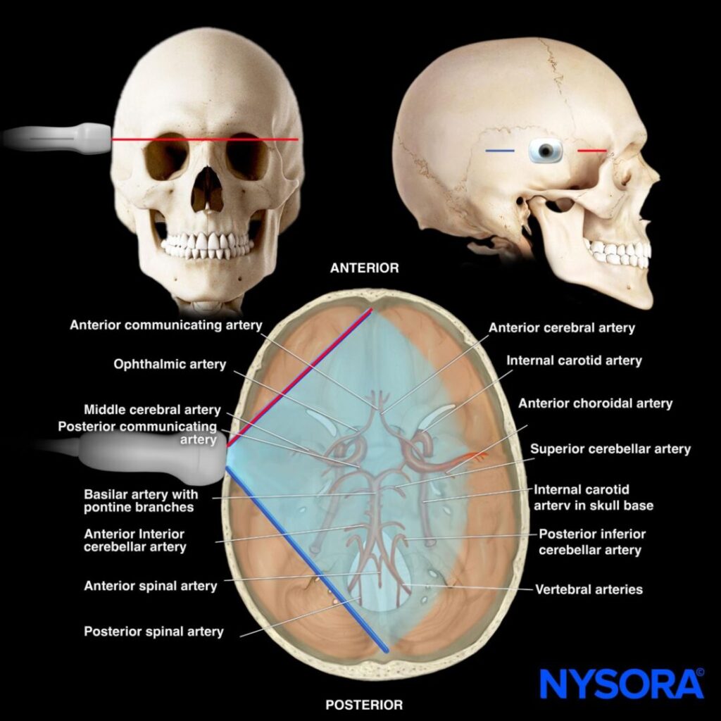
Functional Anatomy and Machine Setup
Anatomy:
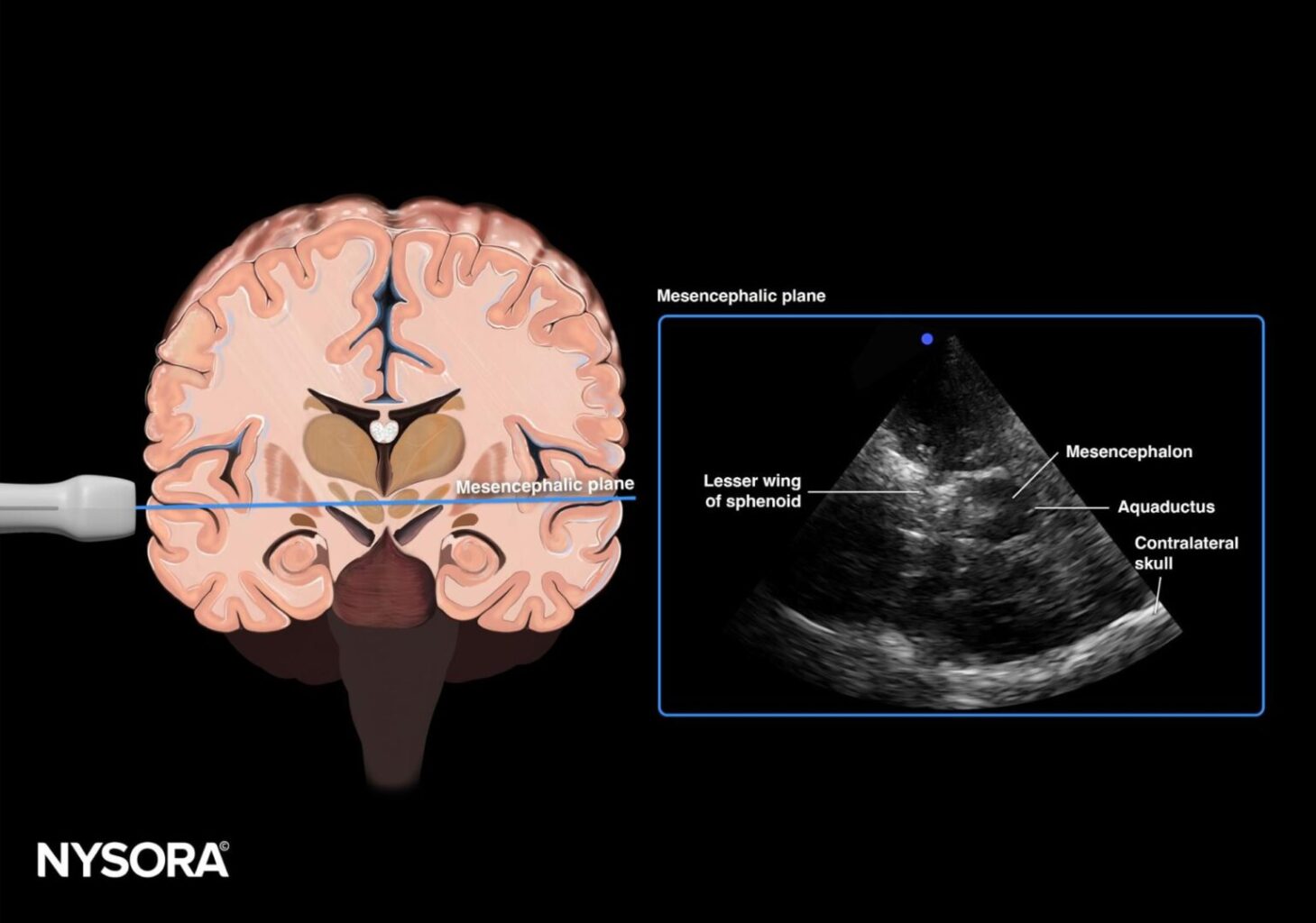
- Key structures include the circle of Willis and intracranial arteries.
- The mesencephalic plane is critical for vascular assessment.
Machine Setup:
- Transducer: Phased array
- Preset: Transcranial (or cardiac)
- Orientation: Index marker toward the frontal bone/orbital
- Depth: 15 cm
Patient Positioning:
- Patient positioned supine with the head of the bed elevated to 30 degrees.
- Landmarks include the ear and temporomandibular joint.
- Transducer placed 2-3 cm above the temporomandibular joint at the level of the temporal bone.
Scanning Plane:
- Mesencephalic plane: Visualizes the middle cerebral artery (MCA) with red flow toward the transducer. Use pulsed wave Doppler to measure cerebral blood flow velocities.
Assessment Using TCD
- Pulsatility Index (PI): Calculated using the formula:
PI = (PSV – EDV)/ MFV
Where PSV is peak systolic velocity, EDV is end diastolic velocity, and MFV is mean flow velocity.
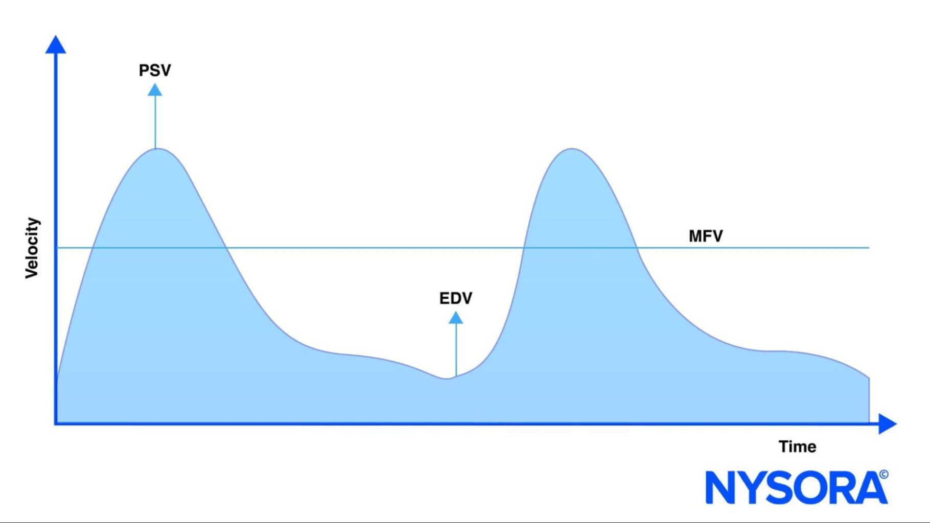
- A normal PI ranges from 0.5 to 1. A PI > 1.2 indicates increased ICP. The estimated ICP approximates PI x 10.
Findings:
- The patient’s PI was 1.4, suggesting raised ICP.
- Immediate intervention with measures to reduce ICP was initiated.
Conclusion
Transcranial Doppler is a valuable, non-invasive technique for assessing intracranial hypertension. By understanding cerebral anatomy and utilizing proper ultrasound techniques, healthcare providers can make rapid and accurate diagnoses, improving patient outcomes.
For more in-depth information on TCD and advanced applications, consider downloading NYSORA’s POCUS App for detailed resources, illustration, animations, and more.