Nerve Blocks App
 Pain Medicine Assistant App
 POCUS App
 IV Access App
 MSK Knee App
 VetRA App
 Nerve Block Manual
 Regional Anesthesia Updates
 Anesthesiology Manual
 Anesthesiology Review
 Anesthesia Updates 2025
 Anesthesia Updates 2026
 Pediatric Anesthesia Updates
 Airway Management Updates
 US Interventional Pain Manual
 Pain Medicine Updates
 Mastering Difficult IV Access
 PACU Nursing Manual
 RA Veterinary Manual
 About
About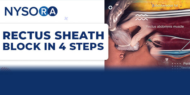

Postoperative pain management in patients having open abdominal surgery through a midline incision must include an interventional analgesia technique. For midline laparotomy, there are many choices, ranging from thoracic epidural analgesia, to TAP, quadratus lumborum variety of blocks, ESP, paravertebral blocks, among others. However, nothing beats the simplicity and efficacy of the rectus sheath block. Therefore, we dug into NYSORA’s Compendium of Regional Anesthesia to outline 4 steps to accomplish this very effective block.
A bilateral rectus sheath block provides analgesia to the anteromedial abdominal wall and periumbilical area (spinal dermatomes T9, T10, and T11). The technique blocks the anterior cutaneous branches of the intercostal nerves, and therefore, it is well suited for postoperative analgesia for midline abdominal incisions. First described in 1899 by Schleich, initially, it was performed as a blind, loss-of-resistance technique. Before the introduction of ultrasound, the block was not used often due to the concerns of accidentally puncturing vascular structures in the way of the needle path. However, the introduction of ultrasound improved the identification of the rectus abdominis muscle, layers of the rectus sheath, and important vascular structures to avoid injury, thereby increasing the technique’s popularity.
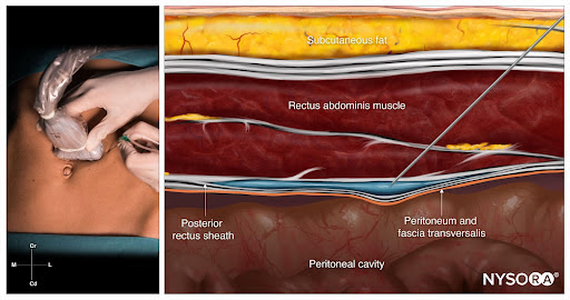
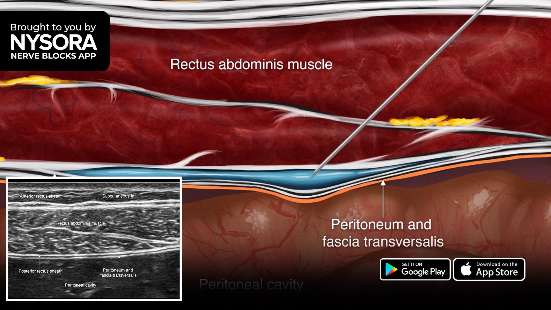
Rectus sheath block; Reverse Ultrasound Anatomy with needle insertion in-plane and local anesthetic spread (blue) between the rectus abdominis muscle and posterior rectus sheath.
Here are 4 steps to successfully perform a rectus sheath block:
- Place the transducer in a transverse orientation above the umbilicus, 1 cm lateral to the midline.
- Identify the rectus abdominis muscle and posterior rectus sheath.
- Insert the needle in-plane through the rectus abdominis muscle until the tip reaches the space between the muscle and posterior rectus sheath, and inject 10-15 mL of local anesthetic (e.g., 0.5-0.375% ropivacaine).
- Repeat on the contralateral side. This block should be performed bilaterally
The quickest way to learn the sonoanatomy required for effective Rectus Sheath Block is to watch the NYSORA’s Reverse Ultrasound Anatomy© Animation that takes the viewer from ultrasound image to illustration and back.
To gain access to the complete reference guide to practical Regional Anesthesia make sure to subscribe to NYSORA’s Compendium of Regional Anesthesia. The COMPENDIUM is the most comprehensive and practical curriculum on Regional Anesthesia from A to Z, featuring NYSORA’s Premium content. The Compendium is continuously updated with NYSORA’s newest videos, animations, and visual content.
Join thousands of your peers on the NYSORA LMS, an interactive platform made so that you never learn alone.
By joining, get FREE access to:
- A bundle of the most popular step-by-step nerve block infographics
- NYSORA’s printable poster collection for your clinic
- An exclusive video addressing the 5 most commonly asked questions in Regional Anesthesia