Nerve Blocks App
 Pain Medicine Assistant App
 POCUS App
 IV Access App
 MSK Knee App
 VetRA App
 Nerve Block Manual
 Regional Anesthesia Updates
 Anesthesiology Manual
 Anesthesiology Review
 Anesthesia Updates 2025
 Anesthesia Updates 2026
 Pediatric Anesthesia Updates
 Airway Management Updates
 US Interventional Pain Manual
 Pain Medicine Updates
 Mastering Difficult IV Access
 PACU Nursing Manual
 RA Veterinary Manual
 About
About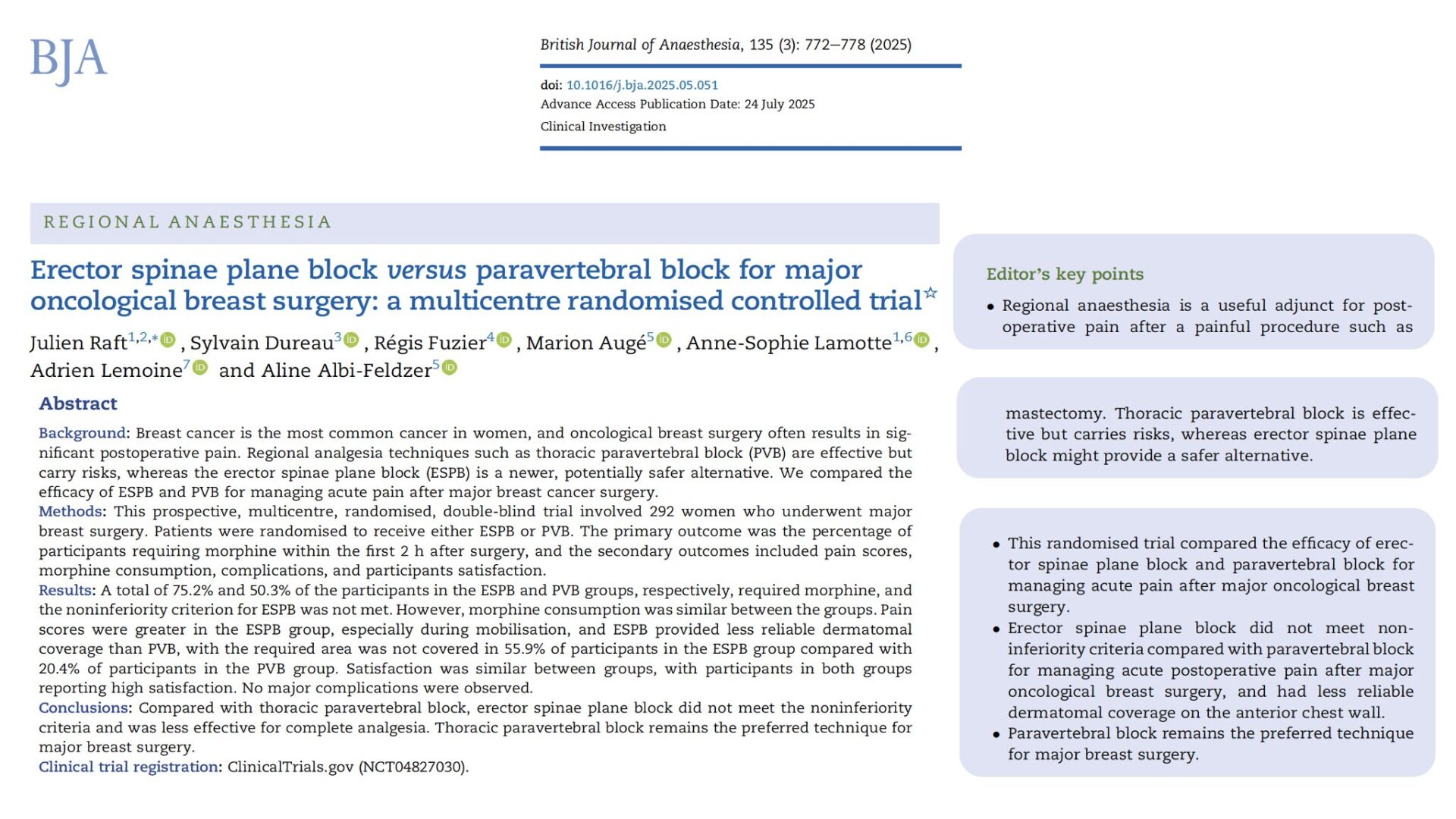
Breast surgery is common, painful, and frequently performed in fast-track pathways where predictable regional analgesia can make or break early recovery. For decades, the thoracic paravertebral block (PVB) has been the reference technique for oncologic breast procedures because it reliably covers ventral rami (T2–T6) and the axilla. But PVB is performed near pleura and neuraxis, raising real—if uncommon—concerns about pneumothorax or neuraxial spread. The erector spinae plane block (ESPB) has surged in popularity as a technically simple, superficial fascial plane alternative with a perceived wider safety margin and easy ultrasound windows. Mechanistically, ESPB consistently captures dorsal rami; ventral rami coverage—and therefore the sternal–anterior chest wall and axillary fields—remains variable.
Small single-center trials and meta-analyses have reported mixed results, often underpowered for patient-centered outcomes and complicated by heterogeneous surgical mixes and open-label designs. The question many of us face in daily practice is straightforward: can ESPB stand shoulder-to-shoulder with PVB for major oncologic breast surgery when blinding, allocation, and outcomes are handled rigorously? A new multicenter, randomized, double-blind trial answers that question directly by testing the noninferiority of ESPB versus PVB in women undergoing major breast cancer surgery (primarily mastectomy ± axillary surgery).
Study objective and methods
The primary goal was to determine whether ESPB is noninferior to PVB for acute analgesia after major oncologic breast surgery, using “need for morphine in the first 2 postoperative hours” as the primary readout. Noninferiority margin: 20% absolute difference.
- Study design: Prospective, multicenter, randomized, double-blind, parallel-group controlled trial (five French centers).
- Participants: 292 women (ASA I–III), unilateral major breast procedures (total mastectomy ± axillary dissection or SLNB; selected breast-conserving surgery with axillary dissection). Key exclusions: Recent opioid use, prior ipsilateral surgery, bilateral cases.
- Randomization: 1:1 ESPB vs PVB, stratified by center and surgery type. Blocks were performed by an independent anesthesiologist; sensory testing and outcome assessment were blinded.
Technique specifics
- ESPB: Single-shot at T3 transverse process; 0.5% ropivacaine, 0.6 mL/kg (max 30 mL) between erector spinae muscle and transverse process.
- PVB: Single-shot at T2–T3 interspace via intercostal (costotransverse ligament) approach; same dose/concentration of ropivacaine.
- Hydrodissection confirmed needle position in both groups. Sensory exam (ice) along the nipple line at ~ 15 minutes. No surgical infiltration.
- Standardized general anesthesia with remifentanil/propofol; multimodal non-opioid prophylaxis; PACU protocolized morphine titration for VAS > 3. Post-op paracetamol + ketoprofen; rescue tramadol.
Outcomes
- Primary: % requiring morphine in PACU (0–2 h).
- Secondary: Pain scores (rest/mobilization) 0–4 h and at 24 h; total PACU morphine; dermatomal coverage (T2–T6 and axilla); PONV; QoR-15 at 24 h; adverse events; satisfaction (patient & anesthesiologist). Analyses by intention-to-treat; pre-specified noninferiority; exploratory superiority post hoc.
Key findings
-
Primary endpoint: Noninferiority failed for ESPB
PACU morphine needed in 75.2% (ESPB) vs 50.3% (PVB); absolute difference 24.8%. ESPB did not meet noninferiority; exploratory analysis favored PVB (P < 0.001).
-
Pain scores: PVB modestly better, especially with movement
Mobilization VAS was higher with ESPB on PACU arrival and at 30 and 60 minutes. Resting pain also trended higher at several time points. Differences were statistically significant but numerically small (≈ 0.6–0.8 VAS units).
-
Morphine amounts similar: Despite more frequent need with ESPB
- Among patients who required opioids, total PACU morphine consumption did not differ (≈ 4.5–4.8 mg over 2 h; P = 0.4).
- Interpretation: PVB reduced the proportion needing opioids; once opioids were needed, the dose was comparable.
-
Coverage reliability: Clear win for PVB
- Complete T2–T6 coverage: 23.8% (PVB) vs 4.1% (ESPB).
- Partial coverage (any T2–T6): 55.8% (PVB) vs 40.0% (ESPB).
- No coverage: 20.4% (PVB) vs 55.9% (ESPB).
- PVB achieved complete or partial coverage in ~ 80% vs ~ 44% for ESPB (P < 0.001). This aligns with the known limitation of ESPB for ventral rami/anterior chest wall.
-
Satisfaction high and similar
Patients and anesthesiologists rated both techniques highly (means ≈ 8.4–8.5/10). The trial teams were experienced, which may narrow perceived ease-of-use differences often touted for ESPB.
-
Safety: No major complications; low signal differences
Overall complications were infrequent and similar. Notably, there was no excess of serious events in either group in this sample. Prior literature worries about pneumothorax/neuraxial spread with PVB remain relevant, but were not seen here.
Conclusion
In a rigorous, multicenter, double-blind head-to-head comparison for major oncologic breast surgery, ESPB did not achieve noninferiority to PVB for early PACU analgesia and produced less reliable dermatomal coverage of the anterior chest wall and axilla. Although absolute pain score differences were small, more ESPB patients needed opioids and coverage failures were common. Within a standardized, non-infiltration regimen, PVB remains the preferred technique when you need consistent, procedure-wide analgesia.
Future research
- Technique optimization for ESPB: Multilevel or bi-level ESPB, volume adjustments, or adjuncts to improve ventral rami reach; imaging/cadaver correlation to define reproducible spread to T2–T6.
- Combination strategies: Hybrid plans (e.g., targeted pectoral/serratus or intercostal adjuncts) to “patch” anterior chest wall gaps after ESPB.
- Patient-centered outcomes beyond pain: Chronic pain at 3–6 months, QoR trajectories, return to baseline activity; subgroup analyses by surgical extent and axillary procedure.
- Safety at scale: Large, registry-based comparisons quantifying pneumothorax/neuraxial events with modern ultrasound PVB and real-world ESPB failure/rescue rates.
Clinical implications
If your goal is predictable, comprehensive coverage for unilateral mastectomy ± axillary surgery without surgical infiltration, the current best-supported single-shot choice is thoracic PVB. Expect fewer PACU opioid triggers and more reliable T2–T6/axillary analgesia than ESPB, with similar patient satisfaction under expert hands. ESPB remains attractive for its superficial approach and perceived safety margin, but its ventral rami reach is inconsistent, leading to higher early opioid needs and more incomplete blocks in this context. In centers or scenarios where PVB risk tolerance, skill mix, or resources argue against routine PVB, ESPB may still be reasonable—with eyes open to frequent rescue requirements or the need to supplement with additional blocks targeting the anterior chest wall or axilla.
Clinical pearls
- ESPB failed noninferiority; more patients needed PACU morphine.
- PVB delivered superior T2–T6/axillary coverage.
- Mobilization VAS favored PVB early; differences were small.
- Total morphine (if required) was similar between groups.
- Satisfaction and safety were comparable under expert hands.
Practical tip: For unilateral mastectomy ± axillary dissection without infiltration, use single-shot PVB at T2–T3 for the most reliable coverage.
For more detailed information, refer to the full article in BJA.
Raft J. et al. Erector spinae plane block versus paravertebral block for major oncological breast surgery: a multicentre randomised controlled trial. Br J Anaesth. 2025;135:772-778.
Master the thoracic PVB and ESPB step-by-step with structured guides in the NYSORA Nerve Blocks App.