Nerve Blocks App
 Pain Medicine Assistant App
 POCUS App
 IV Access App
 MSK Knee App
 VetRA App
 Nerve Block Manual
 Regional Anesthesia Updates
 Anesthesiology Manual
 Anesthesiology Review
 Anesthesia Updates 2025
 Anesthesia Updates 2026
 Pediatric Anesthesia Updates
 Airway Management Updates
 US Interventional Pain Manual
 Pain Medicine Updates
 Mastering Difficult IV Access
 PACU Nursing Manual
 RA Veterinary Manual
 About
About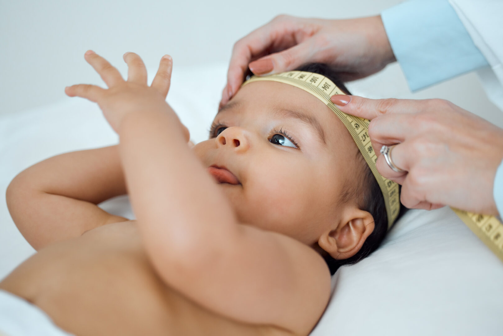
Learning objectives
- Definition and examples of craniofacial dysostosis
- Perioperative management of craniofacial dysostosis
Definition and mechanisms
- Craniofacial dysostosis or craniosynostosis is a condition in which premature fusion of one or more of the cranial sutures occurs, leading to abnormal skull development and head shape
- Abnormal premature fusion of one or several of these sutures results in restricted growth of the skull perpendicular to the affected suture
- Compensatory bone growth occurs parallel to the affected suture in order to allow for continued brain growth and results in distinct clinical skull characteristics
- Children may present with a broad range of conditions requiring correction, from otherwise well children with single suture craniosynostosis (80% of cases) to syndromic children with multiple synostoses with other cranial and extracranial anomalies
- Syndromes most frequently associated with craniosynostosis include:
- Crouzon syndrome:
- The most common craniofacial dysostosis syndrome
- Apert syndrome:
- Is similar to Crouzon syndrome but is more severe
- Craniofacial characteristics of Crouzon syndrome and syndactyly (webbing of finger and toe) in addition
- Intellectual disability is also more common in Apert syndrome
- Pfeiffer syndrome:
- Craniofacial characteristics and hand and feet abnormalities are also present (wide and deviated thumbs or big toes)
- Mental and neurological problems are more likely
- Crouzon syndrome:
- Caused by an inherited or spontaneous autosomal dominant mutation in the fibroblast growth factor receptor 2 (FGFR2) located on chromosome 10
- Incidence is 1 in 2500 live births
- Correction may require extensive surgery that is commonly performed at a young age
Signs and symptoms
- Wide-set eyes (hypertelorism)
- Bulging eyeballs (proptosis)
- Crossed eyes (strabismus)
- Protruding forehead
- Small, beak-shaped nose
- Underdeveloped jaw
- Cleft lip and/or palate
- Abnormal head shape
- Insufficient growth of the midface
Complications
- Vision problems
- Dental problems
- Hearing loss
- Breathing problems
- Hydrocephalus
- Rarely, intellectual disability
Diagnosis
- Physical appearance
- MRI/CT
- Genetic testing
Treatment
- Surgical reconstruction (LeFort osteotomies) to prevent the closure of sutures of the skull from damaging the brain’s development
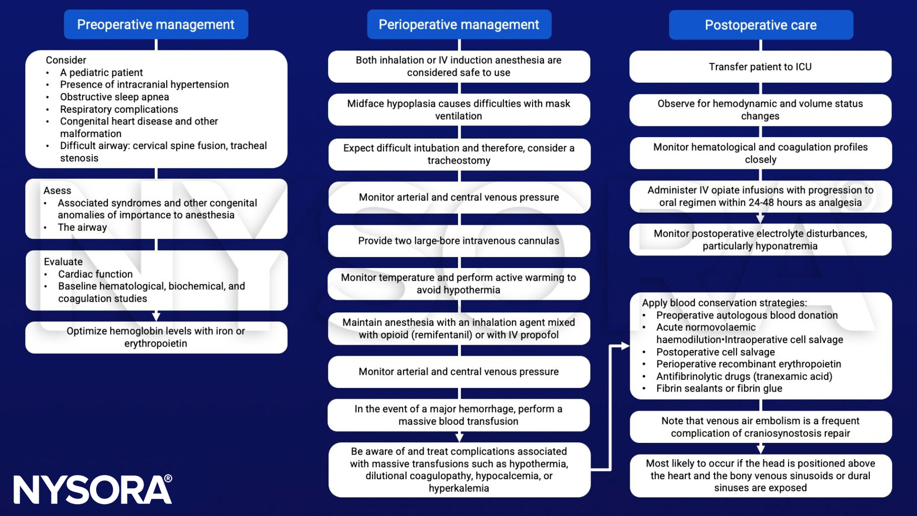
Anesthetic management
Suggested reading
- Pollard BJ, Kitchen, G. Handbook of Clinical Anaesthesia. Fourth Edition. CRC Press. 2018. 978-1-4987-6289-2.
- Pearson, A., Matava, C.T., 2016. Anaesthetic management for craniosynostosis repair in children. BJA Education 16, 410–416.
- Posnick JC, Ruiz RL. The craniofacial dysostosis syndromes: current surgical thinking and future directions. Cleft Palate Craniofac J. 2000;37(5):433.