What is ultrasound?
Let’s simplify this: as the name implies, ultrasound = sound wave. Sound waves travel as mechanical longitudinal waves, with back-and-forth particle motion parallel to their direction of travel. Unlike the sound we hear, ultrasound is a high-frequency sound we do not hear, as it is > 20 kHz.
The ultrasound transducer sends the sound waves and detects them as they bounce back after being reflected by the tissue through which they travel. Incidence refers to the angle between the ultrasound beam and the tissue plane. For reflection, the angle of incidence is less than 90 degrees; reflection occurs at tissue boundaries and tissue interfaces.
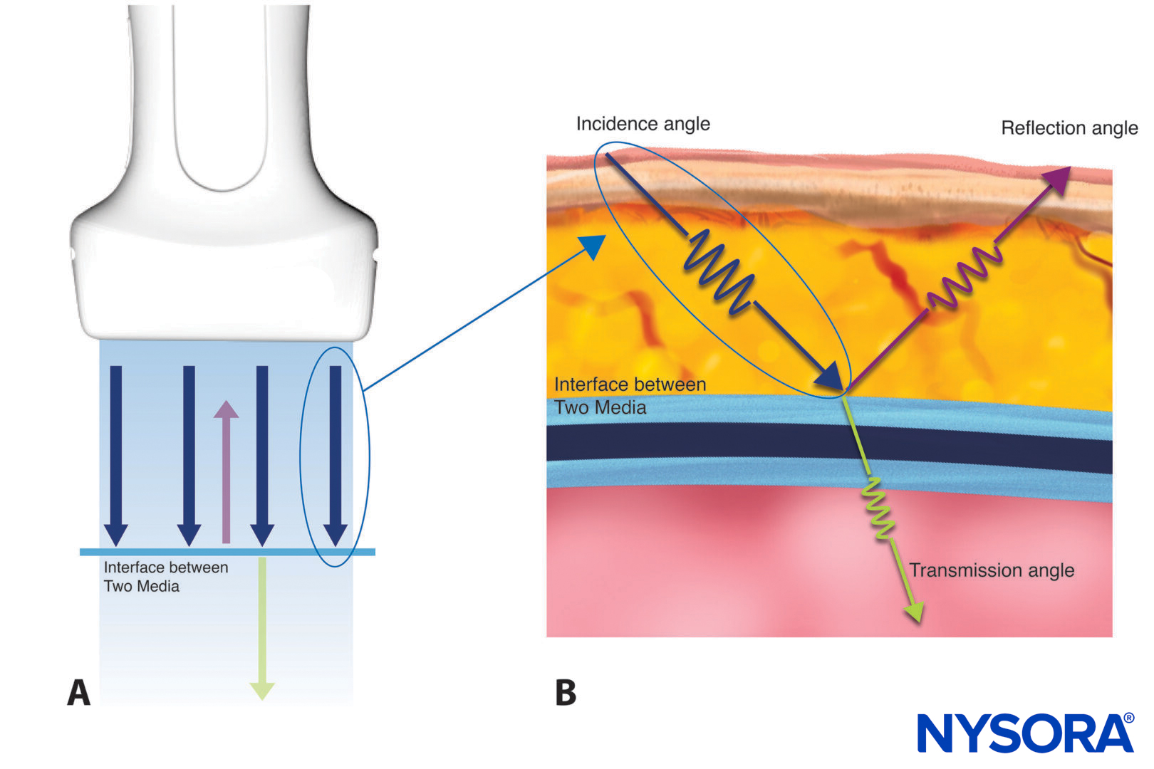
Here’s how the ultrasound is reflected by tissue A: Sound waves emitted (blue) by an ultrasound transducer and reflected by a medium (purple). B. Detailed image of a single sound wave with its incidence, reflection, and transmission.
The reflected waves are then translated into ultrasound images. Strong reflections result in hyperechogenic (bright, white) structures, and weak reflections in hypoechogenic (dark gray and black) structures.
Sound wave
Ultrasound consists of a beam of sound waves. Let’s zoom in on the physical properties of an individual sound wave.
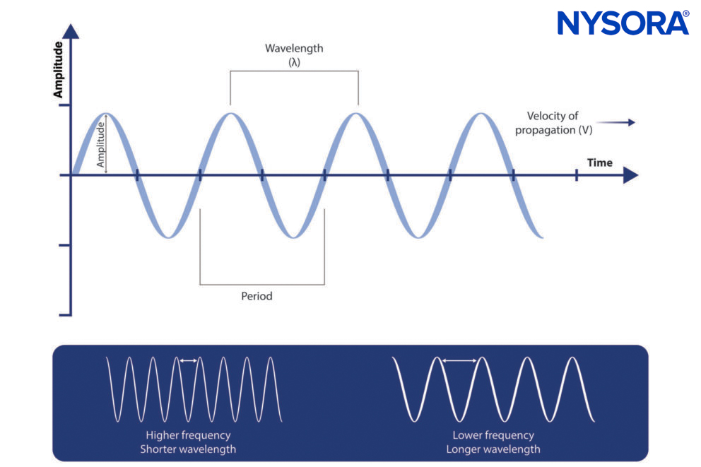
This is what an (ultra)sound wave looks like. Bottom: Difference between a high- and low-frequency sound wave.
- Amplitude: The height of the wave indicates its strength or energy.
- Wavelength: The length of space over which one cycle occurs; the travel distance from the beginning to the end of one cycle. In practical terms, the cycle is important for the resolution between two points in space.
- Period: The time for a sound wave to complete one cycle; it is measured in microseconds (µs).
- Frequency: The number of cycles repeated per second, measured in hertz (Hz).
Interesting fact
The human ear, in ideal circumstances, can hear sound from 20 to 20.000 Hz. Since ultrasound is above this frequency range, it is inaudible.
Acoustic velocity
Acoustic velocity is the speed at which a sound wave travels through a certain medium. Since the ultrasound machine measures the time for the ultrasound to return, it can calculate the spatial position (depth) of the tissues from which it is reflected back to the transducer.
- Formula: c = f x λ
- Acoustic velocity: c, frequency: f, wavelength: λ.
- Formula: c = (κ/ρ)½
- Acoustic velocity: c, density: ρ, stiffness: κ.
Let’s understand the factors that influence ultrasound speed and travel: tissue density and stiffness.
- Density: The concentration of a medium (e.g., tissue) through which the ultrasound travels.
- Stiffness: The resistance of a medium (e.g., tissue) to compression.
Interesting facts
- Propagation speed increases with increased stiffness or decreased density.
- The average propagation speed in soft tissues is 1540 m/s (ranges from 1400 to 1640 m/s).
- Ultrasound cannot penetrate lung or bone tissues.
Acoustic impedance
Acoustic impedance is the degree of difficulty for a sound wave to travel through a medium or the resistance/impediment to its travel. Sound waves reflect when there is a change in density and thus acoustic impedance. Increasing density differences will result in more reflections.
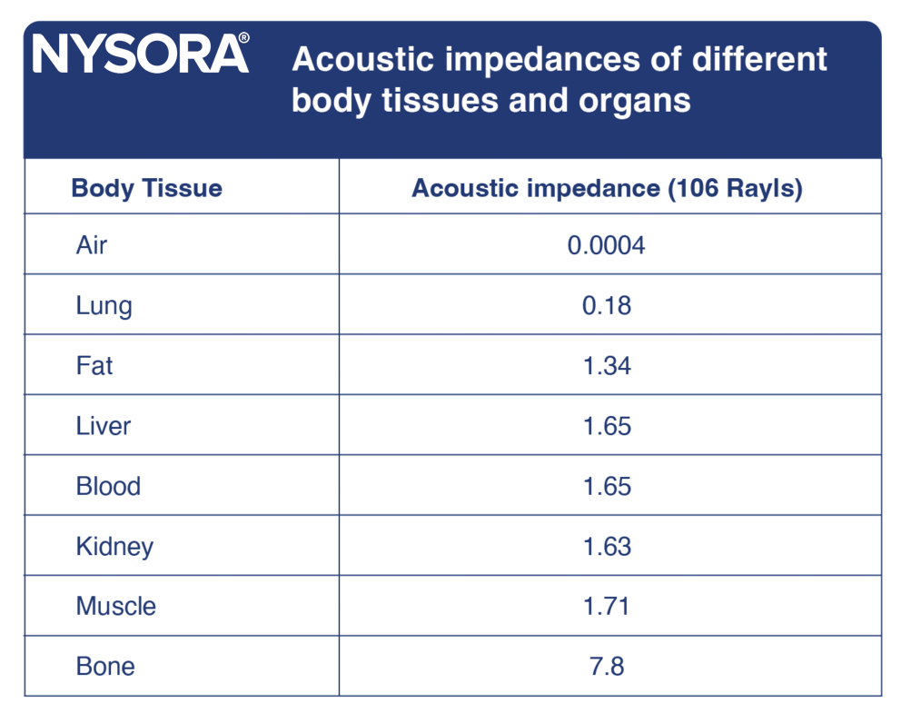
Acoustic impedances of human tissues.
- Formula: z = ρc
- Acoustic impedance: z, density: ρ, acoustic velocity: c.
Interesting facts
- Homogeneous tissues have only a few interfaces and, thus, exhibit low acoustic impedance and little reflection. Result: The US image appears as hypoechoic structures.
- Acoustic impedance increases when the propagation speed or the medium’s density increases, e.g., by increasing the pressure on the ultrasound transducer. Result: Applying pressure on the ultrasound transducer may improve the image resolution.
- Bone reflects or absorbs most of the ultrasound waves. Result: Dark shadows or image dropout behind the bone.
Attenuation coefficient
The attenuation coefficient is the parameter used to estimate the decrease in the amplitude of ultrasound waves after reflection from the tissues. The attenuation coefficient increases with increasing frequency.
Result: The ability to image tissue at increasing depths decreases as the frequency increases.
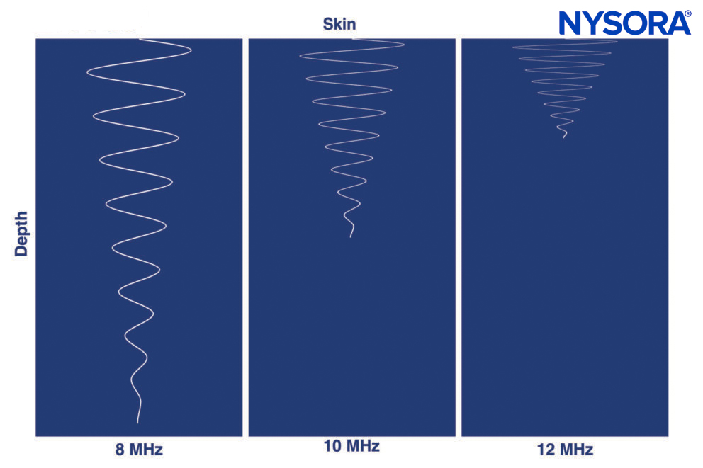
Attenuation increases with the frequency of the ultrasound wave, and the distance traveled by the ultrasound wave decreases. Note that the 8 MHz wave travels deeper than the 10 MHz or 12 MHz waves.