Indications
- Postrenal obstruction
- Urinary catheter position
- Bladder volume
- Bladder or urinary pathology
Tip
Ultrasound screening for hydronephrosis should always include a bladder scan.
Functional anatomy
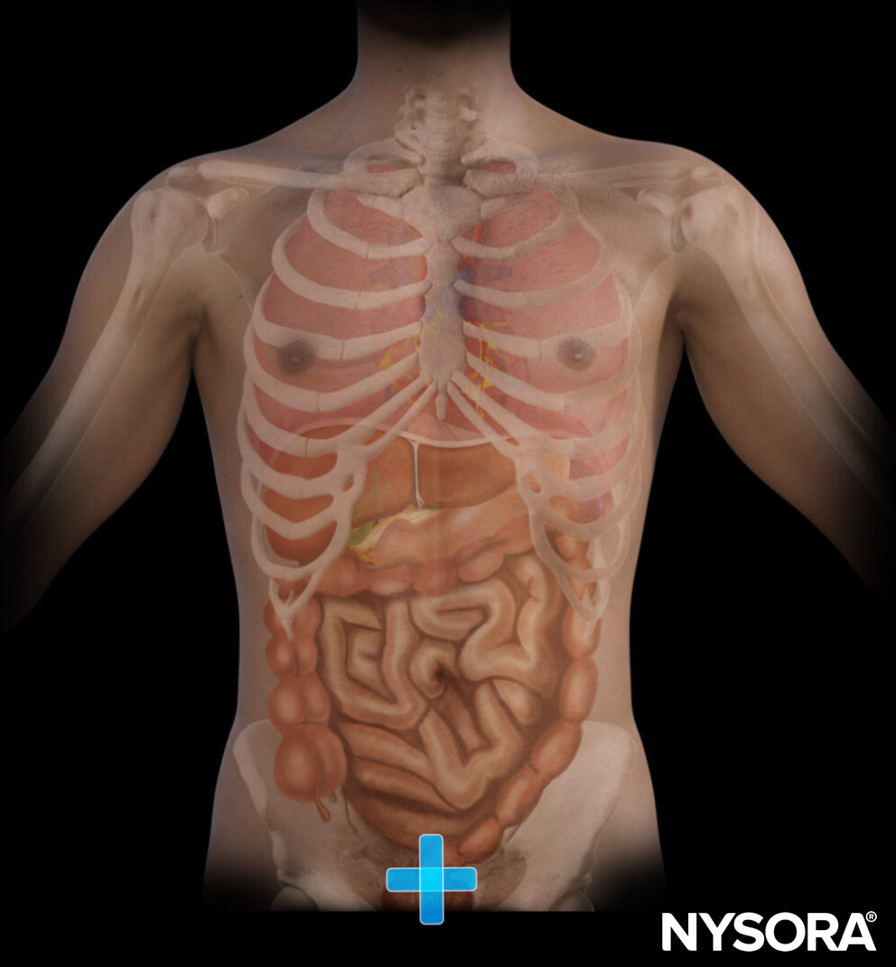
Anatomy of the abdomen
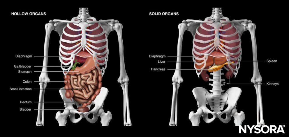
Hollow and solid organs of the abdomen.
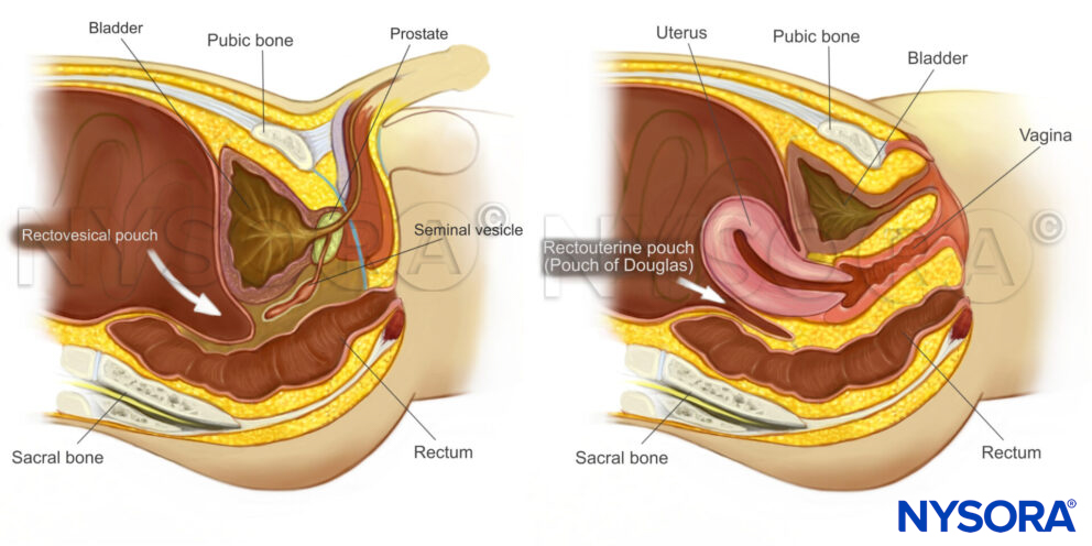
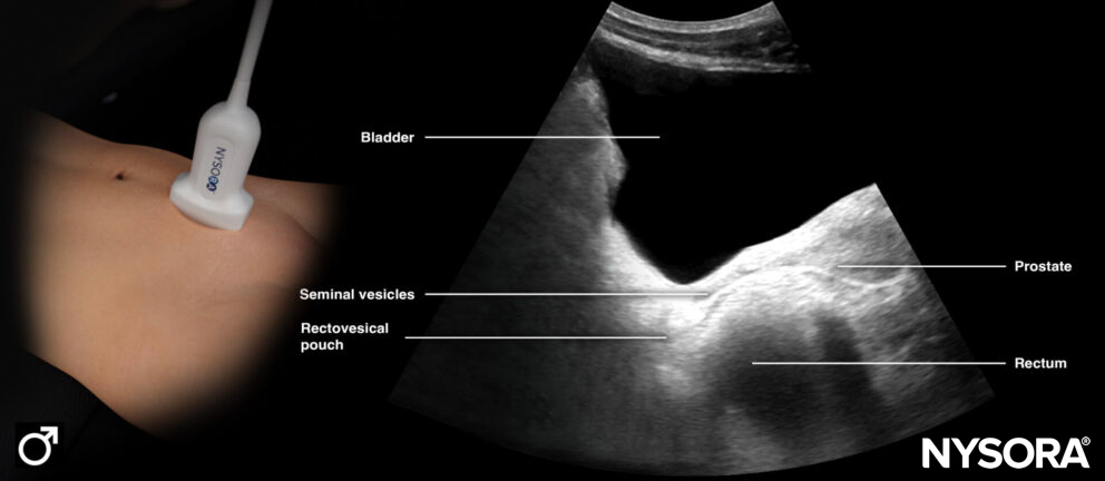
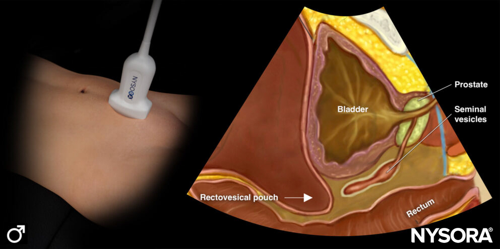
Sagittal section through the pelvis.
Anatomy of the bladder
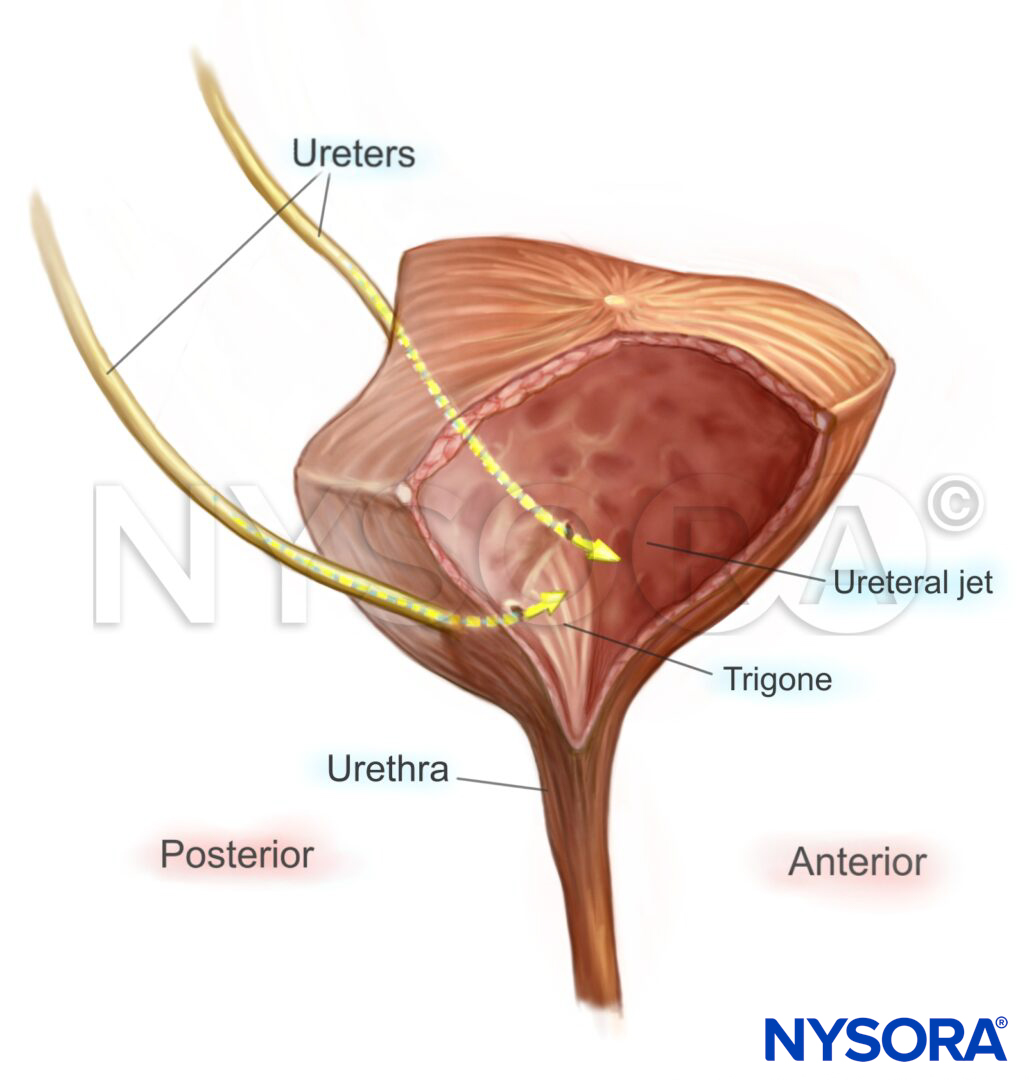
Anatomy of the bladder.
Ultrasound machine setup
- Transducer: curvilinear (or a linear transducer in children or slim adults)
- Ultrasound preset: abdominal
- Orientation: index mark towards the right side of the patient
- Depth: 5 – 10 cm
Patient position
Position the patient supine and flat for a bladder ultrasound.
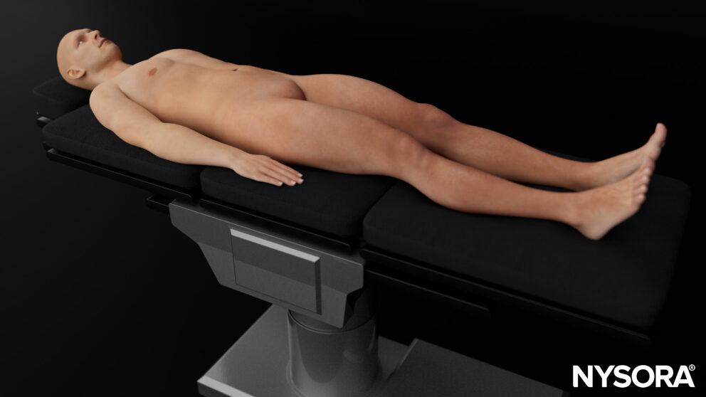
Patient position for a bladder ultrasound.
Landmarks
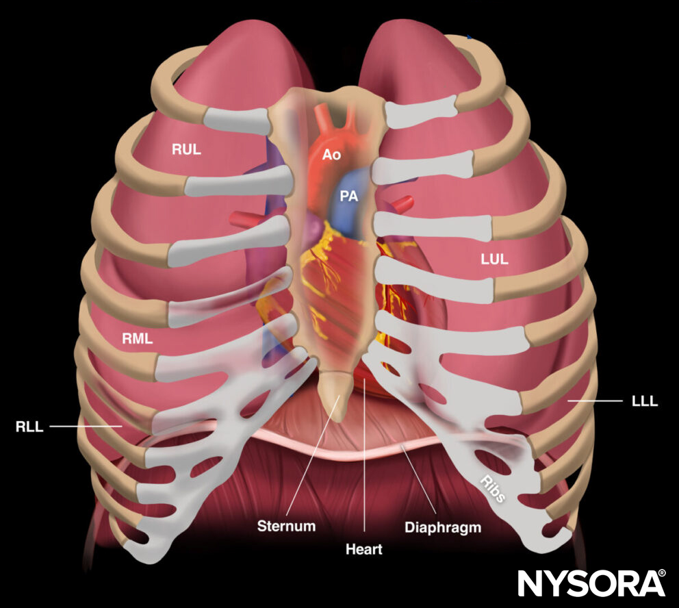
- Costal margin: ribs in the upper abdomen protect the upper abdominal organs but may limit the acoustic window to the liver, spleen, and kidneys
- Xiphoid process: the upper border of the abdomen
- Linea alba: midline of the abdomen that separates the rectus abdominis muscles and connects the xiphoid process with the pubic bone.
- Umbilicus: virtually separates the abdomen into four quadrants
- Pubic bone: a bony structure and the lower margin of the abdomen. The pelvis starts at the level of the pubic bone.
- Anterior superior iliac spine: a bony structure that forms the lateral border of the pelvis
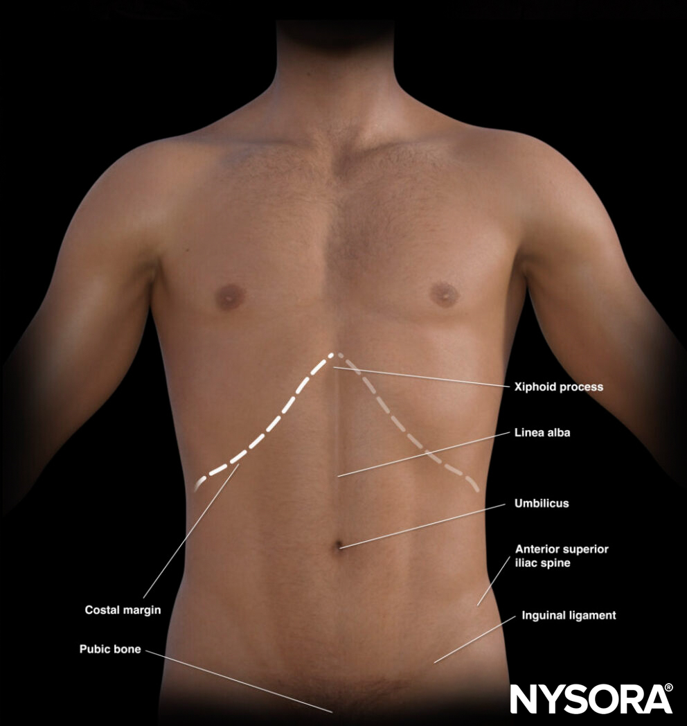
External landmarks of the abdomen.
The umbilicus divides the abdomen into different quadrants that allocate different organs.
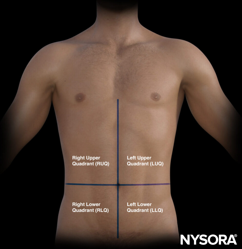
The quadrants of the abdomen.
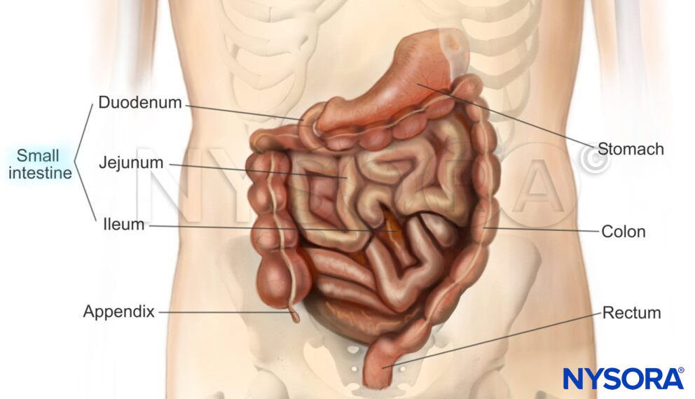
- Right upper quadrant (RUQ): Liver, gallbladder, kidney
- Left upper quadrant (LUQ): Stomach, spleen, kidney
- Pelvic/paracolic region (lower quadrants): Colon, small intestines, rectum, bladder, male or female reproductive organs
Transducer position
Place the transducer on the linea alba, just above the pubic bone. A bladder scan is first performed in longitudinal orientation, then in transverse orientation.
Transducer positioning for a bladder ultrasound: longitudinal and transverse approach.
Tip
Avoid the acoustic shadow of the pubic bone.
Scanning
Longitudinal scan
- The transducer is positioned on the midline above the symphysis pubis with the indicator pointing cranially.
- Tilt or fan the transducer in both directions to visualize all borders and the posterior wall of the bladder.
Longitudinal ultrasound view of the bladder (male).
Longitudinal Reverse Ultrasound Anatomy of the bladder (male).
Tip
Bowel gas can sometimes compromise ultrasound imaging of the superior border of the bladder.
Transverse scan
- For the transverse scan, rotate the probe 90° counterclockwise from the longitudinal position with the indicator towards the patient’s right side.
- Tilt or fan the transducer in both directions to visualize all borders and the posterior wall of the bladder.
Transverse ultrasound view of the bladder (male).
Longitudinal Reverse Ultrasound Anatomy of the bladder (male).
Tips
- Sometimes the bladder is positioned more behind the pubic bone, and a constant caudal tilt is required for scanning.
- There is no difference in bladder anatomy for a male or female, except for the underlying anatomy.
Bladder volume assessment
The combination of longitudinal and transverse ultrasound imaging allows estimation of the bladder volume:
- Measure the maximum length (L) en maximum depth (D) in the transverse view.
- Measure the maximum distance between the cranial and caudal point of the bladder, i.e., the height (H), in the longitudinal view.
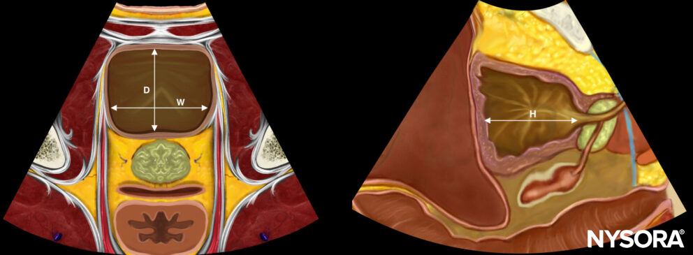
Estimation of bladder volume requires measuring the width (W), depth (D), and height (H) of the bladder.
Max Width (cm) x Depth (cm) x Height (cm) x 0.72 (correction factor) = Bladder volume (mL)
In healthy adults, the entire bladder volume should be less than 300-400 mL, and the post-void residual volume should be less than 50-100mL.
Tip
Based on the 3-dimensional shape of the bladder, other correction factors can be used to improve the accuracy of bladder volume estimation (triangular prism: 0.66; cylinder: 0.81; spherical: 0.52; cuboid: 0.89).
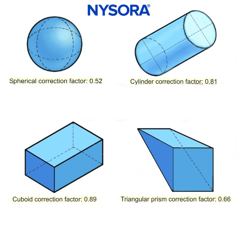
Correction factors for different bladder shapes.
Foley catheter position assessment
A correctly positioned urinary catheter will result in an empty bladder or a non-distended bladder with an intraluminal balloon. If the balloon is surrounded by hypoechogenic fluid, the Foley may be obstructed.
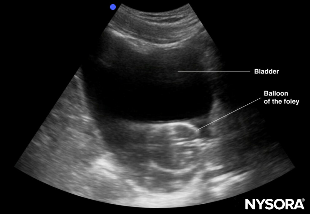
Endoluminal balloon in an obstructed Foley catheter.
Tip
Ultrasound of the bladder can guide troubleshooting for difficult catheterization or when there is no urinary flow.
Bladder or urinary pathologies
By tilting and scanning the bladder wall, it is possible to visualize bladder wall abnormalities as masses, diverticulosis, or bladder stones.
Tips
- It is not the goal of POCUS to search for bladder pathology; it may, however, be possible to witness this pathology during a bladder assessment.
- Bladder stones will always present on the posterior wall of the bladder and typically create an acoustic shadow.
- Bladder masses often have an irregular shape.
- Blood clots can behave like bladder masses but are often mobile.
Ureteral jets
The ureters insert into the trigone of the bladder. By scanning the posterior wall of the bladder with Doppler (color or power) and in the transverse orientation, the ureteral jets can be identified. The presence of jets indicates patent ureteral flow.
Tips
The absence of urethral jets does not indicate an obstruction.