 Nerve Blocks App
 Anesthesia Assistant App
 Ultrasound Pain App
 Pain Medicine Assistant App
 POCUS App
 IV Access App
 MSK Knee App
 VetRA App
 Nerve Block Manual
 Regional Anesthesia Updates
 Anesthesiology Manual
 Anesthesiology Review
 Anesthesia Updates 2025
 Anesthesia Updates 2026
 Pediatric Anesthesia Updates
 Airway Management Updates
 US Interventional Pain Manual
 Pain Medicine Updates
 Mastering Difficult IV Access
 PACU Nursing Manual
 RA Veterinary Manual
 Books
Books About
About
 Back to News
 1 min read

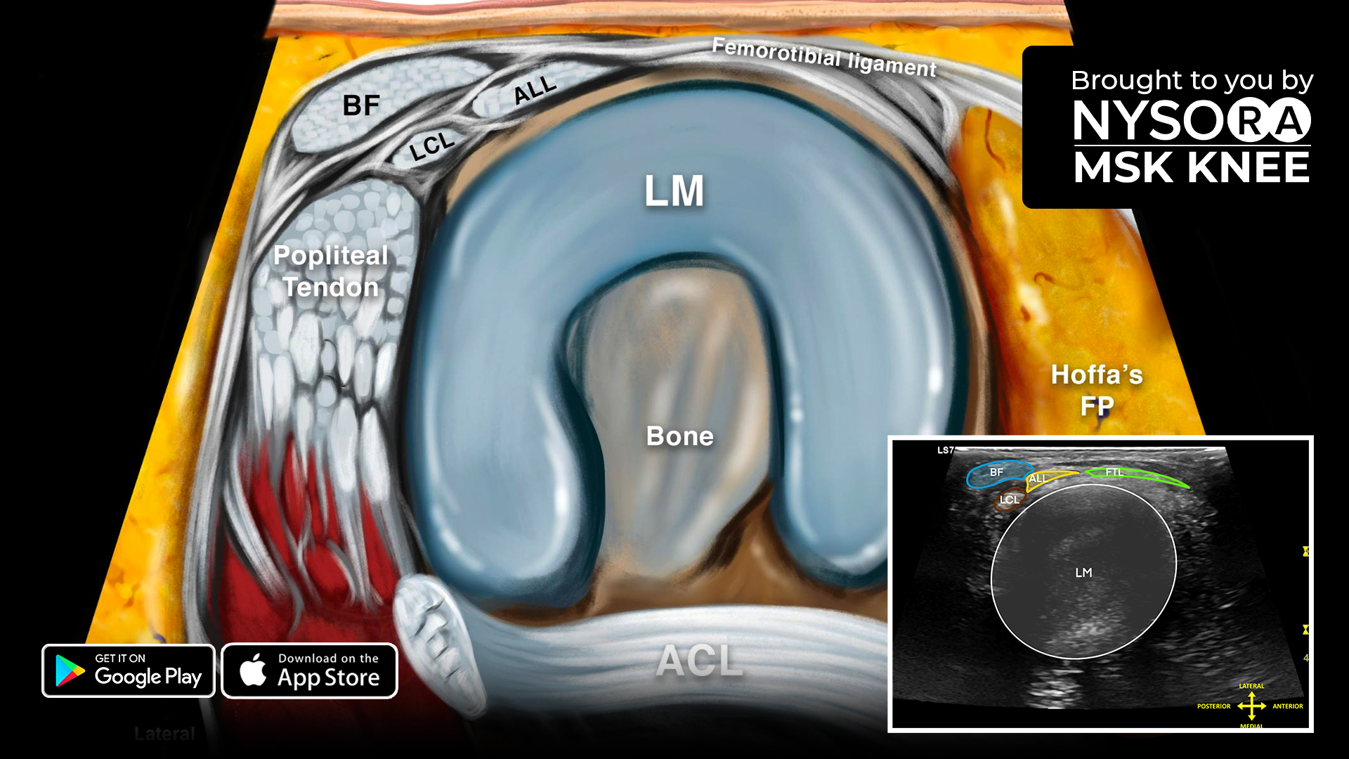
Knee injuries involve trauma to one or more tissues that make up the knee joint: ligaments, tendons, cartilage, bones, and muscles. This makes scanning the knee the critical step in diagnosing and treating the patient. This week, we are focusing on the anterolateral ligament.
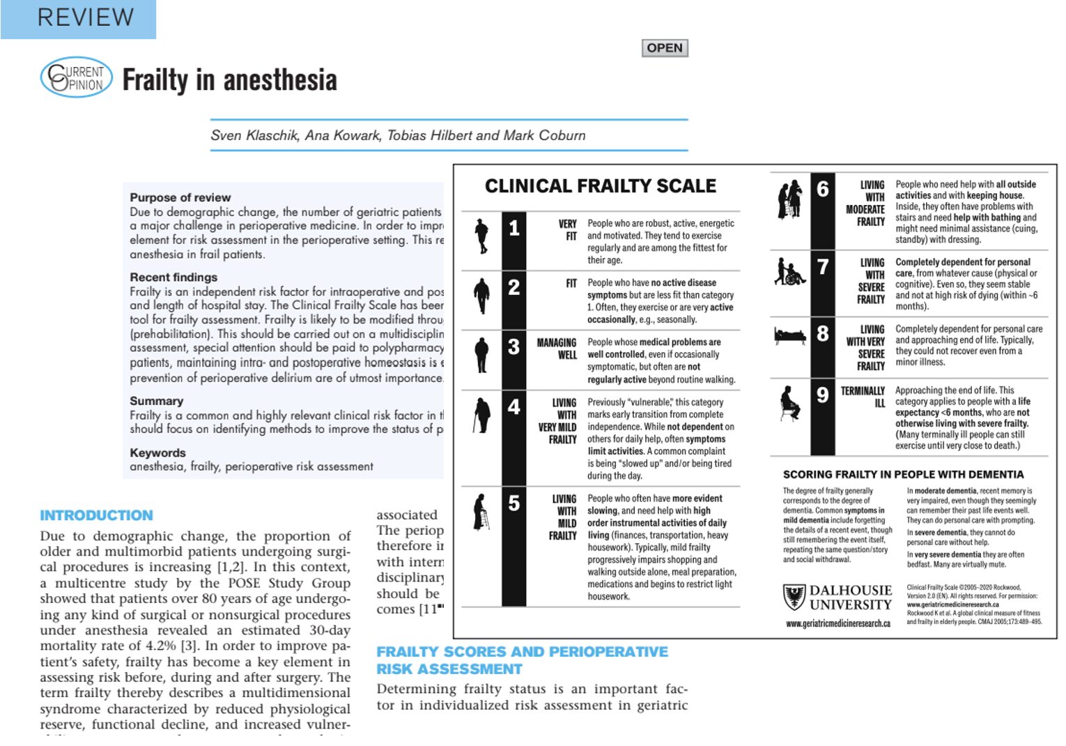
The anterolateral ligament (ALL) is also known as the lateral femorotibial ligament or lateral retinaculum. It originates at the femoral condyle and tucks underneath the iliotibial band.
Here are 4 top tips for scanning the anterolateral ligament
- Place the patient in a supine position with the knee flexed 90°.
- Bridge the transducer over the lateral femoral condyle and tibia.
- Identify the ALL underneath the iliotibial band.
- The lateral collateral ligament and popliteus tendon surround the ALL at its origin on the lateral femoral condyle.
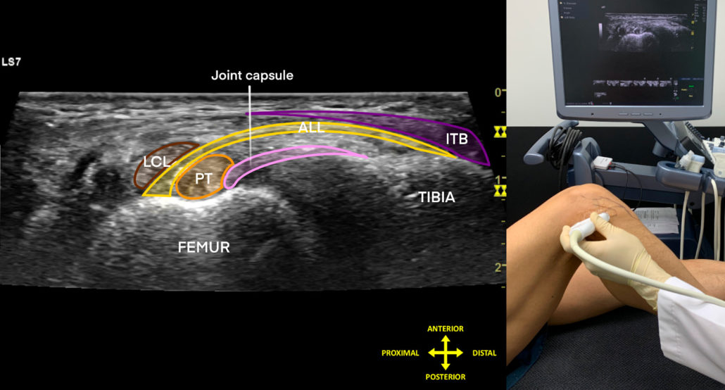
Sonoanatomy of the anterolateral ligament. ALL, anterolateral ligament; ITB, iliotibial band; LCL, lateral collateral ligament; PT, popliteus tendon.
Download the MSK App for more tips and the most practical and applicable techniques in musculoskeletal ultrasound anatomy and regenerative therapy of the knee.