Nerve Blocks App
 Pain Medicine Assistant App
 POCUS App
 IV Access App
 MSK Knee App
 VetRA App
 Nerve Block Manual
 Regional Anesthesia Updates
 Anesthesiology Manual
 Anesthesiology Review
 Anesthesia Updates 2025
 Anesthesia Updates 2026
 Pediatric Anesthesia Updates
 Airway Management Updates
 US Interventional Pain Manual
 Pain Medicine Updates
 Mastering Difficult IV Access
 PACU Nursing Manual
 RA Veterinary Manual
 About
About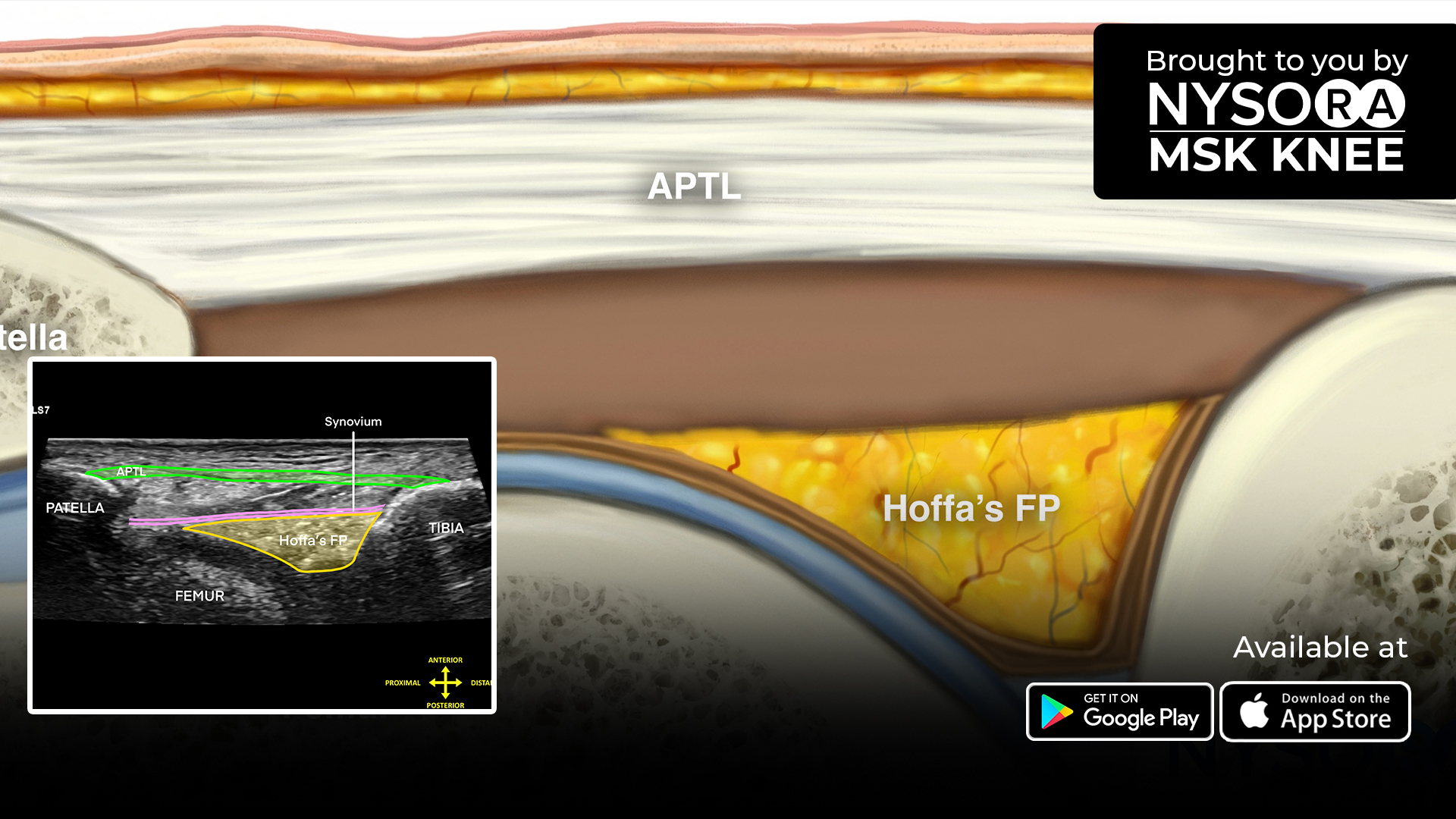


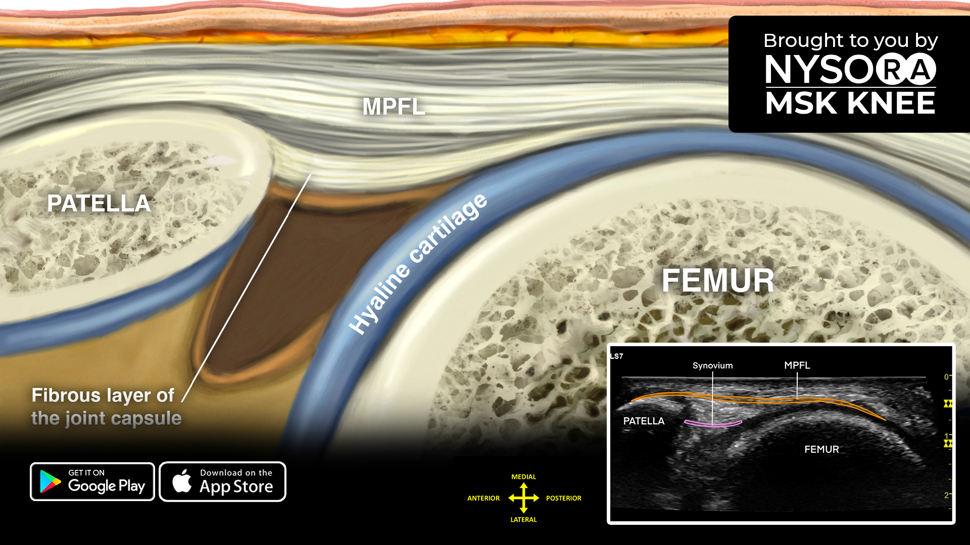
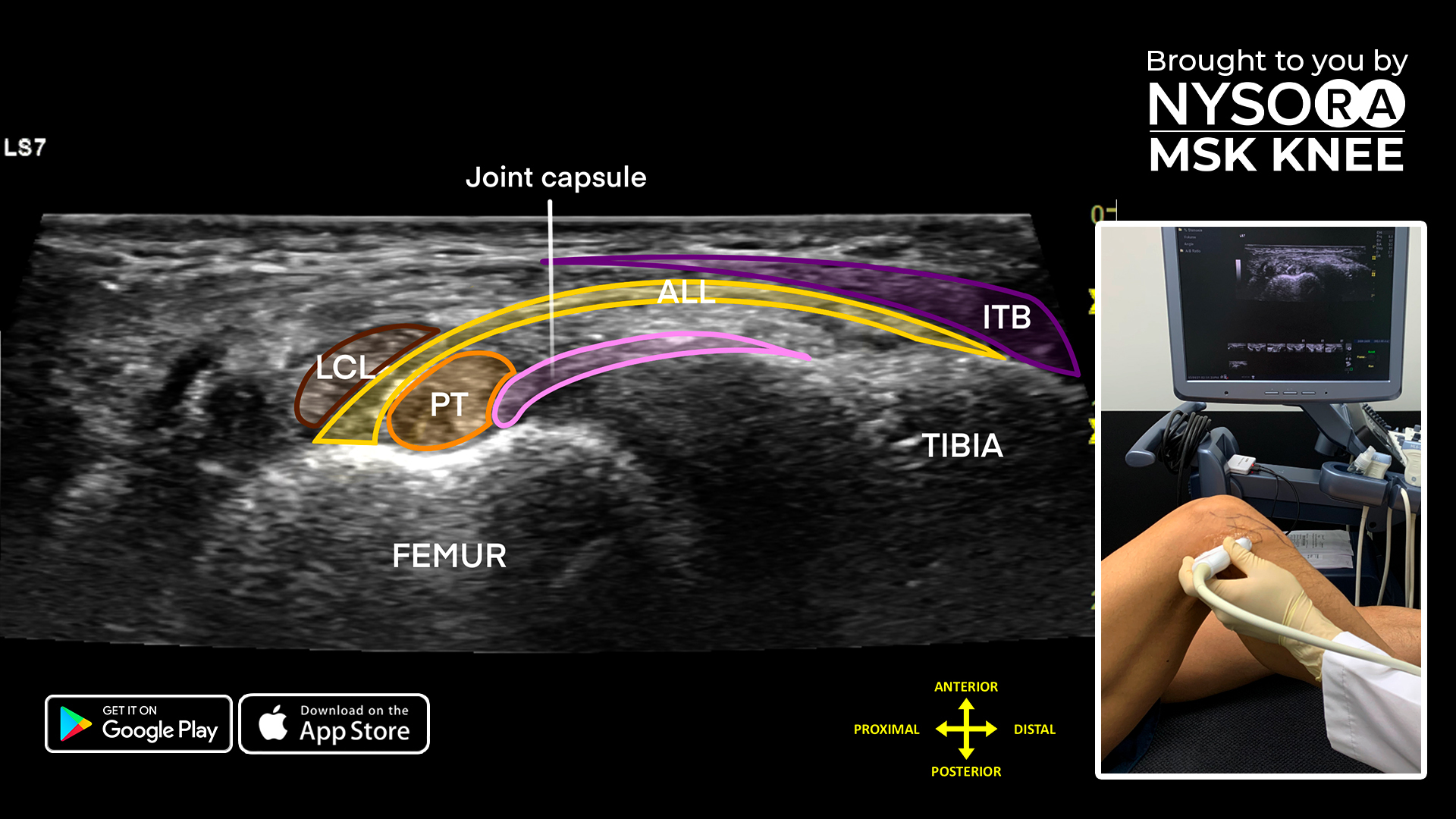
Patients with persistent lateral knee pain and instability should always be evaluated closely, so today we’re sharing a simple 4-step guide to scanning the lateral patellotibial ligament.
- Place the patient in a supine position with the knee flexed 90°.
- Position the transducer longitudinal on the lateral aspect of the knee, bridging the patella and tibia.
- Identify the lateral patellotibial ligament and its trilaminar appearance running from the patella to the tibia.
- Other anatomical structures in this view are the synovium and Hoffa’s fat pad, which is located underneath the patellotibial ligament.
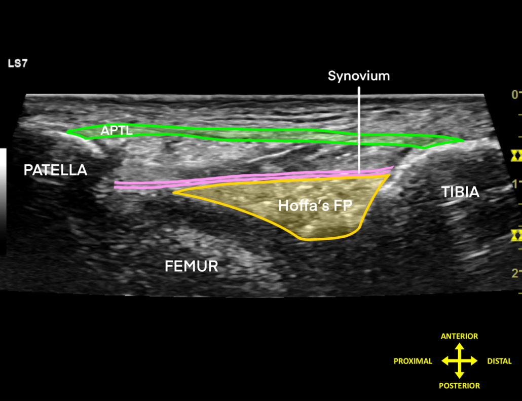
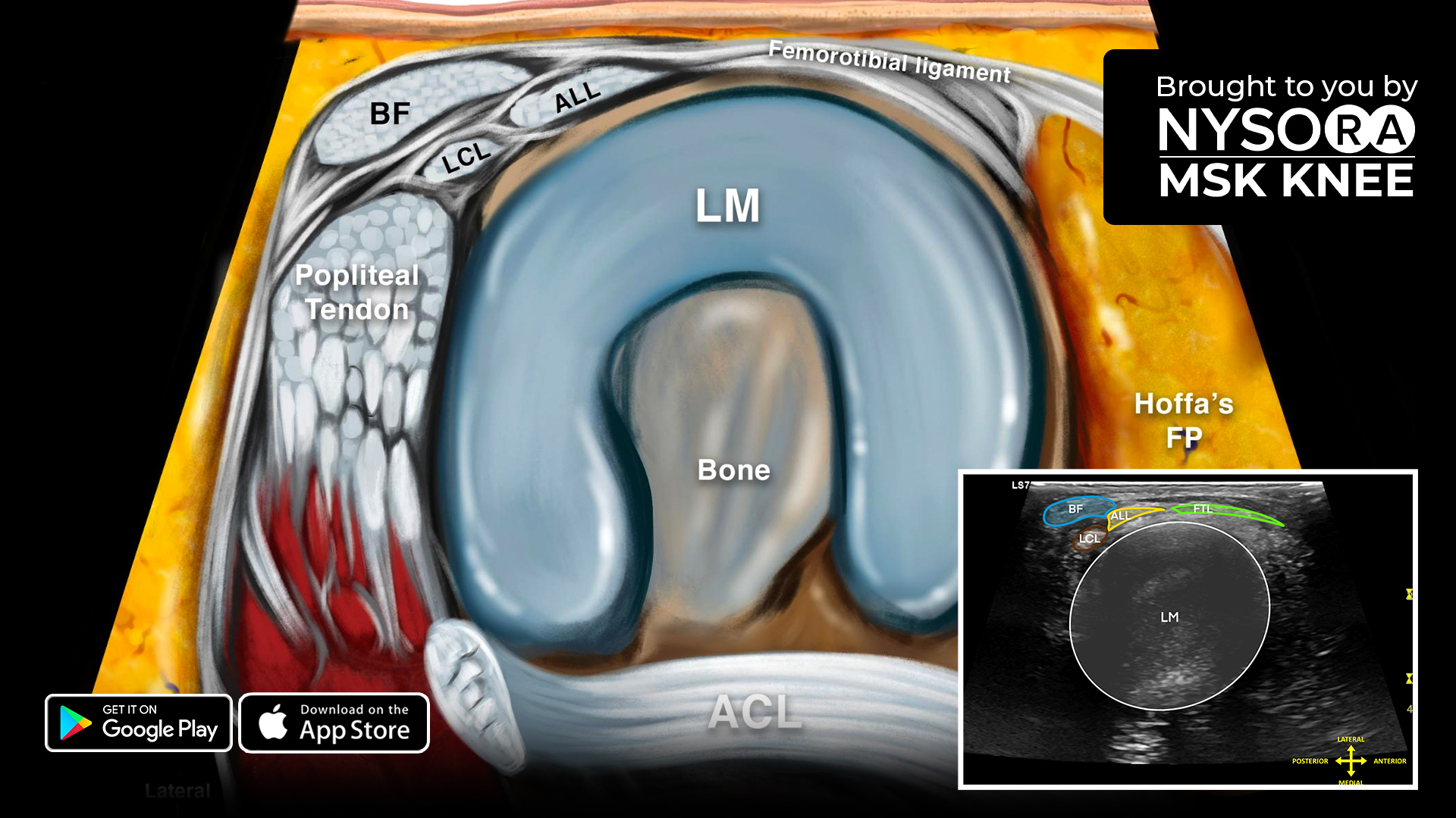
Sonoanatomy
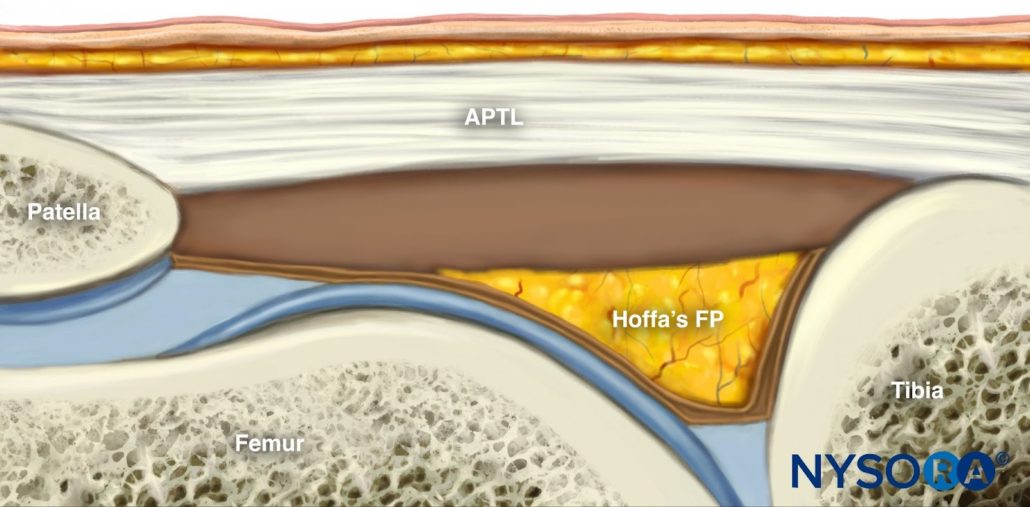
Reverse Ultrasound Anatomy
Comparison of sonoanatomy and reverse ultrasound anatomy of the lateral patellotibial ligament.
Download the MSK App for more tips, and check out the all-new reverse ultrasound anatomy illustration and slider image added in “Sonoanatomy of the Lateral Knee > Patellotibial Ligament,” as well as the most practical and applicable techniques in musculoskeletal ultrasound anatomy and regenerative therapy of the knee.