 Nerve Blocks App
 Anesthesia Assistant App
 Ultrasound Pain App
 Pain Medicine Assistant App
 POCUS App
 IV Access App
 MSK Knee App
 VetRA App
 Nerve Block Manual
 Regional Anesthesia Updates
 Anesthesiology Manual
 Anesthesiology Review
 Anesthesia Updates 2025
 Anesthesia Updates 2026
 Pediatric Anesthesia Updates
 Airway Management Updates
 US Interventional Pain Manual
 Pain Medicine Updates
 Mastering Difficult IV Access
 PACU Nursing Manual
 RA Veterinary Manual
 Books
Books About
About

Learning objectives
- Define hydrocephalus
- Describe the age-related signs and symptoms of hydrocephalus
- Anesthetic management of a patient with hydrocephalus
Definition and mechanisms
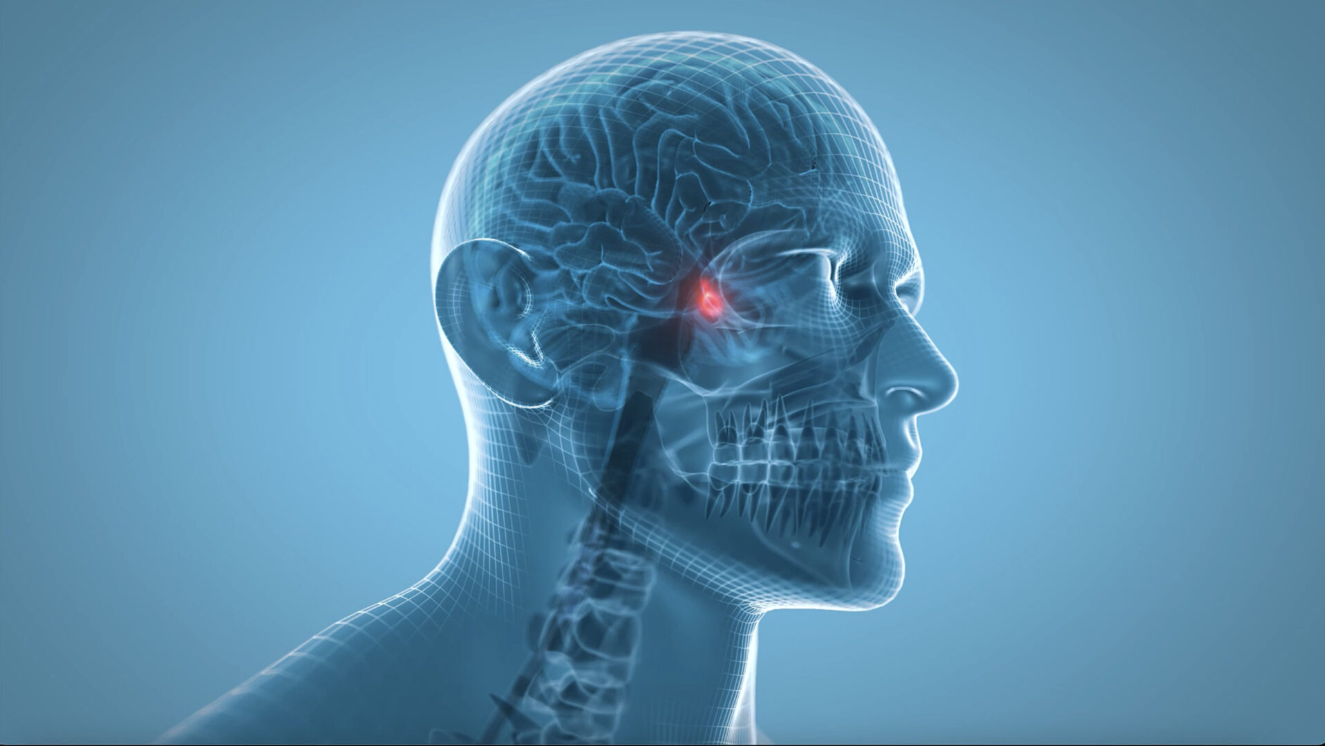
- Hydrocephalus is the excess accumulation of cerebrospinal fluid (CSF) in the ventricular system of the brain, resulting in increased intracranial pressure (ICP)
- It is caused by obstruction to CSF flow in the ventricles or subarachnoid space, or by excess CSF production from a congenital malformation blocking normal drainage or from complications of head injuries or infections
- Hydrocephalus can happen at any age but occurs more frequently among infants and older adults (>60 years)
Signs and symptoms
The signs and symptoms vary by the age of onset
Infants
- Changes in the head
- Unusually large head
- Rapid increase in the size of the head
- Bulging or tense soft spot (fontanel) on the top of the head
- Physical signs and symptoms
- Nausea and vomiting
- Sleepiness or lethargy
- Irritability
- Poor eating
- Seizures
- Reduced upward gaze (Parinaud’s syndrome)
- Problems with muscle tone and strength
Toddlers and older children
- Physical signs and symptoms
- Headache
- Blurred or double vision
- Abnormal eye movements
- Abnormal enlargement of the head
- Sleepiness or lethargy
- Nausea or vomiting
- Unstable balance
- Poor coordination
- Poor appetite
- Loss of bladder control or frequent urination
- Behavioral and cognitive changes
- Irritability
- Change in personality
- Decline in school performance
- Delays or problems with previously acquired skills (e.g., walking, talking)
Young and middle-aged adults
- Headache
- Lethargy
- Loss of coordination or balance
- Loss of bladder control or frequent urination
- Vision problems
- Decline in memory, concentration, and other thinking skills
Older adults (>60 years)
- Loss of bladder control or frequent urination
- Memory loss
- Progressive loss of other thinking or reasoning skills
- Gait disturbance
- Poor coordination or balance
Causes
Noncommunicating hydrocephalus: Obstruction of CSF outflow
- Space-occupying lesion
- Subarachnoid or intraventricular hemorrhage
- Spina bifida
- Arnold-Chiari malformation
- Dandy-Walker malformation
- Head injury
- Aqueductal stenosis
- Tumors
Communicating hydrocephalus: Failure of absorption of CSF by the arachnoid villi
- Subarachnoid or intraventricular hemorrhage
- Meningitis
- Head injury
- Achondroplasia
- Craniofacial syndromes
- Other skull base deformities
- Choroid plexus papilloma or carcinoma
Risk factors
Newborns
- Abnormal development of the central nervous system that obstructs the CSF flow
- Bleeding within the ventricles (i.e., intraventricular hemorrhage), a possible complication of premature birth
- Infection in the uterus (e.g., rubella or syphilis) during pregnancy may cause inflammation in the fetal brain tissues
Other contributing factors
- Lesions or tumors of the brain or spinal cord
- Central nervous system infections (e.g., bacterial meningitis or mumps)
- Bleeding in the brain from a stroke or head injury
- Other traumatic injury to the brain
Treatment
- Short-term (acute and emergency situations): External ventricular drain or ventriculostomy
- Inserted into the frontal horn of the lateral ventricle
- Drain reduces the ICP and allows to measure ICP
- Only short-term management due to the risk of infection
- Long-term: Cerebral shunt
- Allows drainage of CSF to distal sites
- Placement of a ventricular catheter into the cerebral ventricles to bypass the flow obstruction/malfunctioning arachnoid villi and drain the excess CSF into other body cavities from where it can be resorbed
- Most shunts drain fluid into the peritoneal cavity (ventriculoperitoneal shunt), alternative sites include the right atrium (ventriculoatrial shunt), pleural cavity (ventriculopleural shunt), and gallbladder
- The shunt can also be placed in the lumbar space of the spine to redirect the CSF to the peritoneal cavity (lumboperitoneal shunt)
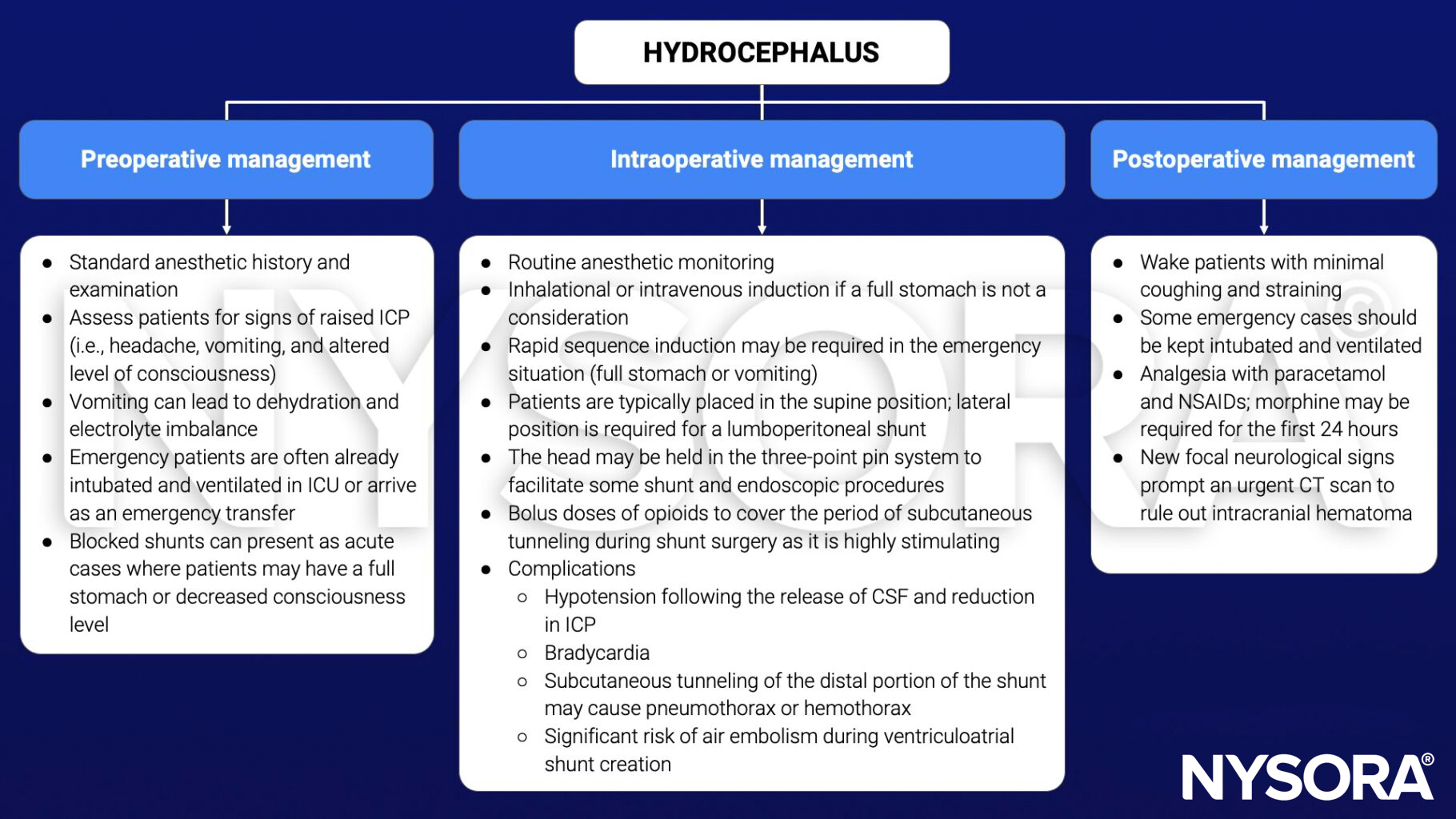
Management
Suggested reading
- Krovvidi H, Flint G, Williams AV. Perioperative management of hydrocephalus. BJA Educ. 2018;18(5):140-146.
- Pollard BJ, Kitchen G. Handbook of Clinical Anaesthesia. 4th ed. Taylor & Francis group; 2018. Chapter 14 Neurosurgery, Chapman E.