Nerve Blocks App
 Pain Medicine Assistant App
 POCUS App
 IV Access App
 MSK Knee App
 VetRA App
 Nerve Block Manual
 Regional Anesthesia Updates
 Anesthesiology Manual
 Anesthesiology Review
 Anesthesia Updates 2025
 Anesthesia Updates 2026
 Pediatric Anesthesia Updates
 Airway Management Updates
 US Interventional Pain Manual
 Pain Medicine Updates
 Mastering Difficult IV Access
 PACU Nursing Manual
 RA Veterinary Manual
 About
About
Indications
These are a few of the most common indications for vascular ultrasound:
- Difficult intravenous access
- Need for invasive blood pressure monitoring
- Intravenous drug administration, suspicion
- Deep venous thrombosis
Essential info
- A sterile technique should be used at all times.
- Needle insertions can be performed with an in-plane or out-of-plane technique.
- Ultrasound is an aid for vascular access, but anatomy knowledge is essential to avoid complications.
Ultrasound machine setup
- A linear transducer can be used for most vascular ultrasound applications.

- Preset: Vascular.
- Depth: According to the anatomical structure of interest.
Transducer orientation
In-plane
In-plane is a long-axis approach of the needle. For an optimal image, the transducer is aligned perfectly with the plane wherein the needle is advanced.
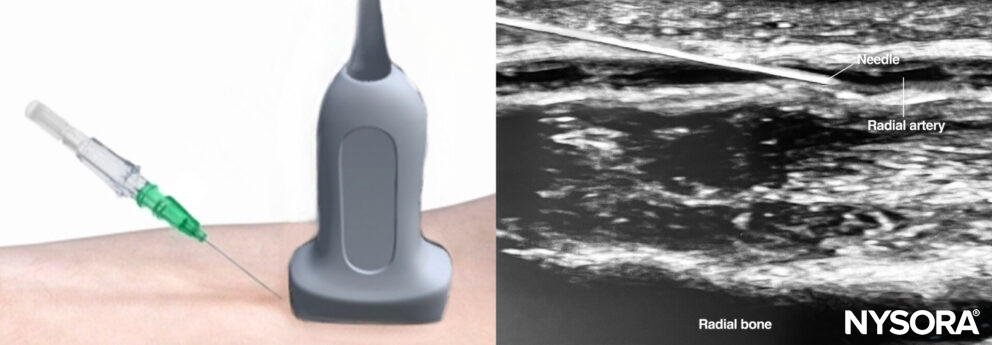
In-plane insertion of a needle with ultrasound.
Out-of-plane
The out-of-plane technique positions the transducer in a transverse, or short-axis, orientation relative to the needle.
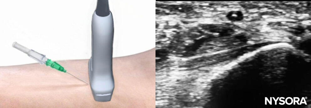
Out-of-plane insertion of a needle with ultrasound.
Clinical tips
- Most vascular access is performed with an out-of-plane technique, and the transducer is transversely oriented toward the structure.
- Make sure the transducer marker and screen marker are oriented the same way.
- Use enough gel and ensure the transducer makes full contact with the skin.
- Applying excessive pressure to the transducer can deform underlying structures, making vascular access more difficult.
- For difficult visualization of the needle tip, move the transducer instead of the needle.
Scanning
How to improve needle visualization
Using the creep technique, it is possible to track the needle tip during out-of-plane techniques and to improve needle tip visualization and, thus, safety.
- The needle is inserted out-of-plane, and advancement is stopped when the needle is visualized.
- The transducer is then advanced slightly distant from the needle tip until the tip disappears from the ultrasound image.
- Then the needle is advanced until the tip is visualized again.
- Repeating these steps allows the needle to be directed without losing control of the needle tip.
Angle optimization and tilting are important for achieving optimal beam reflections and thus visualizing the needle. When the needle is not visible, it is also described as anisotropy or the impossibility of visualizing the desired structure of interest due to the loss of sound waves by decreased reflection towards the transducer.
Optimization of the ultrasound conditions
- Easy access and exposure of the region of interest.
- Ergonomic position.
- Adjust the gain, depth, and focus.
Analyze the area
- The vessel location relative to other structures (artery, nerve, …)
- Anomalies (clot in the vessel lumen, aberrant anatomy, …)
Interesting fact
A thrombosed vessel is not compressible.
Vein vs. artery
Veins and arteries can be difficult to distinguish from each other. These characteristics will allow you to differentiate them:
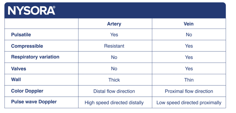
Characteristics of arteries and veins.