Nerve Blocks App
 Pain Medicine Assistant App
 POCUS App
 IV Access App
 MSK Knee App
 VetRA App
 Nerve Block Manual
 Regional Anesthesia Updates
 Anesthesiology Manual
 Anesthesiology Review
 Anesthesia Updates 2025
 Anesthesia Updates 2026
 Pediatric Anesthesia Updates
 Airway Management Updates
 US Interventional Pain Manual
 Pain Medicine Updates
 Mastering Difficult IV Access
 PACU Nursing Manual
 RA Veterinary Manual
 About
AboutIndications
- Intracranial hypertension
- Diagnosis of cerebral circulatory arrest
- Vasospasm
- Identification of midline shift
Advanced indications (not included in this course):
- Thrombosis or emboli
- Evaluation of thrombolysis efficacy
- Sickle cell hyperemia
- Shunts
Essential info
- Advantages of transcranial Doppler (TCD):
- Trending is possible
- ‘Real-time’ information
- Bedside examination
- TCD is NOT better than a computerized tomography (CT) scan
- Only Doppler of the middle carotid artery (MCA) will be assessed in this course
- A POCUS TCD should never replace a formal TCD study by a certified neurosonologist.
- Transorbital, submandibular, and transforaminal TCD are more advanced methods and will not be discussed in this course.
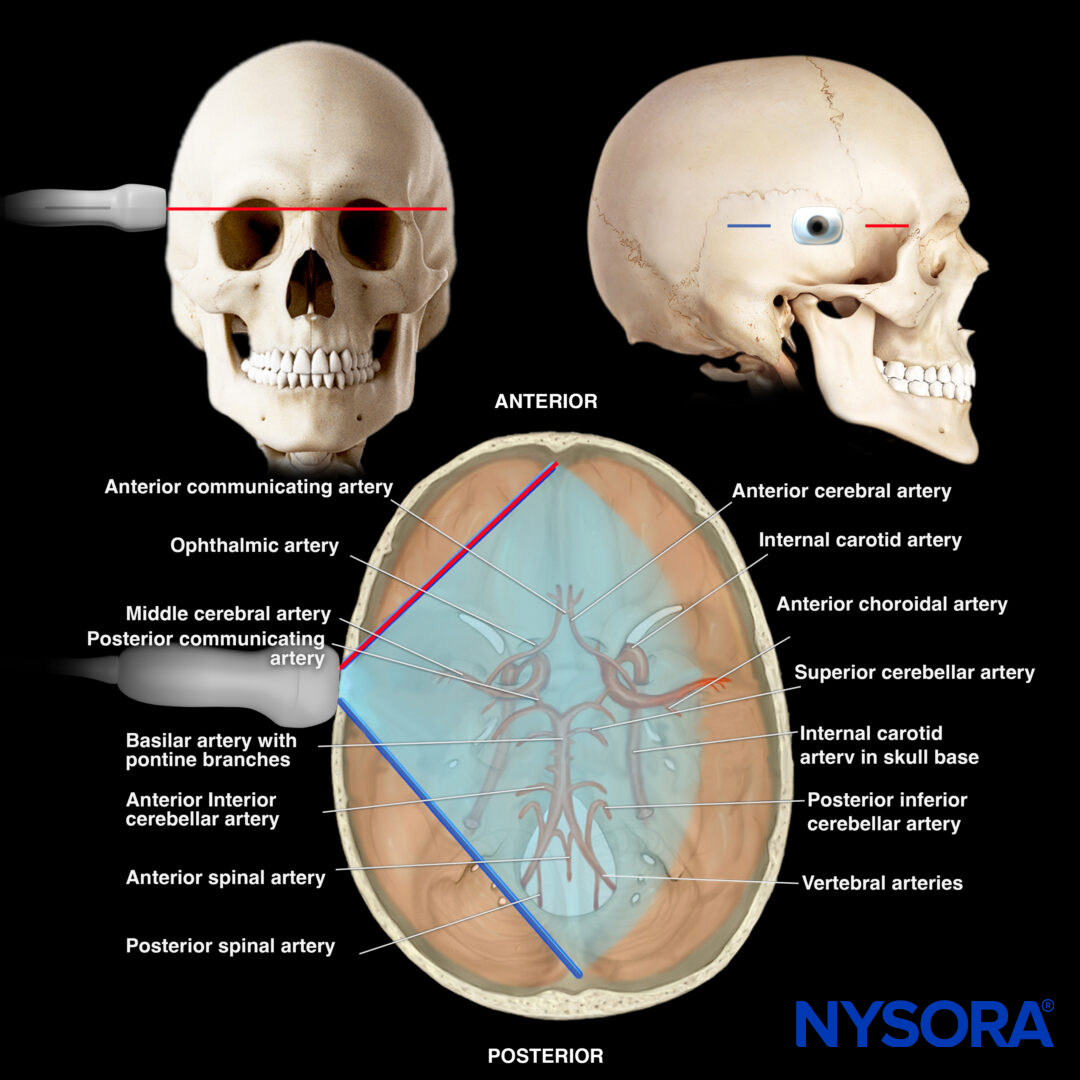
Functional anatomy

Circle of Willis and intracranial arteries.
Coronal section and relevant anatomical structures.
Ultrasound machine setup
- Transducer: Phased array
- Preset: Transcranial (or cardiac)
- Orientation: Index marker towards the frontal bone/orbital
- Depth: 15 cm
Tips
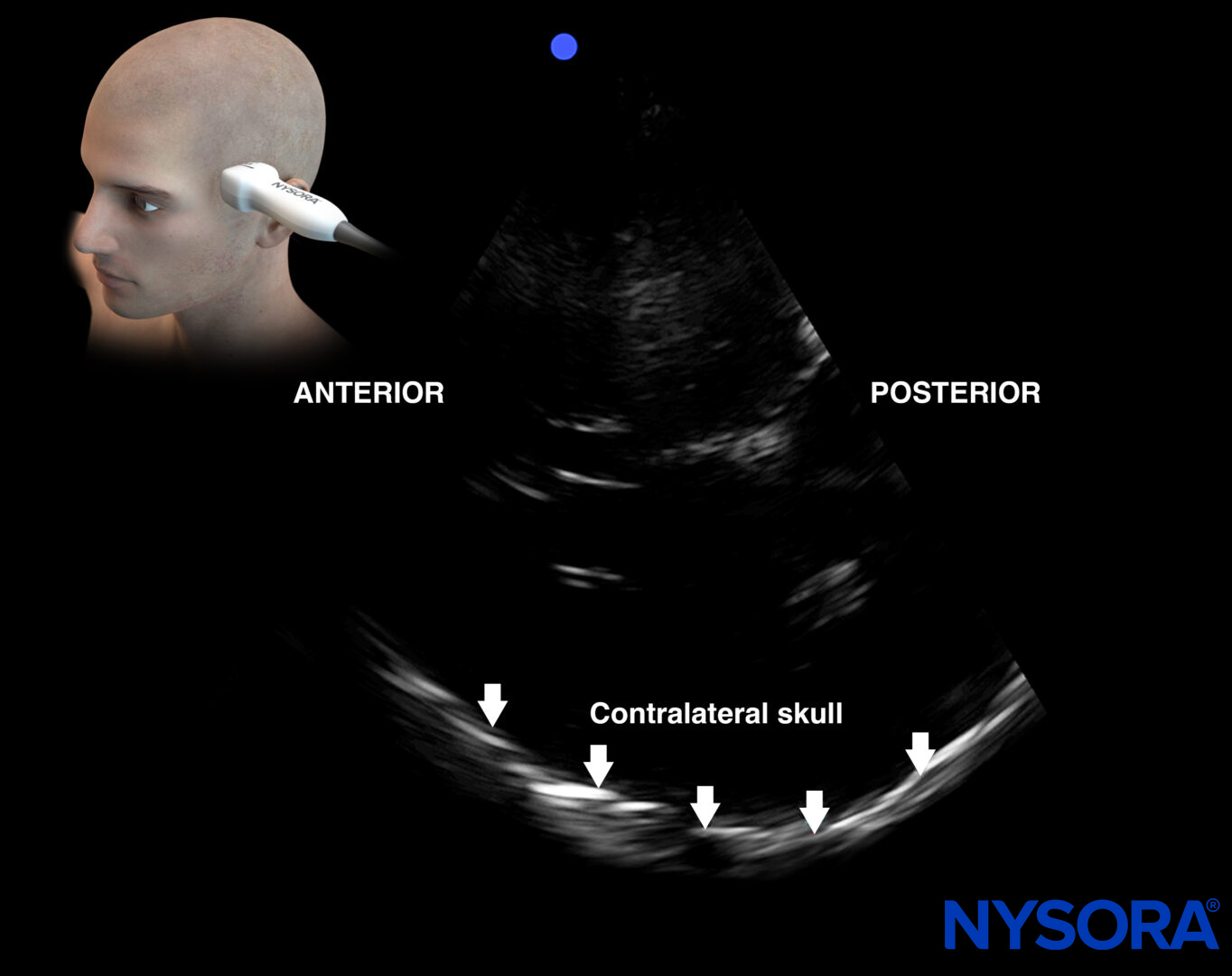
- The contralateral skull must be visualized to demonstrate the temporal acoustic window.
- Always perform a left and right transtemporal assessment.
Index mark orientation for transcranial settings.
Note
For the transcranial settings, the index mark will be oriented towards the frontal bone or nose, while the index mark will point towards the occiput for the cardiac preset.
Patient position
Position the patient supine with the head of the bed elevated 30 degrees.
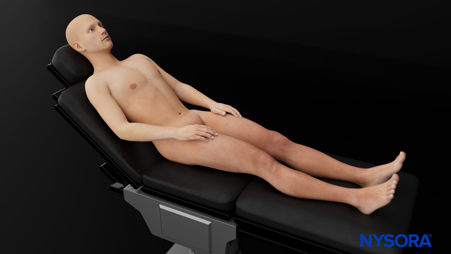
Patient position for a transcranial Doppler examination.
Landmarks
- Ear
- Temporomandibular joint
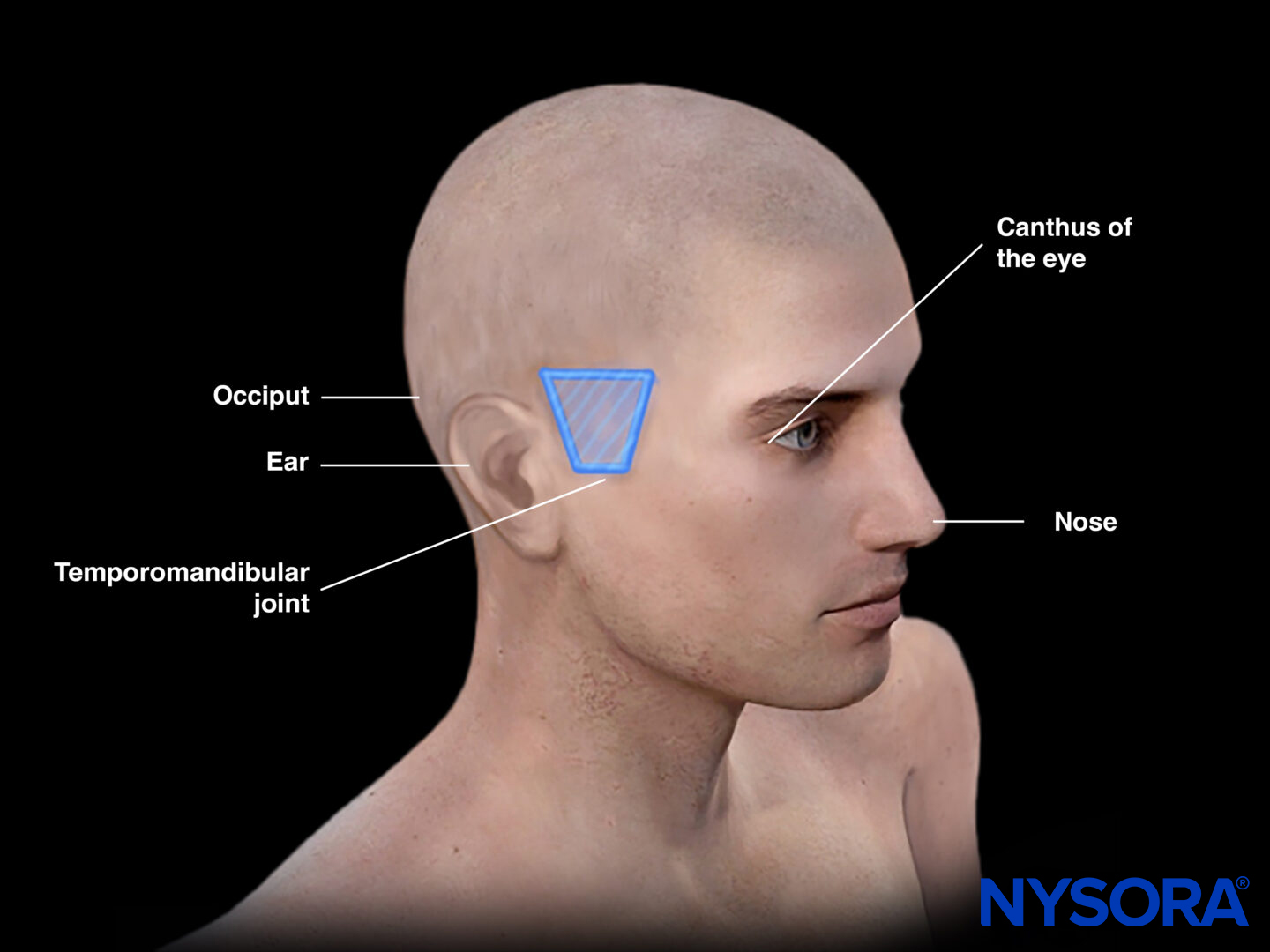
External landmarks for a transcranial Doppler examination.
Tip
The line on which the transducer is positioned connects the external auditory meatus and the outer canthus of the eye and is called the orbitomeatal line.
Transducer position
The transducer is positioned 2-3 cm above the temporomandibular joint at the level of the temporal bone (pterion).
Transducer position for a transcranial Doppler examination.
Tips
- Always use a sufficient amount of ultrasound gel to establish good transducer-patient contact.
- Make small circular movements when it is difficult to find an adequate window for TCD.
- Patients with a thick skull may not be good candidates for TCD.
View and relevant anatomy in the transtemporal window. The dark blue line illustrates the frontal orientation of the ultrasound beam.
Scanning
Tilting the phased array transducer in the transtemporal position results in different planes:
- Mesencephalic plane
- Diencephalic plane
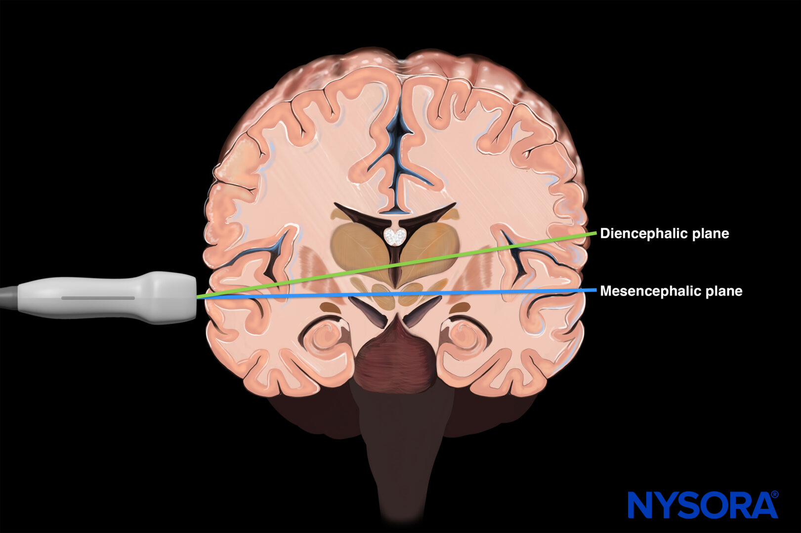
By tilting the transducer, two planes are obtained: The diencephalic and mesencephalic planes.
Mesencephalic plane
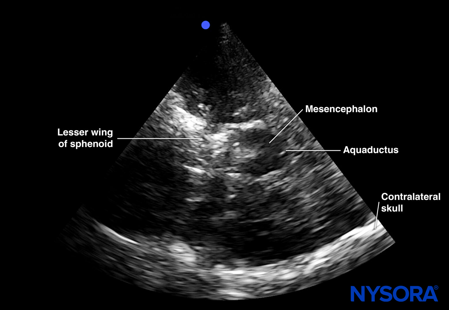
Ultrasound view of the mesencephalic plane.
The mesencephalic plane is the most basal plane. It comprises the mesencephalon on the midline, which has a typical butterfly shape due to the peduncles.
Use:
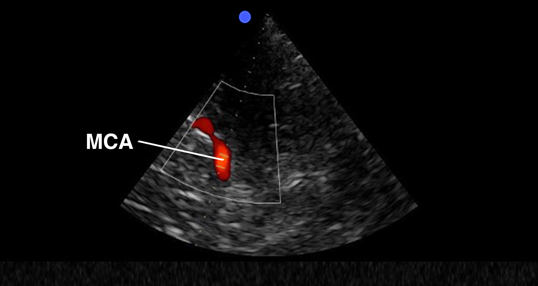
Since the mesencephalic plane cuts through the circle of Willis, it is specifically interesting for cerebral vascular assessment. Identification of the ipsilateral MCA (M1 segment) is one of the most common applications of TCD. It can be visualized anterior to the peduncles using color or pulsed-wave Doppler, with red flow towards the transducer. Its depth is usually between 3 and 6 cm.
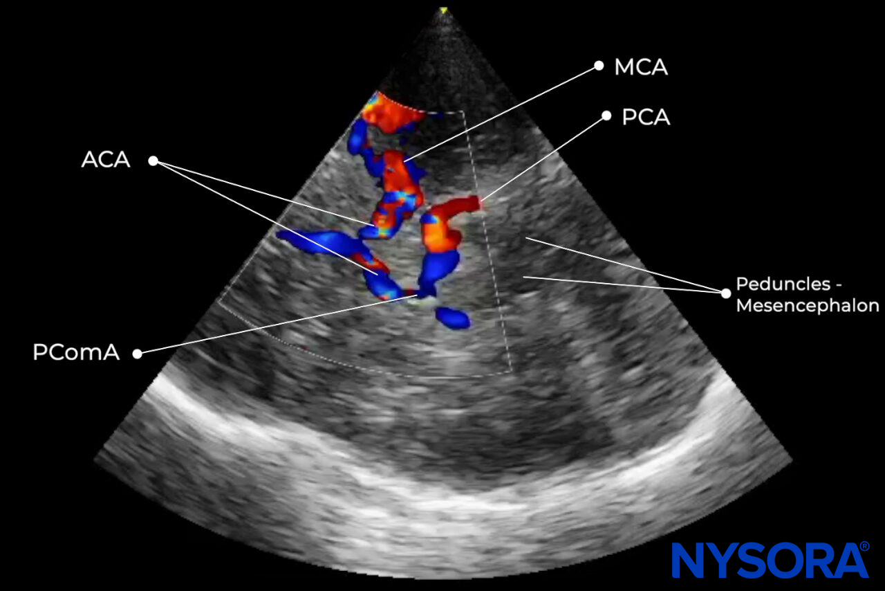
Color Doppler of the circle of Willis.
Tips
- The wings of the butterfly, which represent the mesencephalon, are oriented anteriorly.
- Aliasing can be identified in the color Doppler of the circle of Willis and occurs when flow velocities exceed the Nyquist limit.
Note
Ultrasound Doppler waves are (nearly) parallel to the MCA flow so no angle correction is needed.
Diencephalic plane
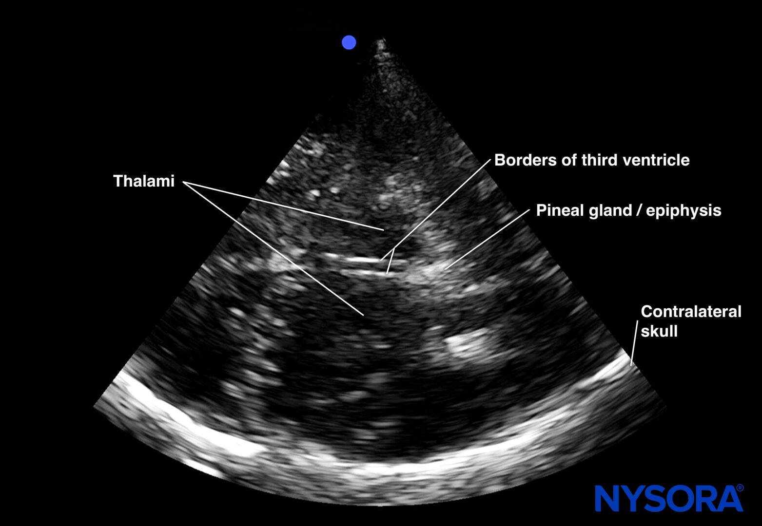
Ultrasound view of the diencephalic plane.
The diencephalic plane can be observed by tilting the transducer approximately 10° cranially. The third ventricle is often visualized first in the diencephalic plane as 2 parallel lines on the midline. Lateral to the third ventricle, the thalami appear as hypoechoic areas. Posterior to the third ventricle, the pineal gland can be identified.
Use:
This view can be used to assess midline shift.
Tips
- The distance between the walls of the third ventricle is usually less than 10 mm.
- This distance is an indicative measure for hydrocephalus and/or brain atrophy
Overview
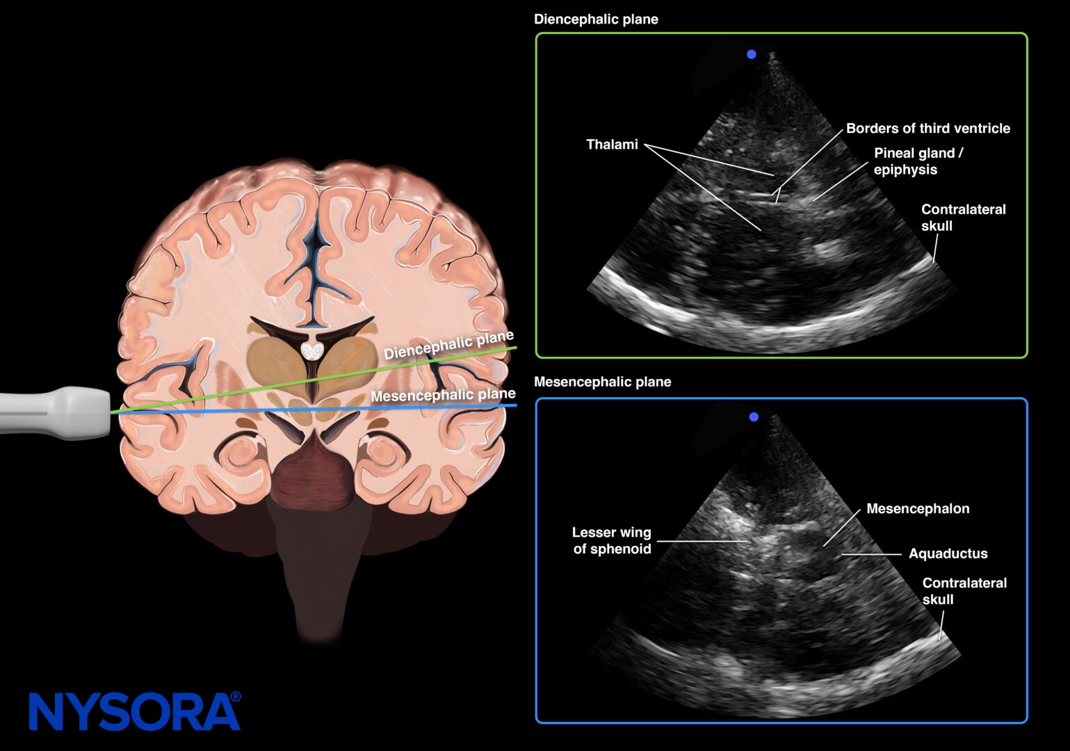
Overview of the diencephalic and mesencephalic planes.