Explore NYSORA knowledge base for free:
Explore NYSORA knowledge base for free:

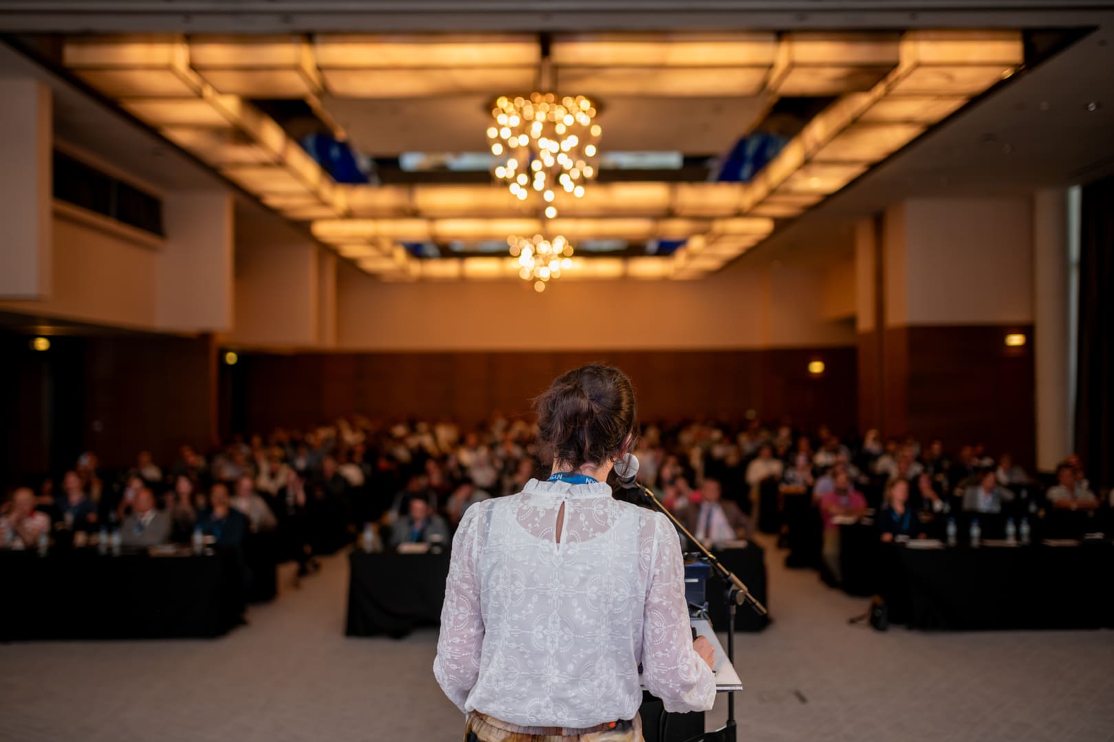
A comprehensive, case-driven, educational experience designed for anesthesiologists committed to advancing their expertise in regional anesthesia, perioperative medicine, and ultrasound-guided techniques. This conference delivers practical clinical updates, contemporary management strategies, and clinically focused education aimed at improving daily practice and patient outcomes.
Cutting-Edge Clinical Updates
The program focuses on the most relevant developments shaping modern anesthesia practice. Faculty present practical insights on topics such as spinal versus general anesthesia for joint replacement, complications of nerve blocks and how to avoid them, emerging regional anesthesia techniques, perioperative anticoagulation management, and the evolving role of fascial plane blocks.
Participants will gain clarity on updated protocols, current evidence, and how these developments influence clinical decision-making in everyday practice.
Practical Techniques & Applied Clinical Learning
Through focused lectures and case-based discussions, faculty present practical strategies for performing reliable nerve blocks, interpreting ultrasound anatomy, and optimizing perioperative analgesia.
Sessions highlight:
• Essential ultrasound anatomy from the neck to the lower extremity
• Updated techniques for upper and lower extremity blocks
• Truncal and fascial plane block strategies
• Catheter techniques and perioperative pain management approaches
• Strategies to prevent complications and improve block reliability
The program emphasizes practical knowledge and clinical insights that can be directly applied to daily anesthesia practice.
NYSORA Protocols & Structured Clinical Approaches
The course introduces structured NYSORA methodologies for performing nerve blocks, managing perioperative analgesia, and optimizing anesthesia workflows. Participants will learn standardized, reproducible techniques designed to enhance safety, efficiency, and consistency in regional anesthesia practice.
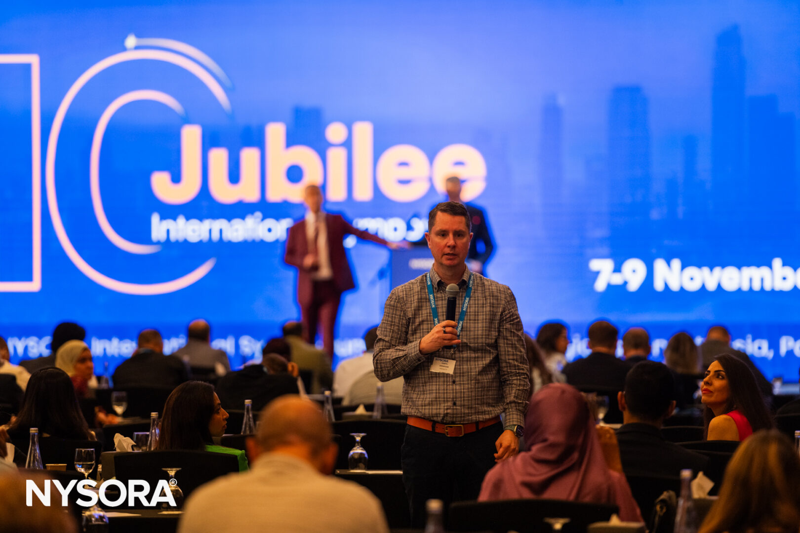
Interactive Learning & Expert Faculty
Interactive Q&A sessions and discussion periods are integrated throughout the program. International faculty share practical insights from daily clinical practice, offering strategies to manage complex cases and avoid common pitfalls.
The format encourages open discussion, peer learning, and meaningful exchange of experience among participants and experts.
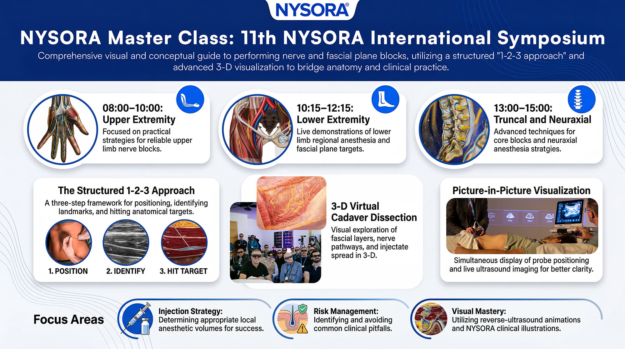
Optional Sunday Demonstration Masterclass
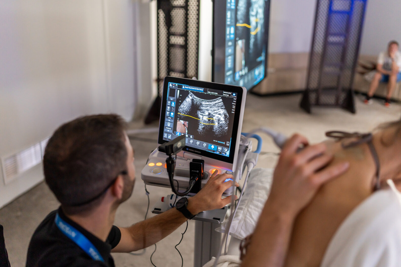
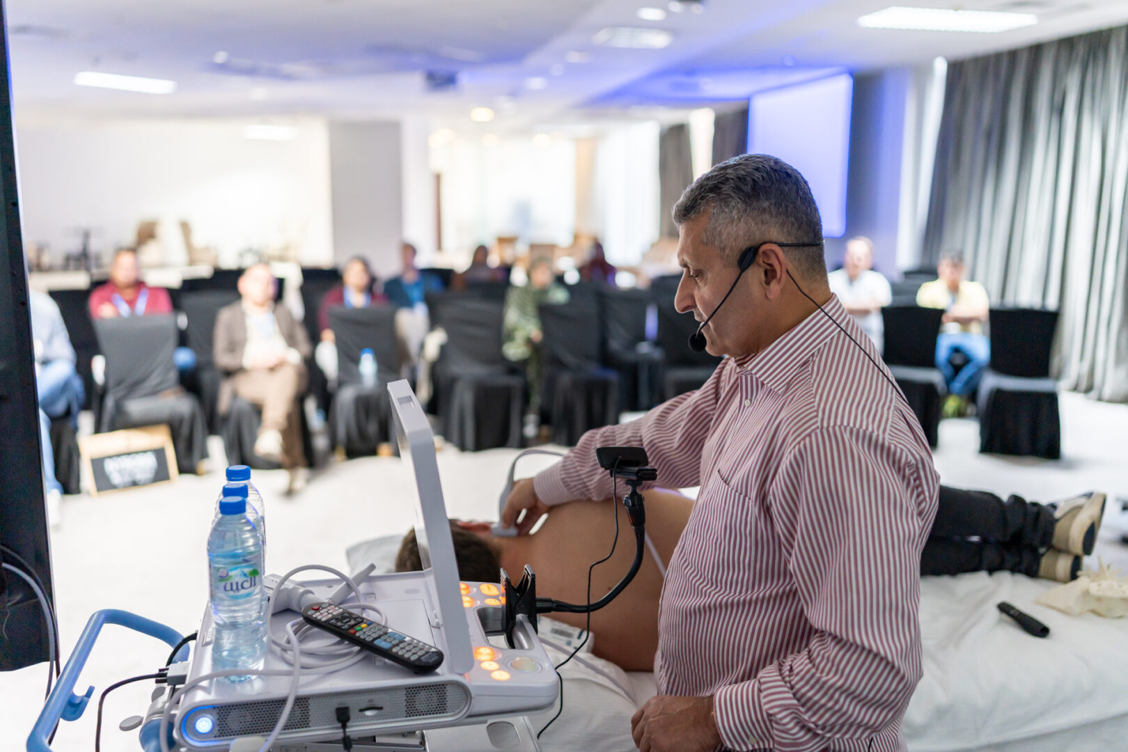
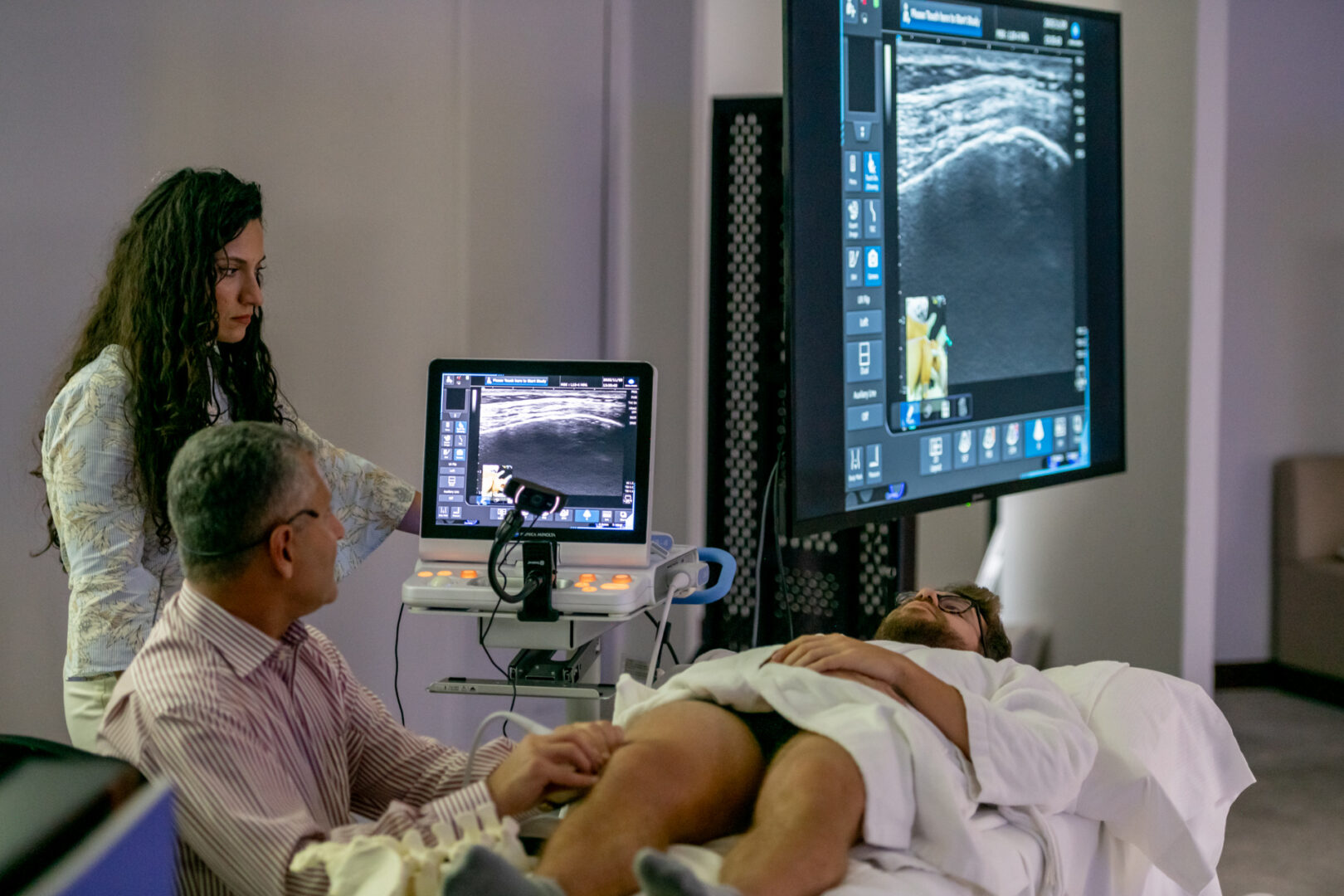
An optional Sunday masterclass provides an in-depth visual exploration of regional anesthesia techniques through live ultrasound-guided demonstrations of upper extremity, lower extremity, truncal, and neuraxial blocks.
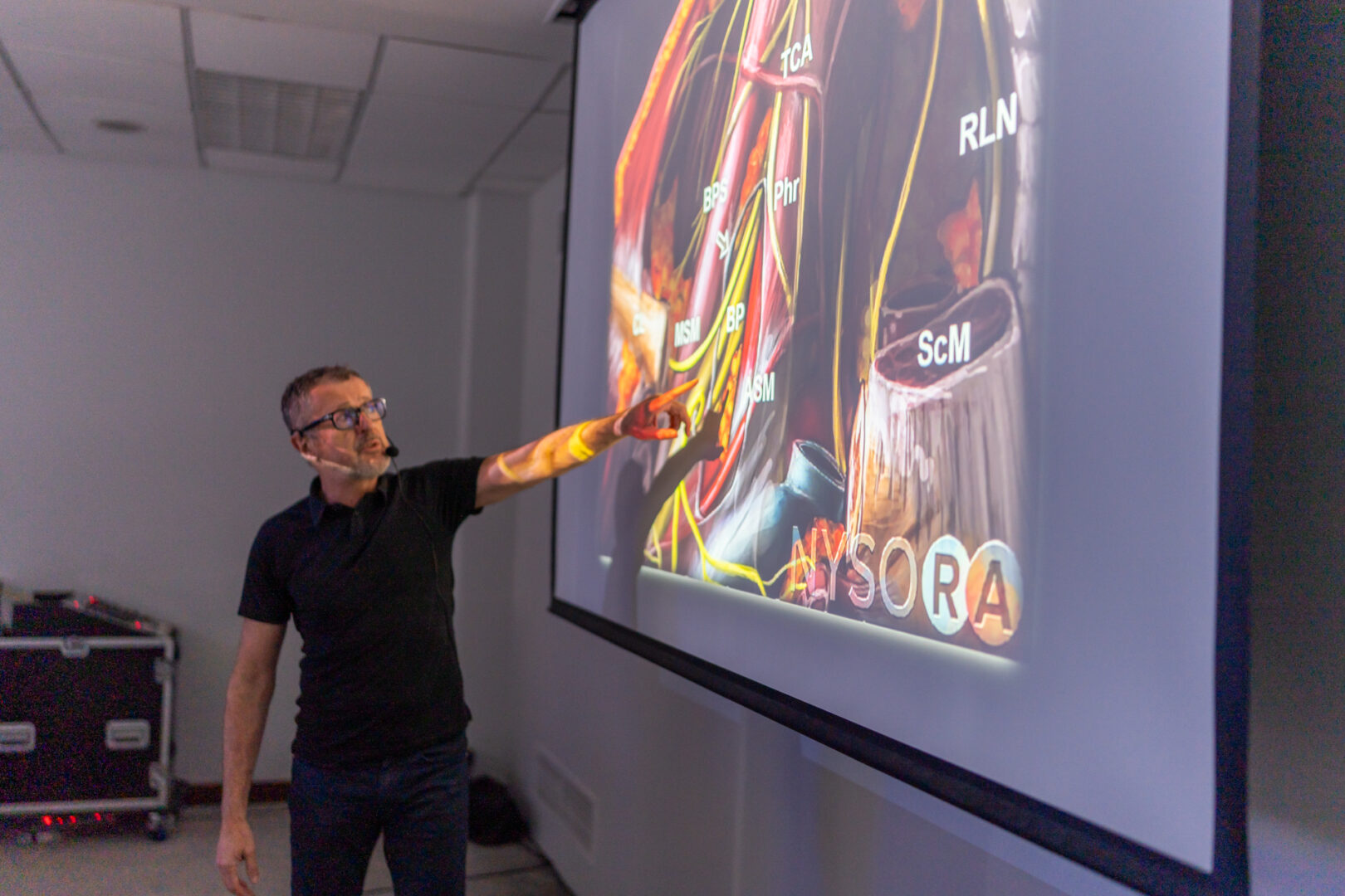
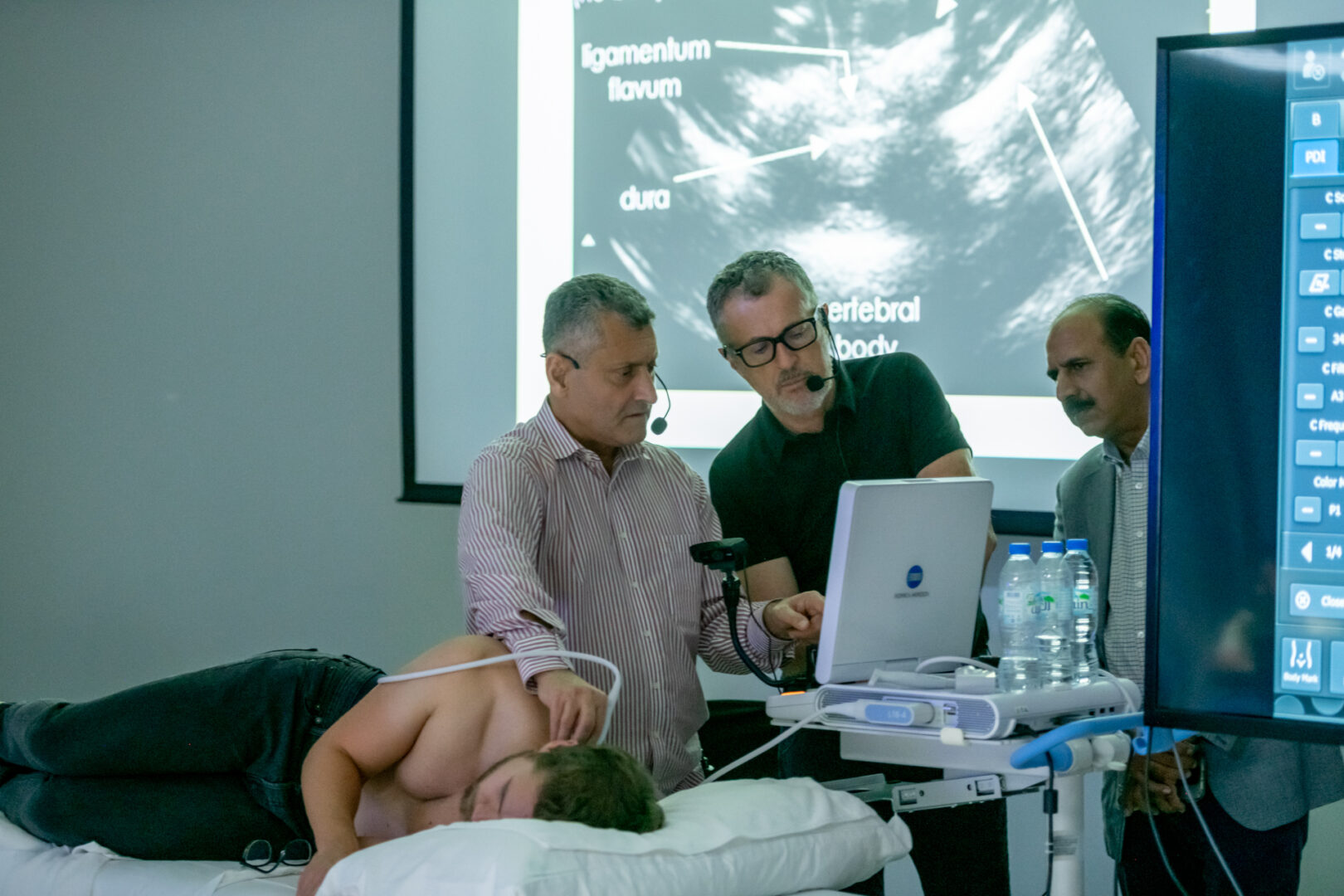
This master-class style program focuses on the practical strategy behind performing reliable nerve blocks and fascial plane blocks. Through simultaneous live ultrasound scanning demonstrations, faculty will present NYSORA’s structured 1-2-3 approach to regional anesthesia, highlighting probe positioning, surface anatomy landmarks, and the key anatomical targets required for block success.
Teaching is delivered in a dynamic picture-in-picture format, allowing participants to see both probe position and ultrasound images simultaneously, complemented by clinical videos, NYSORA illustrations, and reverse-ultrasound animations.
A unique highlight of the session is the 3-D virtual cadaver dissection, enabling participants to explore fascial layers, nerve pathways, and injectate spread in three dimensions. This immersive visualization bridges the gap between anatomy, ultrasound imaging, and block technique.
Faculty will emphasize clinical decision-making, including injection strategies, appropriate local anesthetic volumes, and common pitfalls to avoid.
Designed to accommodate a large audience seeking an in-depth technique masterclass, this program does not include hands-on training but provides a comprehensive visual and conceptual understanding valuable for clinicians at all levels of experience.


Afternoon Session
14:00 - 17:30
Morning Session
09:00 - 12:40
Afternoon Session
12:40 - 17:10
Morning Session
08:00 - 12:15
Afternoon Session
13:00 - 15:00


No Registration Info.
Extend your learning beyond lectures with immersive, faculty-led demonstrations designed to translate directly into clinical practice.
Note: Participation in the Sunday live demonstrations is available at an additional fee of $180.
Director – NYSORA, Anesthesiologist, Professor of Anesthesiology, Department of Anesthesiology, ZOL, Genk, Belgium

Consultant Anesthesia & Pain Management
Abu Dhabi Stem Cell Centre – Yas Clinic – Abu Dhabi – UAE
Founder of Gulf Pain School
Adjunct Clinical Professor at Lerner Medical College – Ohio – USA
President World Society of Pain Clinician (WSPC)
Anesthesiologist at Ziekenhuis Oost-Limburg (ZOL), Genk, Belgium
This symposium is suitable for anesthesiology professionals including consultants, trainees, CRNAs, anesthesia nurses, and perioperative team members, as well as . Educators and researchers focused on improving anesthesia knowledge and patient outcomes.
The earlybird discount is $ enter price. Registration must be made prior to enter date.
Please check your spam or junk folder first. If the confirmation still hasn’t arrived, reach out to us at [email protected] and our team will be happy to help.
All cancellations have to be either made using the view or modify link on the bottom of your confirmation email, or in writing to [email protected]. Cancellation terms:
All registered participants will receive an email with access details for their complimentary 3-month subscription.
The group access email is sent approximately one month before the event and includes everything you need to log in and start exploring the course materials.
If you do not receive this email or experience any login issues, please contact us for assistance.
If we ever need to cancel a course, we will let you know as early as possible. You will be able to choose between a full refund of your registration fee or a transfer to another NYSORA program. In addition we would offer an extension of your complimentary subscription. Please note that we cannot reimburse travel or accommodation costs, so we strongly recommend considering travel insurance to protect any non-refundable expenses.
Every now and then, we may need to update the schedule or faculty lineup. Any adjustments will always be reflected on the agenda page of our website. It’s a good idea to check in from time to time to stay up to date with the latest details.
Email us at [email protected] with the subject line “Assistance with [event name]” and include a few details about what you need. One of our team will get back to you as quickly as possible.
As in past events, a professional photographer/videographer may take photos and/or videos of participants at the event programs.


Join our mailing list and get weekly educational updates delivered straight to your inbox.