Learn
Anesthesiology
Regional Anesthesia
Pain Management
IV Access
POCUS
Anesthesia Information for Patients
NYSORA360
Apps
Anesthesia
Nerve Blocks App
Anesthesia Assistant App
Pain Medicine
US Pain Management App
Pain Medicine Assistant App
Imaging & Access
POCUS App
IV Access App
MSK Knee App
Veterinary
VetRA App
View all apps
Books
Regional Anesthesia
Nerve Block Manual
Regional Anesthesia Updates
Anesthesiology
Anesthesiology Manual
Anesthesiology Review
Anesthesia Updates 2025
Anesthesia Updates 2026
Pediatric Anesthesia Updates
Airway Management Updates
View all books & manuals
Pain Medicine
US Interventional Pain Manual
Pain Medicine Updates
Nursing
Mastering Difficult IV Access
PACU Nursing Manual
Veterinary
RA Veterinary Manual
Events
Conferences
Workshops
Boot Camps
View all events
News
Education
Industry
Media
Partners
Industry Marketing
Institutions
About
For Clinicians
Community
Fellowship
×
Learn
Anesthesiology
Regional Anesthesia
Pain Management
IV Access
POCUS
Anesthesia Information for Patients
NYSORA360
Apps
Nerve Blocks App
Anesthesia Assistant App
View all apps
Books
Events
Conferences
Workshops
Boot Camps
View all events
News
Education
Industry
Media
Partners
Industry Marketing
Institutions
About
For Clinicians
Community
Fellowship
Profile
Log In
POCUS
Midline shift
Cerebral vasospasm
Cerebral circulatory arrest
Intracranial hypertension
Essential transcranial Doppler
eFAST
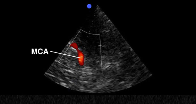
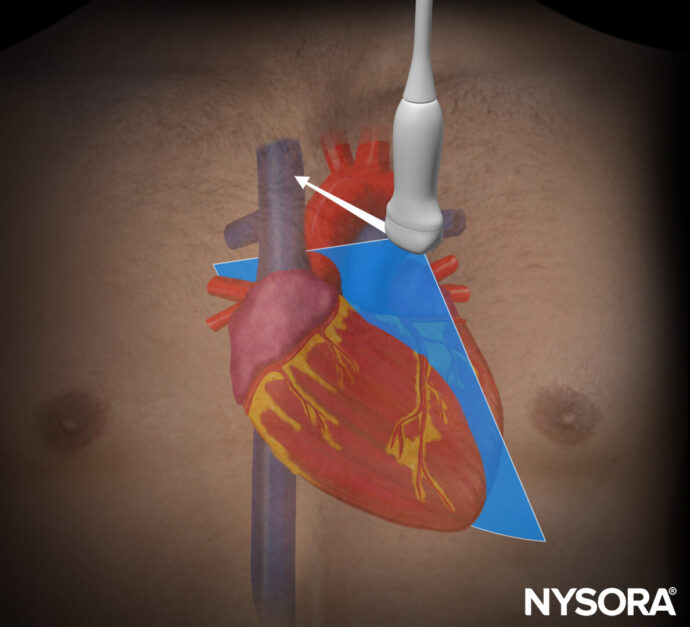
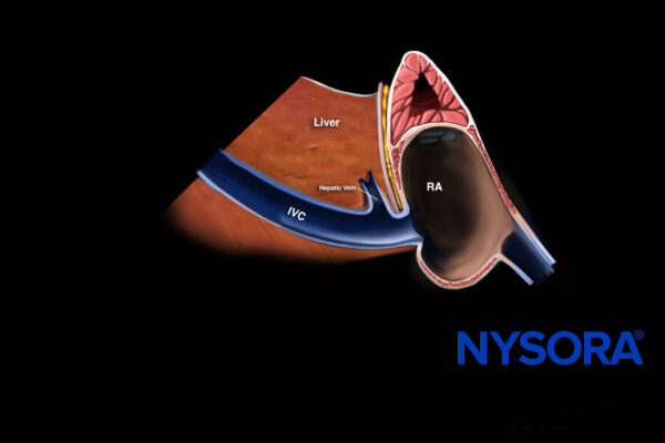
Central venous catheter position – Rapid atrial swirl sign (RASS)
Basic valve assessment
Volume status assessment
Pericardial effusion – tamponade
1
2
3
...
>

 VetRA App

 VetRA App
 Nerve Blocks App
 Pain Medicine Assistant App
 POCUS App
 IV Access App
 MSK Knee App
 Nerve Block Manual
 Regional Anesthesia Updates
 Anesthesiology Manual
 Anesthesiology Review
 Anesthesia Updates 2025
 Anesthesia Updates 2026
 Pediatric Anesthesia Updates
 Airway Management Updates
 US Interventional Pain Manual
 Pain Medicine Updates
 Mastering Difficult IV Access
 PACU Nursing Manual
 RA Veterinary Manual
 About
About