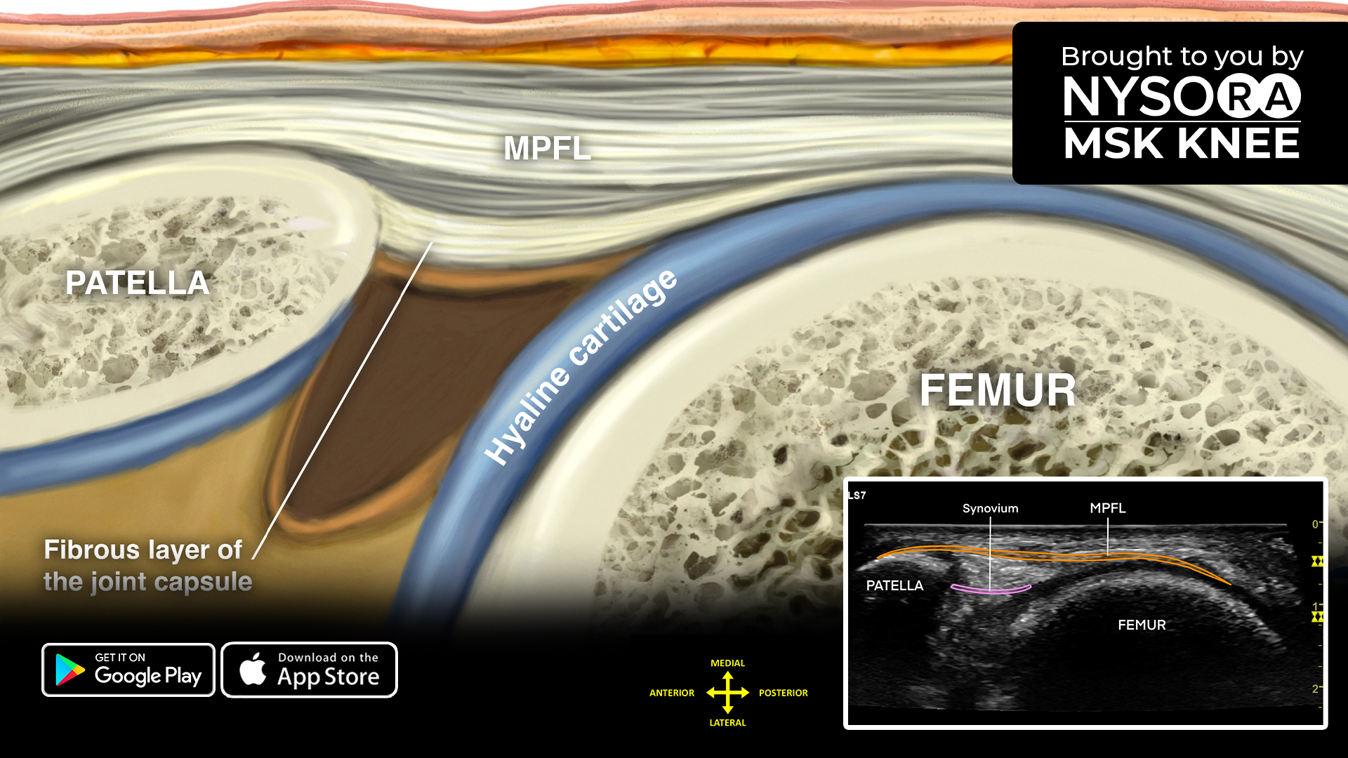


The medial patellofemoral ligament is a part of the complex network of soft tissues that stabilize the knee and as such requires particular attention when scanned.
Here are 4 top tips for scanning the medial patellofemoral ligament
- Place the patient in a supine position with the knee flexed 90°.
- Bridge the transducer over the medial edge of the patella and medial femoral condyle.
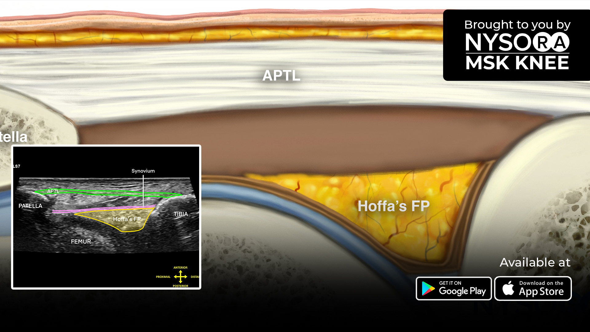
- Identify the medial patellofemoral ligament. Another anatomical structure in this view is the synovium, which is located inside the joint capsule.
- Pay attention to the integrity of the medial patellofemoral ligament. Since the knee is flexed, the ligament will be tightened.
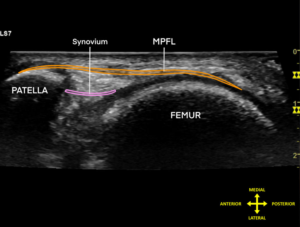
Sonoanatomy
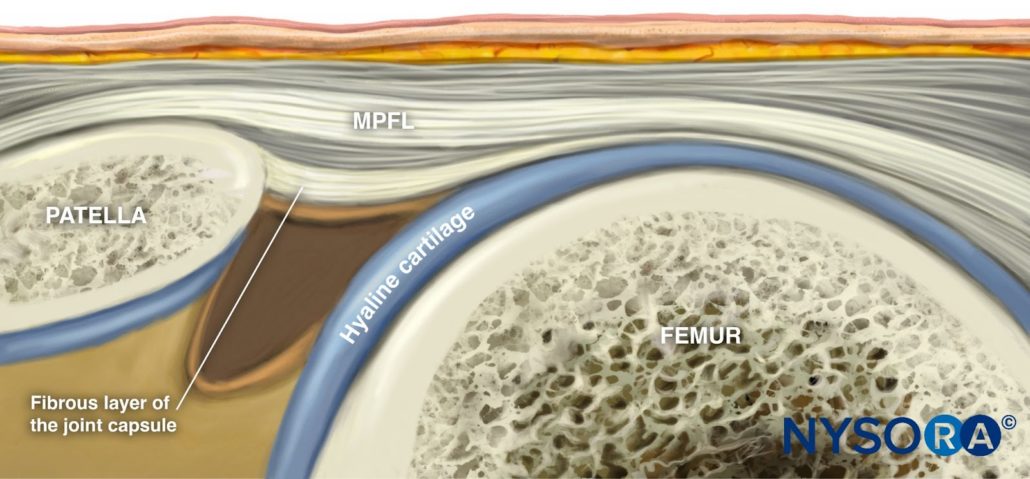
Reverse Ultrasound Anatomy
Comparison of sonoanatomy and reverse ultrasound anatomy of the medial patellofemoral ligament.
Download the MSK App for more tips, and check out the all-new reverse ultrasound anatomy illustration and slider image added in “Sonoanatomy of the Medial Knee > Medial Patellofemoral Ligament”, as well as the most practical and applicable techniques in musculoskeletal ultrasound anatomy and regenerative therapy of the knee.