Important surface anatomy landmarks that should be identified prior to sonographic assessment of the spine include:
Spinous processes to ascertain midline and to assess for abnormal spine curvature: Due to delayed fusion in neonates and infants, these may be palpable as two adjacent bony landmarks.
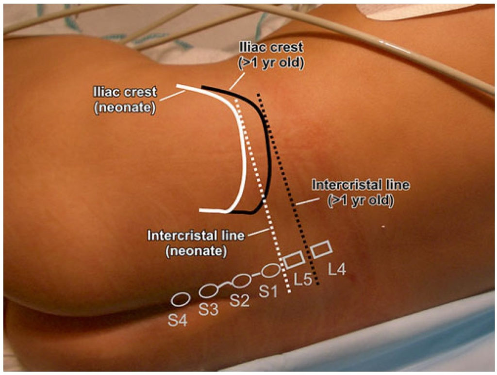
Iliac crests: An imaginary line between the anterior iliac crests, commonly known as the intercristal (or Truffier’s) line, will cross the L5–S1 interspace in neonates and infants less than 1-year-old and L4–L5 in older children (Fig. 2).
Shoulders: Ensure that the left shoulder is not rotated forward and that both shoulders remain square to the bed. This will help ensure effective upper trunk flexion and alignment of the thoracic and lumbar spine, which may help with eventual dural puncture success.
Fig. 2 Surface anatomy for pediatric spinal anesthesia. White and black lines indicate positions of iliac crests and intercristal line for neonates and children over 1-year-old, respectively.
Pediatric Atlas of Ultrasound and Nerve Stimulation-Guided Regional Anesthesia
Enriched with NextLevel CME™ technology: – Make notes in seconds and never lose them – Insert your own images, infographics – Add and watch videos inside your notes – Attach PDFs, articles, website links – Listen to the audio