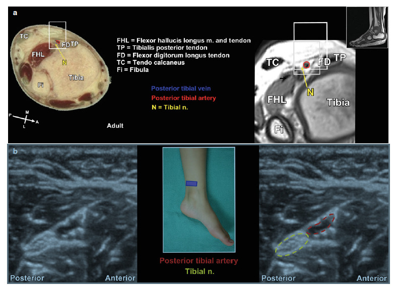
Posterior tibial nerve: The probe is positioned in transverse (short) axis to the nerve just posterior and inferior to the medial malleolus. Alternatively, the nerve can be identified in the distal quarter of the lower leg above the medial malleolus (Fig. 3).
Color Doppler is helpful to localize the nerve at the above locations since in each one the nerve lies posterior and deep to the posterior tibial artery. The nerve should be localized before it branches into the medial and lateral plantar nerves.
Fig. 3 (a) VHVS and MRI images of major anatomical structures surrounding the tibial nerve. (b) Ultrasound image of major anatomical structures surrounding the tibial nerve.
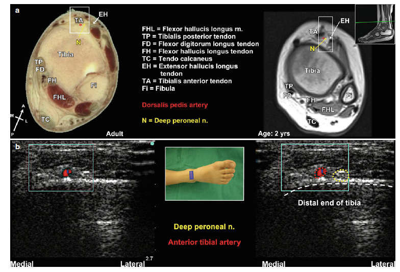
Deep peroneal nerve: The probe is placed in transverse (short) axis to the nerve at the anterior surface of the ankle joint. Alternatively, the nerve can also be found in the distal quarter of the lower leg above the ankle joint (Fig. 5). However, the nerve itself can be difficult to see, and only the artery can be consistently located.
Color Doppler can be used at both locations to illuminate the anterior tibial artery lying medial to the nerve.
Fig. 5 (a) VHVS and MRI images of major anatomical structures surrounding the deep peroneal nerve. (b) Ultrasound image of major anatomical structures surrounding the deep peroneal nerve.
Pediatric Atlas of Ultrasound and Nerve Stimulation-Guided Regional Anesthesia
Enriched with NextLevel CME™ technology: – Make notes in seconds and never lose them – Insert your own images, infographics – Add and watch videos inside your notes – Attach PDFs, articles, website links – Listen to the audio