NYSORA MSK KNEE App - NYSORA

Explore NYSORA knowledge base for free:

Musculoskeletal Ultrasound Anatomy and Regenerative Therapy for the Knee

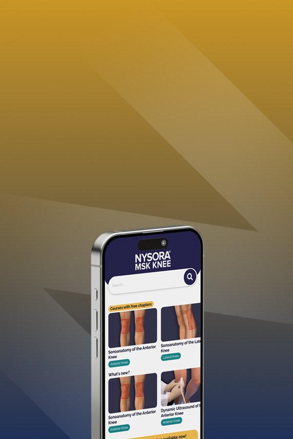
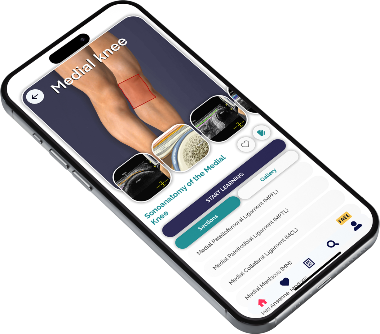
The most practical App on MSK ultrasound examinations for the knee

The Most Practical and Applicable Techniques in Musculoskeletal Ultrasound Anatomy and Regenerative Therapy for the Knee.
Key Benefits

NYSORA Knee Ultrasound Quality

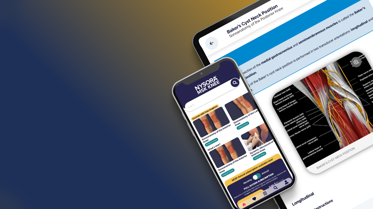
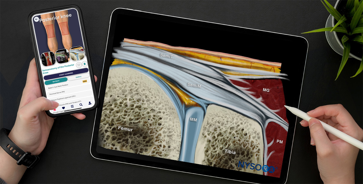
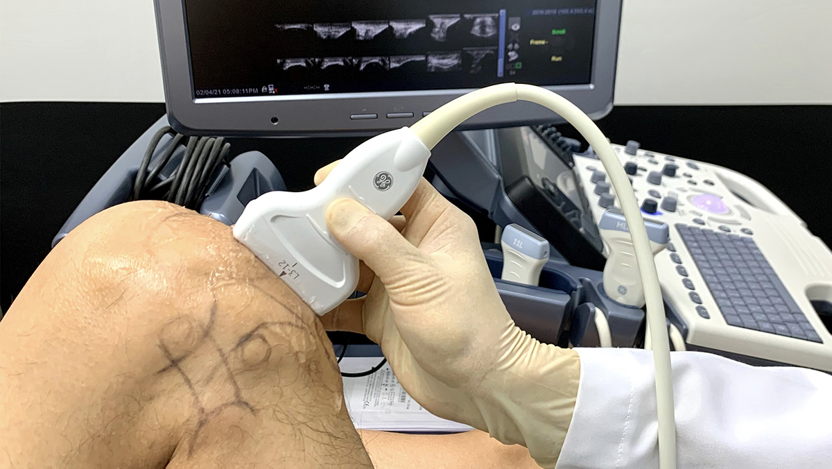
Clear ultrasound images, illustrations, functional anatomy, dynamic tests, animations, and ultrasound-guided MSK procedures.

Step-by-Step Approach

Sonoanatomy of the: anterior, lateral, medial, and posterior knee, varus and valgus tests, and flexion and extension tests in different patient positions.

Optimized App for Mobile Phones & Tablets

Get ready for a mobile app covering all of the latest innovations in musculoskeletal ultrasound and regenerative injection therapy.

Discover the benefits
Detailed Insights
Visual tools that aid in thorough MSK knee evaluations.
Pathology Insights
Detailed scans and explanations for common knee pathologies.
Start Your Free 7-Day Trial of the NYSORA MSK Ultrasound Knee App Today and Transform Your Musculoskeletal Ultrasound Skills!

Dr. Stanley Lam

Comprehensive Ultrasound Image Gallery
Regularly enhanced by NYSORA’s illustrations and animations;
Regular Updates
Regular content updates and user experience upgrades… And more!
NYSORA Quality
Gold-Standard Content
Free Trial
Test it out with a 7-day free trial!
Curious about which techniques are included in the app? Take a look at the detailed list below!

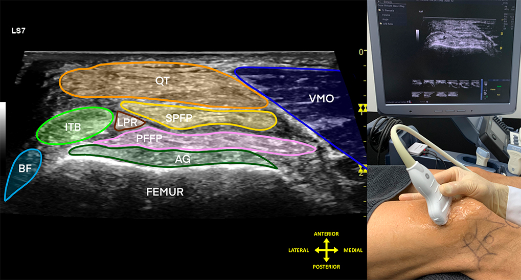
Sonoanatomy of the anterior knee

  • Quadriceps tendon (QT)
  • Patella
  • Patellar tendon (PT)
  • Superior ⅓ of the femoral condyle
  • Inferior ⅓ of the femoral condyle
  • Anterior cruciate ligament (ACL)

Dynamic ultrasound of the anterior knee

  • Patellofemoral joint stability or friction test
  • Dynamic visualization of the ACL
  • Flexion and extension of the knee:
    Standing and sitting
    Walking

Sonoanatomy of the lateral knee

  • Bony landmarks and related anatomical structures
  • Patellotibial ligament (PTL)
  • Patellofemoral ligament (PFL)
  • Iliotibial band (ITB)
  • Lateral collateral ligament (LCL)
  • Biceps femoris muscle (BFM) and tendon
  • The “LCL – ITB – LCL – biceps femoris dance”
  • Anterolateral ligament (ALL)
  • Popliteal origin
  • Lateral meniscus (LM)
  • Proximal tibiofibular ligament (TFL)

Dynamic tests of the lateral knee

  • Introduction
  • Varus and valgus stress test of the lateral collateral ligament (LCL)
  • Varus and valgus stress test of the lateral meniscus (LM)
  • Flexion and extension of the knee:
    Supine
  • Flexion and extension of the knee:
    Seated
  • Flexion and extension of the knee:
    Semi-squat
  • Flexion and extension of the knee:
    Stepping down
    Walking

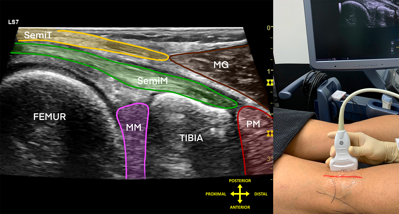
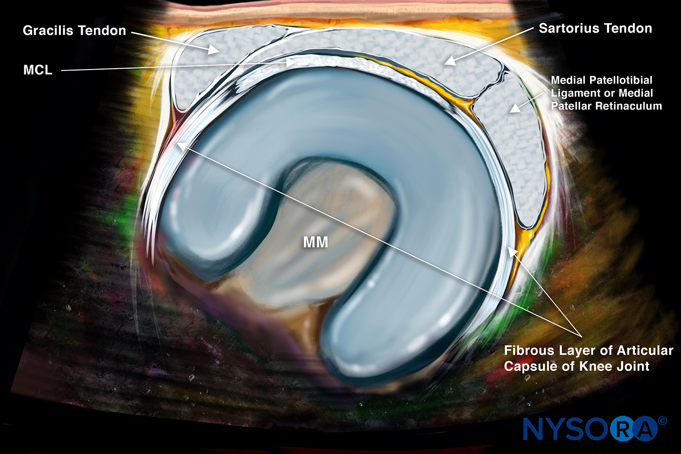
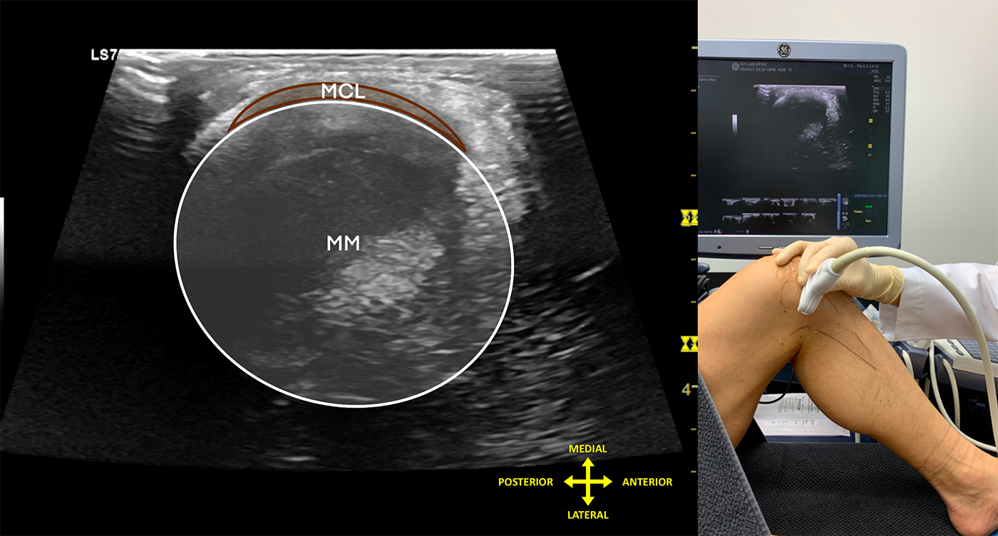
Sonoanatomy of the medial knee

  • Medial patellofemoral ligament (MPFL)
  • Medial patellotibial ligament (MPTL)
  • Medial collateral ligament (MCL)
  • Medial meniscus (MM)
  • Pes anserine tendons

Dynamic tests of the medial knee

  • Varus and valgus stress test of the medial collateral ligament (MCL)
  • Varus and valgus stress test of the medial meniscus (MM)
  • Varus and valgus stress test of the lateral meniscus (LM)
  • Flexion and extension of the knee:
    Supine
  • Flexion and extension of the knee:
    Seated
  • Flexion and extension of the knee:
    Semi-squat
    Walking

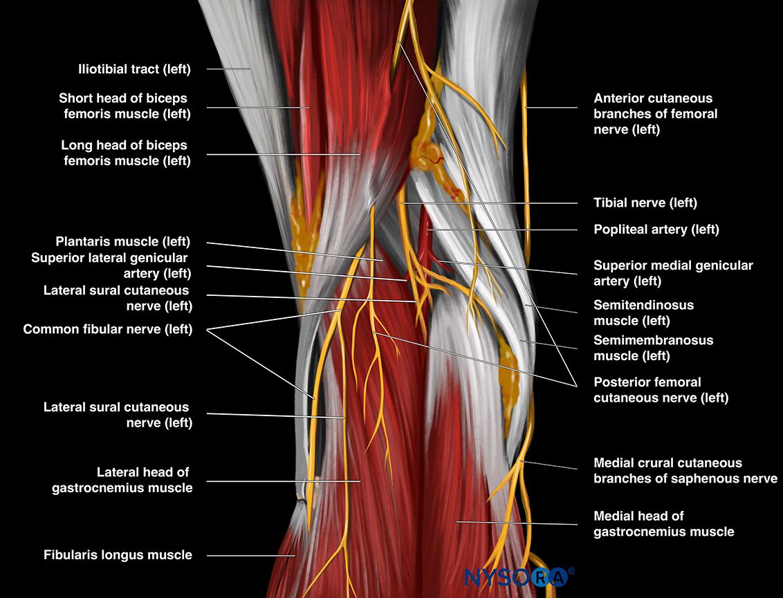
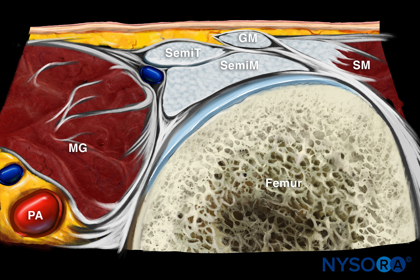
Sonoanatomy of the posterior knee

  • Baker’s cyst neck position
  • Peroneal nerve (PN)
  • Oblique popliteal ligament (OPL)
  • Popliteus muscle (PM) and tendon (PT)
  • Arcuate ligament (AL)
  • Lateral gastrocnemius (LG) origin
  • Meniscofemoral ligament (MFL) and posterior cruciate ligament (PCL)

Dynamic ultrasound of the posterior knee

  • Dynamic visualization of the PCL
  • Varus and valgus stress test of the medial meniscus (MM)
  • Varus and valgus stress test of the lateral meniscus (LM)
  • Flexion and extension of the knee:
    Semi-squat
    Walking

Extensive Image Gallery​

App Reviews

Download MSK App and access the full version for 7 days!

Name and Surname Anesthesiologist
Excellent application. You have made Regional anesthesia simple and interesting. Thank you so much.
Name and Surname Anesthesiologist
Fantastic app. Beautiful images. Lot of hard work has gone into making this app.
Name and Surname Anesthesiologist
Beautiful images combined with clear and practical information on all kinds of blocks!
Name and Surname Anesthesiologist
The best resource for information on nerve blocks. I already know I’m going to get a lot of use from this app. Thanks NYSORA!
Name and Surname Anesthesiologist
Excellent application. You have made Regional anesthesia simple and interesting. Thank you so much.
Name and Surname Anesthesiologist
Fantastic app. Beautiful images. Lot of hard work has gone into making this app.
Name and Surname Anesthesiologist
Beautiful images combined with clear and practical information on all kinds of blocks!
Name and Surname Anesthesiologist
The best resource for information on nerve blocks. I already know I’m going to get a lot of use from this app. Thanks NYSORA!
Download MSK App Today and Get Started With a 7-day Free Trial
Frequently asked questions

Musculoskeletal (MSK) ultrasound is a medical imaging technique that uses high-frequency sound waves to produce real-time images of muscles, tendons, ligaments, joints, and soft tissues. It allows for detailed visualization of musculoskeletal structures to aid in diagnosis and treatment planning.

An MSK ultrasound can show various abnormalities or conditions affecting the muscles, tendons, ligaments, joints, and soft tissues. It can reveal injuries such as tears, inflammation, fluid accumulation, cysts, tumors, and structural abnormalities. Additionally, MSK ultrasound can assess blood flow to tissues and guide interventional procedures such as injections or aspirations.

MSK ultrasound can diagnose a wide range of musculoskeletal conditions and injuries, including tendonitis, muscle strains, ligament tears, bursitis, arthritis, nerve entrapments, joint effusions, and injuries.

Learning MSK ultrasound requires specialized training and practice but is certainly attainable with dedication and proper instruction. Healthcare professionals, including physicians, physical therapists, and sonographers, can undergo training programs and hands-on workshops to develop the skills necessary for proficient MSK ultrasound imaging. With continued practice and experience, proficiency in MSK ultrasound can be achieved to accurately diagnose and manage musculoskeletal disorders.