
You are currently viewing a preview of chapter content.
Transducer position
- Place the transducer transversely over the medial aspect of the knee, approximately 2-3 cm above the patella.
Scanning
- Slide the transducer proximally/distally to identify the distal femoral shaft and popliteal artery.
| Tips: |
|---|
|
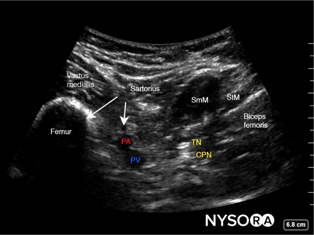
Fig. SmM, semimembranosus muscle; PA, popliteal artery; PV, popliteal vein; TN, tibial nerve; CPN, common peroneal nerve.
Needle insertion
- Insert the needle in-plane, from the anteromedial aspect of the knee, toward the space between the popliteal artery and femur.
- When the posterior aspect of the popliteal artery is reached, inject 2 mL of the local anesthetic to confirm proper needle position.
| Tips: |
|---|
|
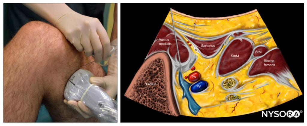
Fig. IPACK block; Reverse Ultrasound Anatomy. SmM, semimembranosus muscle; StM, semitendinosus muscle; PA, popliteal artery; PV, popliteal vein; TN, tibial nerve; CPN, common peroneal nerve.
Alternative approach
- Place the transducer over the popliteal fossa crease in order to visualize the tibial nerve, common peroneal nerve, popliteal artery, and femoral condyles.
- From this location, slide the transducer proximally until the flat posterior aspect of the shaft of the femur becomes visible.
- Insert the needle in-plane from the medial (or lateral) side, toward the space between the popliteal artery and femur.
- Inject 1-2 mL of local anesthetic to confirm correct needle position.
- Complete the block with 15-20 mL.
- This approach can be performed with the patient either in prone or supine position.

NYSORA Compendium of Regional Anesthesia
Enriched with Nysora LMS technology:
– Make notes in seconds and never lose them
– Insert your own images, infographics
– Add and watch videos inside your notes
– Attach PDFs, articles, website links
– Listen to the audio


