
Atlas of Ultrasound-Guided Procedures in Interventional Pain Management - NYSORA

Explore NYSORA knowledge base for free:

Welcome to the
NextLevel NYSORA.

New

Atlas of Ultrasound-Guided Procedures in Interventional Pain Management

Author: Samer Narouze

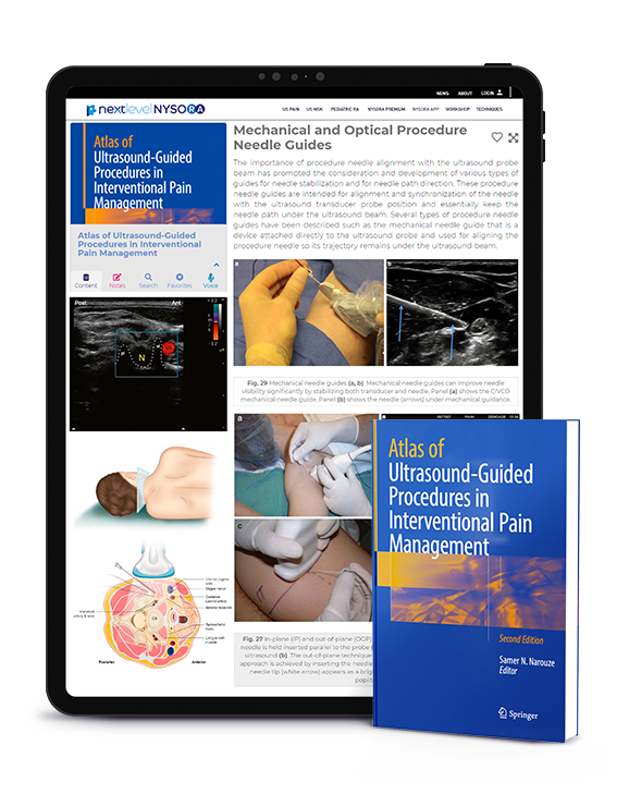
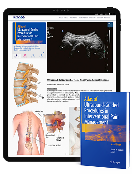
Atlas of Ultrasound-Guided Procedures in Interventional Pain Management is the #1 primer and a must-read for both novices and experts performing invasive procedures for pain relief.
  • PRACTICAL. Step-by-step guidance from the world’s leading USPM experts in their area of subspecialty expertise.
  • UNDERSTAND. Coverage of acute perioperative pain, chronic pain clinic settings, and MSK applications.
  • BE INSPIRED. NextLevel NYSORA will motivate you to study with our proprietary visual cognitive aids for a unique journey through courses and lessons.

Preview

Review

Atlas of Ultrasound-Guided Procedures in Interventional Pain Management , Review

Introduction “Atlas of Ultrasound-Guided Procedures in Interventional Pain Management” Atlas of Ultrasound-Guided Procedures in Interventional Pain Management was a huge undertaking. For much of the past decade, fluoroscopy held sway as the favorite imaging tool of many practitioners performing interventional pain procedures. Quite recently, ultrasound has emerged as a “challenger” to this well-established modality. The growing popularity of ultrasound application in regional anesthesia and pain medicine reflects a shift in contemporary views about imaging for nerve localization and target-specific injections. For regional anesthesia, ultrasound has already made a marked impact by transforming antiquated clinical practice into a modern science. No bedside tool ever before has allowed practitioners to visualize needle advancement in real time and observe local anesthetic spread around nerve structures. For interventional pain procedures, I believe this radiation-free, point-of-care technology will also find its unique role and utility in pain medicine and can complement some of the imaging demands not met by fluoroscopy, computed tomography, and magnetic resonance imaging. And over time, practitioners will discover new benefits of this technology, especially for dynamic assessment of musculoskeletal pain conditions and improving accuracy of needle injection for small nerves, soft tissue, tendons, and joints. Ultrasound application for pain medicine is an evolving subspecialty area Most conventional pain interventionists skilled in fluoroscopy will find it necessary to undertake some special learning and training to acquire a new set of cognitive and technical skills before they can optimally integrate ultrasound into their clinical practices. Although continuing medical educational events help facilitate the learning process and skill development, they are often limited in breadth, depth, and training duration. This is why the arrival of this comprehensive text, Atlas of Ultrasound-Guided Procedures in Interventional Pain Management, is so timely and welcome. To my knowledge, this is the first illustrative atlas of its kind that addresses the
Read More »

Table of contents

  • Introduction
  • C-Arm FDCT
  • Ultrasound
  • Intra-Articular Injections
  • Trigger Point and Muscular Injections
  • Zygapophyseal and Medial Branch Blocks
  • Epidural Blocks
  • Sympathetic Blocks
  • Combined US and CT/Fluoroscopy
  • Conclusion
  • References
  • Basic Principles of B-Mode US
  • Generation of Ultrasound Pulses
  • Ultrasound Wavelength and Frequency
  • Ultrasound-Tissue Interaction
  • Recent Innovations in B – Mode Ultrasound
  • Conclusion
  • References
  • Frequency and Probe Selection
  • Depth
  • Gain
  • Time Gain Compensation
  • Focus
  • Presets
  • Color Doppler
  • Power Doppler
  • Compound Imaging
  • Tissue Harmonic Imaging
  • Optimization Button
  • Freeze Button and Image Acquisition
  • References
  • Training and Phantom Simulation
  • High-Fidelity Simulation
  • Combined Ultrasound and Fluoroscopic Phantom Simulators
  • Procedural Needle: Related Visibility Factors
  • Acoustic Impedance as the Basis for Procedure Needle Visualization
  • Size (Gauge) of the Procedure Needle and Its Echogenicity
  • The Skin Insertion Site Selected and Angle of Procedure Needle Passage
  • Echogenic Procedure Needles
  • Procedure Needle Tip
  • The Ultrasound Device and Procedure Needle Visibility
  • Impact of Various Sonographic Modes on Procedure Needle Visibility
  • Frequency of the Ultrasound Probe (AKA Depth) Acoustic Power and Gain
  • Time Gain Compensation and Harmonic Imaging
  • Brightness, Motion, and Doppler Modes
  • 3D and 4D Ultrasound Imaging
  • Recent Advances in Ultrasound Imaging and Procedure Needle Visibility
  • Needle-Probe Alignment
  • “In-Plane” and “Out-Of-Plane” Needle Approach: Classical Probe-Needle Interpositions
  • Oblique Plane Needle Approach for Ultrasound-Guided Pain Management
  • Biplane Needle Imaging Approach for Ultrasound-Guided Pain Management
  • Mechanical and Optical Procedure Needle Guides
  • Advanced Procedure Needle Positioning Systems
  • The “ART” of Scanning for Better Procedure Needle Visualization
  • Ergonomics for Better Procedure Needle Visibility
  • Enhancement and Techniques to Improve Procedure Needle Localization
  • Enhancement with Priming, Insertion of Stylet or Guide Wire, and Vibration
  • Hydrolocalization of the Procedure Needle
  • Procedure Needle Visibility by Agitated Solutions or with Ultrasound Contrast Agents
  • Localization of the Procedure Needle Tip with the Aid of Nerve Stimulation
  • Summary
  • References
  • Cervical Spine
  • Thoracic Spine
  • Lumbar Spine
  • Sacrum
  • Sonoanatomy of the Cervical Spine: Superficial
  • Sonoanatomy of the Cervical Spine: Deep
  • Sonoanatomy of the Thoracic Spine: Superficial
  • Sonoanatomy of the Thoracic Spine: Deep
  • Sonoanatomy of the Lumbar Spine: Superficial
  • Sonoanatomy of the Lumbar Spine: Deep
  • Sonoanatomy of the Sacrum and Sacroiliac Joint: Superficial
  • Sonoanatomy of the Sacrum and Sacroiliac Joint: Deep
  • Indications for Cervical Medial Branch Block
  • Why Ultrasound-Guided Facet Nerve Block? The Literature and our Experience
  • Possible Advantages of Ultrasound for Cervical Facet Nerve Blocks
  • Limitations of Ultrasound
  • Ultrasound-Guided Technique for TON and Cervical Medial Branch Block
  • Identifying the Correct Level: Method 1
  • Identifying the Correct Level: Method 2
  • Practical Performance of Block
  • Conclusion
  • References
  • Indications for Cervical Zygapophyseal Joint Intra-Articular Injection
  • Literature Review of Ultrasound-Guided Cervical Facet Injections
  • Ultrasound-Guided Technique for Cervical Facet Intra-Articular Injection
  • Posterior Approach
  • References
  • Indications
  • Limitations of the Fluoroscopy-Guided Techniques
  • Literature Review and Advantages of Ultrasound-Guided Cervical Nerve Root Block
  • Identification of Small Critical Vessels
  • Why Ultrasound?
  • Sonoanatomy of the Cervical Spine and Identification of the Cervical Level
  • Ultrasound-Guided Technique for Cervical Selective Nerve Root Block
  • References
  • Anatomy
  • Mechanism of block
  • Techniques of TPVB
  • Ultrasound-Guided TPVB
  • Sonoanatomy Relevant for TPVB
  • Techniques of USG TPVB
  • Conclusion
  • References
  • Anatomy
  • Literature Review
  • Scanning Technique
  • Injection Technique
  • Intraarticular Lumbar Zygapophyseal Joint Injection
  • Limitations of Ultrasound-Guided Zygapophysial Nerve and Joint Injection
  • References
  • Ultrasound-Guided Technique
  • Limitations of the Ultrasound-Guided Technique
  • References
  • History
  • Ultrasound Imaging of the Spine
  • Axis of Scan
  • Spinal Sonoanatomy
  • The Water-Based Spine Phantom
  • Ultrasound Imaging of the Sacrum
  • Ultrasound Imaging of the Lumbar Spine
  • Ultrasound Imaging of the Thoracic Spine
  • Ultrasound-Guided CNB
  • Caudal Epidural Injection
  • Lumbar Epidural Injection
  • Thoracic Epidural Injection
  • Spinal Injection
  • The Evidence
  • Education and Training
  • Conclusion
  • References
  • Indications for Caudal Epidural Injection
  • Limitations of the Landmark “Blind” Technique
  • Literature Review of Ultrasound-Guided Caudal Epidural Injections
  • Ultrasound-Guided Caudal Injection Is Better Than the “Blind” Technique
  • Ultrasound-Guided Caudal Injection Is as Effective as the Fluoroscopy-Guided Technique
  • Ultrasound-Guided Technique for Caudal Epidural Injection
  • Limitations of the Ultrasound-Guided Technique
  • References
  • Indications for SIJ Injection
  • Literature Review on Ultrasound-Guided SIJ Injection
  • Technique of Ultrasound-Guided SIJ Injection
  • Limitations of Ultrasound-Guided SIJ Injection
  • References
  • Anatomy
  • The Classic Approach
  • Ultrasound-Guided Technique
  • Ultrasound-Guided TAP Catheter
  • Summary
  • References
  • Anatomy of the Celiac Plexus
  • Current Techniques of the Celiac Plexus Block
  • The Anterior Percutaneous Approach
  • Advantages of US-Guided Anterior Approach
  • Limitations of US-Guided Anterior Approach
  • Sonoanatomy of the Epigastrium and Relevant Structures
  • Percutaneous Ultrasound-Guided Celiac Plexus Block Technique
  • References
  • Ilioinguinal, Iliohypogastric, and Genitofemoral Neuralgia
  • Anatomy
  • Literature Review on Injection Techniques for Ilioinguinal, Iliohypogastric, and Genitofemoral Nerve Block
  • Ultrasound-Guided Technique of Ilioinguinal, Iliohypogastric, and Genitofemoral Nerve block
  • Genital Branch of Genitofemoral Nerve
  • Piriformis Syndrome
  • Anatomy
  • Literature Review on Piriformis Muscle Injections
  • Ultrasound-Guided Technique for Piriformis Muscle Injection
  • Pudendal Neuralgia
  • Anatomy
  • Literature Review on Pudendal Nerve Injections
  • Ultrasound-Guided Technique for Pudendal Nerve Injection
  • Conclusion
  • References
  • Indications
  • Limitations of the Current Technique
  • Literature Review on Ultrasound-Guided Ganglion Impar Block
  • Technique of Ultrasound-Guided Ganglion Impar Block
  • Limitations of the Ultrasound-Guided Technique
  • References
  • Brachial Plexus Anatomy
  • Interscalene Block
  • Supraclavicular Block
  • Infraclavicular Block
  • Axillary Block
  • Anesthetizing Distal Peripheral Nerves in the Upper Extremity
  • Summary
  • References
  • Femoral Nerve Block
  • Anatomy
  • Preparation and Positioning
  • Ultrasound Technique
  • Single-Injection Technique
  • Continuous Catheter Technique
  • Sciatic Nerve Block
  • Anatomy
  • Preparation and Positioning
  • Ultrasound Technique
  • Sciatic Nerve block in the Popliteal Fossa
  • Anatomy
  • Preparation and Positioning
  • Ultrasound Technique
  • Lumbar Plexus Block
  • Anatomy
  • Preparation and Positioning
  • Ultrasound Technique
  • Obturator Nerve Block
  • Anatomy
  • Preparation and Positioning
  • Ultrasound Technique
  • Lateral Femoral Cutaneous Nerve Block
  • Anatomy
  • Preparation and Positioning
  • Ultrasound Technique
  • Saphenous Nerve Block
  • Anatomy
  • Preparation and Positioning
  • Ultrasound Technique
  • Ankle Block
  • Anatomy
  • Preparation and Positioning
  • Ultrasound Technique
  • block of the Posterior Tibial Nerve
  • block of the Deep Peroneal Nerve
  • block of the Proximal Superficial Peroneal Nerve
  • References
  • Applications
  • Overview of Ultrasound-Guided Perineural Catheter Insertion
  • Nerve in Short Axis: Needle Out-of-Plane Approach
  • Nerve in Long Axis: Needle In-Plane
  • Preparation for Ultrasound-Guided Perineural Catheter Insertion
  • Standard Perineural Catheter Equipment
  • Ultrasound-Guided Perineural Catheter Insertion Techniques for Common Surgical Procedures
  • Infraclavicular CPNB
  • Femoral CPNB
  • Subgluteal Sciatic CPNB
  • Popliteal Sciatic CPNB
  • Transversus Abdominis Plane (TAP) CPNB
  • Conclusions
  • References
  • Scanning Technique
  • Ultrasound-Guided Injection Technique for Superficial Trigeminal Nerves
  • Conclusions
  • References
  • Anatomy and Topography of the GON and Adjacent Structures
  • Traditional Method of GON Block: Limitations and Disadvantages
  • Ultrasound-Guided GON Block: Our Technique and Experience
  • Nerve Block
  • Experience and Outlook
  • Conclusion
  • References
  • Anatomy
  • Existing Techniques
  • Technique for Ultrasound-Guided Injection
  • References
  • Lateral Femoral Cutaneous Nerve Block
  • Anatomy
  • Literature Review of Injection Techniques
  • Ultrasound-Guided Block Technique
  • Suprascapular Nerve Block
  • Anatomy
  • Literature Review of Injection Techniques
  • Ultrasound-Guided Block Technique
  • Intercostal Nerve Block
  • Anatomy
  • Literature Review of Injection Techniques
  • Ultrasound-Guided Block Technique
  • Conclusion
  • References
  • Subacromial/Subdeltoid Bursa
  • Anatomy
  • Clinical Presentation
  • Limitations of the Blind Approach
  • Ultrasound-Guided Technique
  • Biceps Tendon Sheath (Biceps – Long Head)
  • Ultrasound-Guided Technique
  • Acromioclavicular Joint
  • Clinical Presentation
  • Ultrasound-Guided Technique
  • Glenohumeral Joint
  • Clinical Presentation
  • Limitations of the Blind Approach
  • Ultrasound-Guided Technique
  • The Rotator Interval Approach
  • Subscapularis Tendon/Subscapularis Bursa
  • Clinical Presentation
  • Ultrasound-Guided Technique
  • Sternoclavicular Joint
  • Clinical Presentation
  • Ultrasound-Guided Technique
  • Conclusion
  • References
  • Anatomy Ultrasound-Guided Carpal Tunnel Injections
  • Literature Review on Ultrasound-Guided Carpal Tunnel Injections
  • Ultrasound-Guided Technique for Carpal Tunnel Injections
  • Anatomy Ultrasound-Guided Trigger Finger Injections
  • Literature Review on Ultrasound-Guided Trigger Finger Injections
  • Ultrasound-Guided Technique for Trigger Finger Injections
  • Anatomy Ultrasound-Guided Wrist Injections
  • Literature Review on Ultrasound-Guided Wrist Injections
  • Ultrasound-Guided Technique for Wrist Injections
  • Anatomy Ultrasound-Guided Injections for Tendon Dysfunction
  • Tendon Dysfunction
  • Ultrasound-Guided Technique for Tendon Dysfunction
  • Anatomy Ultrasound-Guided Elbow Injections
  • Literature Review on Ultrasound-Guided Elbow Injections
  • Ultrasound-Guided Elbow Injection Technique
  • References
  • Anatomy of the Hip Joint
  • Intra-articular Hip Injections
  • Limitations of the Blind Technique
  • Evidence for Ultrasound-Guided Hip Injections
  • Technique of Ultrasound-Guided Hip Injections
  • Conclusions
  • References
  • Limitations of the Current Surface Landmarks Technique
  • Technique for Ultrasound-Guided Knee Joint Injection
  • Conclusion
  • References
  • Technique
  • Nerve Anatomy
  • Nerve Injury
  • Neuritis
  • Nerve Subluxation
  • Nerve Entrapment/Impingement
  • Nerve Transection/Neuroma
  • Nerve Mass Lesions/Tumors
  • Ultrasound-Guided Perineural Injection
  • References
  • Diagnostic Criteria
  • Etiology
  • Occipital Nerve Entrapment
  • The Role of Ultrasound in Diagnosis and Treatment
  • Interventional Ultrasound
  • References
  • Anatomy of the Atlanto-Axial and AtlantoOccipital Joints
  • Ultrasound-Guided Atlanto-Axial and Atlanto-Occipital Joint Injection Technique
  • References
  • Current Evidence
  • Patient Selection and Role of Neurolysis
  • Anatomical Considerations
  • Radial Nerve Stimulation
  • Ulnar Nerve Stimulation
  • Median Nerve Stimulation
  • Sciatic Nerve at Popliteal Bifurcation
  • Posterior Tibial Nerve
  • Conclusion
  • References
  • Limitations of the Current ONS Technique
  • Anatomy of the GON
  • Occipital Field Stimulation Versus Peripheral Nerve Stimulation
  • Technique of US-Guided ONS Lead Implant
  • References
  • Limitations of the Current Technique
  • Groin Field Stimulation Versus Peripheral Nerve Stimulation
  • Technique of Ultrasound-Guided IL/IH PNS Lead Implant
  • References
  • Limitations of the Fluoroscopically Guided Cervical Diskography
  • Technique of Ultrasound-Assisted Cervical Diskography
  • References

New!

Atlas of Ultrasound-Guided Procedures in Interventional Pain Management is the #1 primer and a must-read for both novices and experts performing invasive procedures for pain relief.