 Nerve Blocks App
 Anesthesia Assistant App
 Ultrasound Pain App
 Pain Medicine Assistant App
 POCUS App
 IV Access App
 MSK Knee App
 VetRA App
 Nerve Block Manual
 Regional Anesthesia Updates
 Anesthesiology Manual
 Anesthesiology Review
 Anesthesia Updates 2025
 Anesthesia Updates 2026
 Pediatric Anesthesia Updates
 Airway Management Updates
 US Interventional Pain Manual
 Pain Medicine Updates
 Mastering Difficult IV Access
 PACU Nursing Manual
 RA Veterinary Manual
 Books
Books About
About
 Back to News
 2 min read

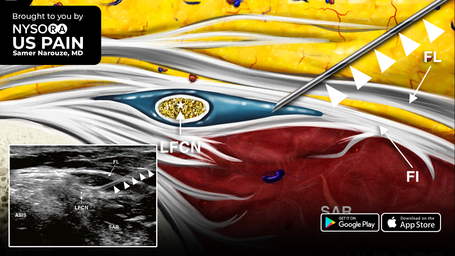
The lateral femoral cutaneous nerve (LFCN) innervates the skin of the anterior and lateral parts of the thigh as far as the knee. An LFCN block is performed for acute pain relief following surgical procedures and diagnosing and treating meralgia paresthetica.
Here are 4 easy-to-apply tips for scanning for an LFCN block
- Place the transducer over the anterior superior iliac spine (ASIS) initially, with the long-axis view of the inguinal ligament. Continue scanning by moving the transducer distally.
- Visualize the ASIS as a hyperechoic structure with posterior acoustic shadowing.
- Identify the sartorius muscle as an inverted triangular-shaped structure.
- The LFCN will appear as one or more hypoechoic structures in the short-axis view superficial to the sartorius muscle. Tip: In some situations, the nerve will be more medial between the fascia lata and the fascia iliaca.
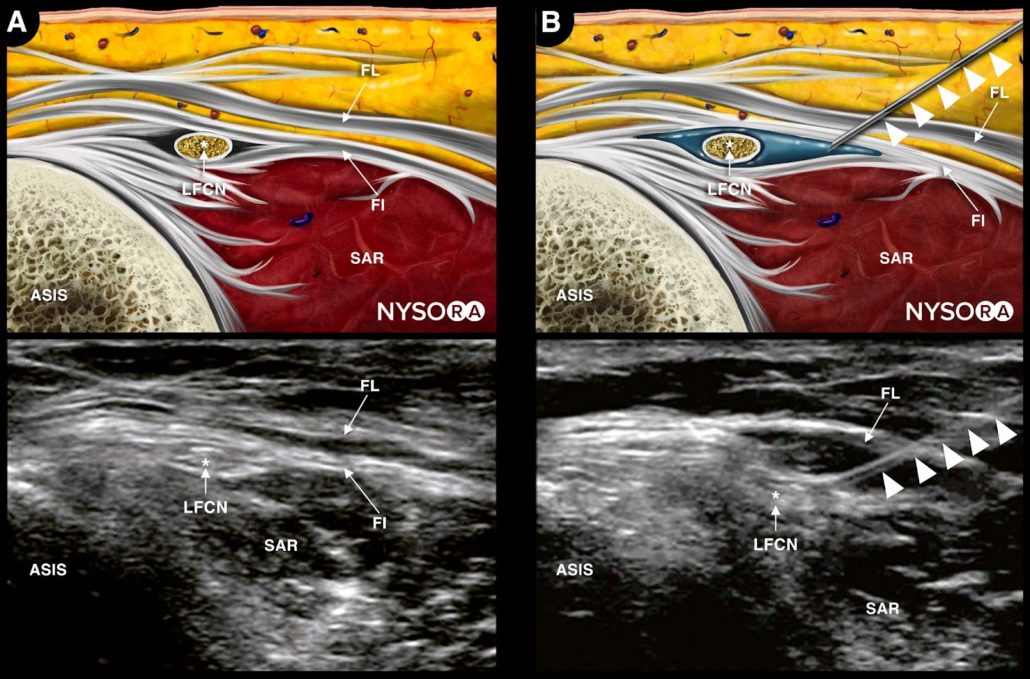
Reverse Ultrasound Anatomy and sonoanatomy of the lateral femoral cutaneous nerve (A) before and (B) after injection. ASIS, anterior superior iliac spine; LFCN, lateral femoral cutaneous nerve; FL, fascia lata; FI, fascia iliaca; SAR, sartorius muscle. The solid arrow heads indicate the needle path.
Check out the all-new reverse ultrasound anatomy illustration added in “Lateral Femoral Cutaneous Nerve Block > Technique” in the Ultrasound Pain App by Samer Narouze for a complete understanding of the principles of the technique.
Download the US Pain App HERE to read other tips on managing acute and chronic pain and to access the complete guide to ultrasound-guided chronic pain blocks.