Nerve Blocks App
 Pain Medicine Assistant App
 POCUS App
 IV Access App
 MSK Knee App
 VetRA App
 Nerve Block Manual
 Regional Anesthesia Updates
 Anesthesiology Manual
 Anesthesiology Review
 Anesthesia Updates 2025
 Anesthesia Updates 2026
 Pediatric Anesthesia Updates
 Airway Management Updates
 US Interventional Pain Manual
 Pain Medicine Updates
 Mastering Difficult IV Access
 PACU Nursing Manual
 RA Veterinary Manual
 About
About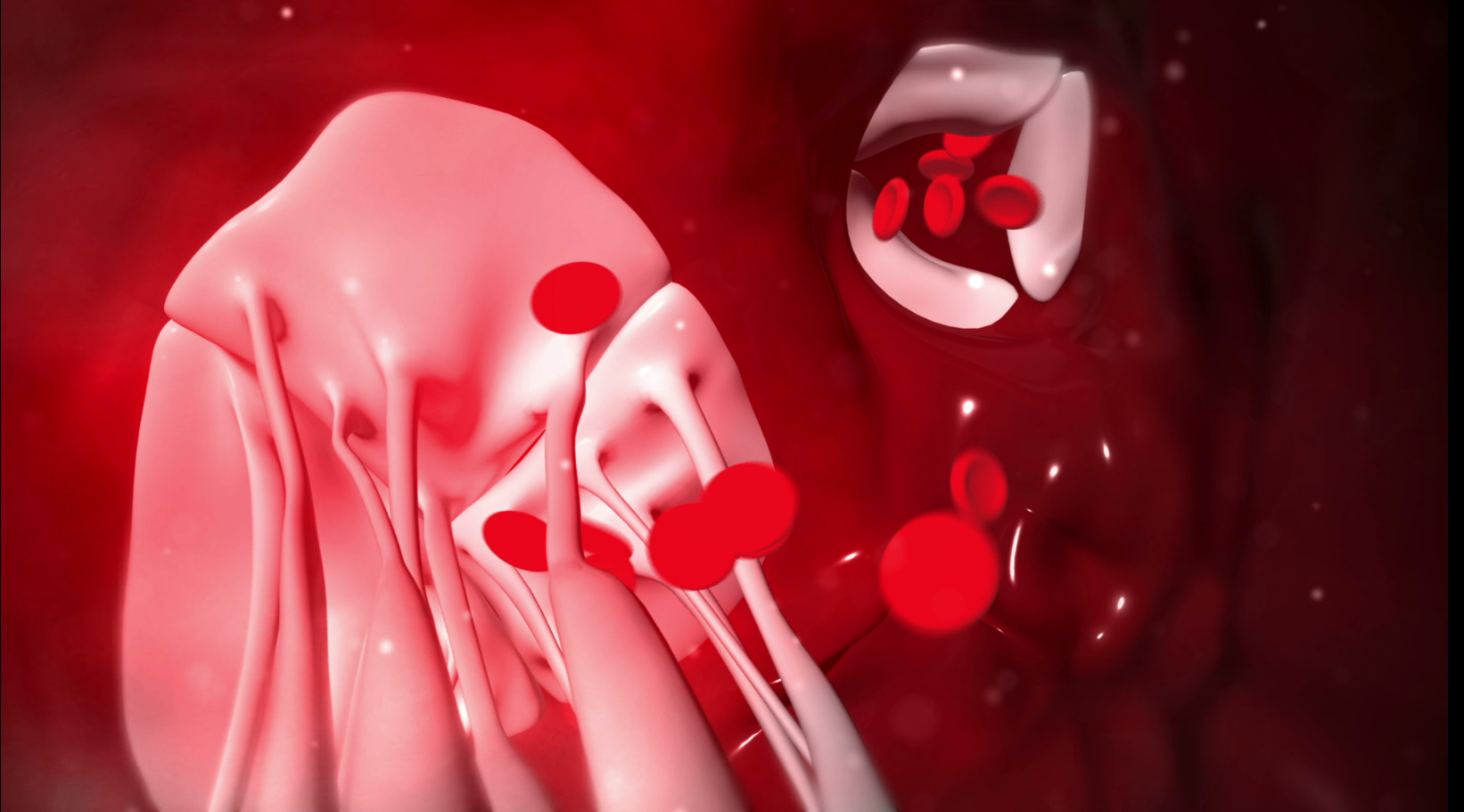
Learning objectives
- Recognize common causes of TR
- Describe the signs and symptoms of TR
- Grade the severity of TR cases
- Anesthetic management of TR
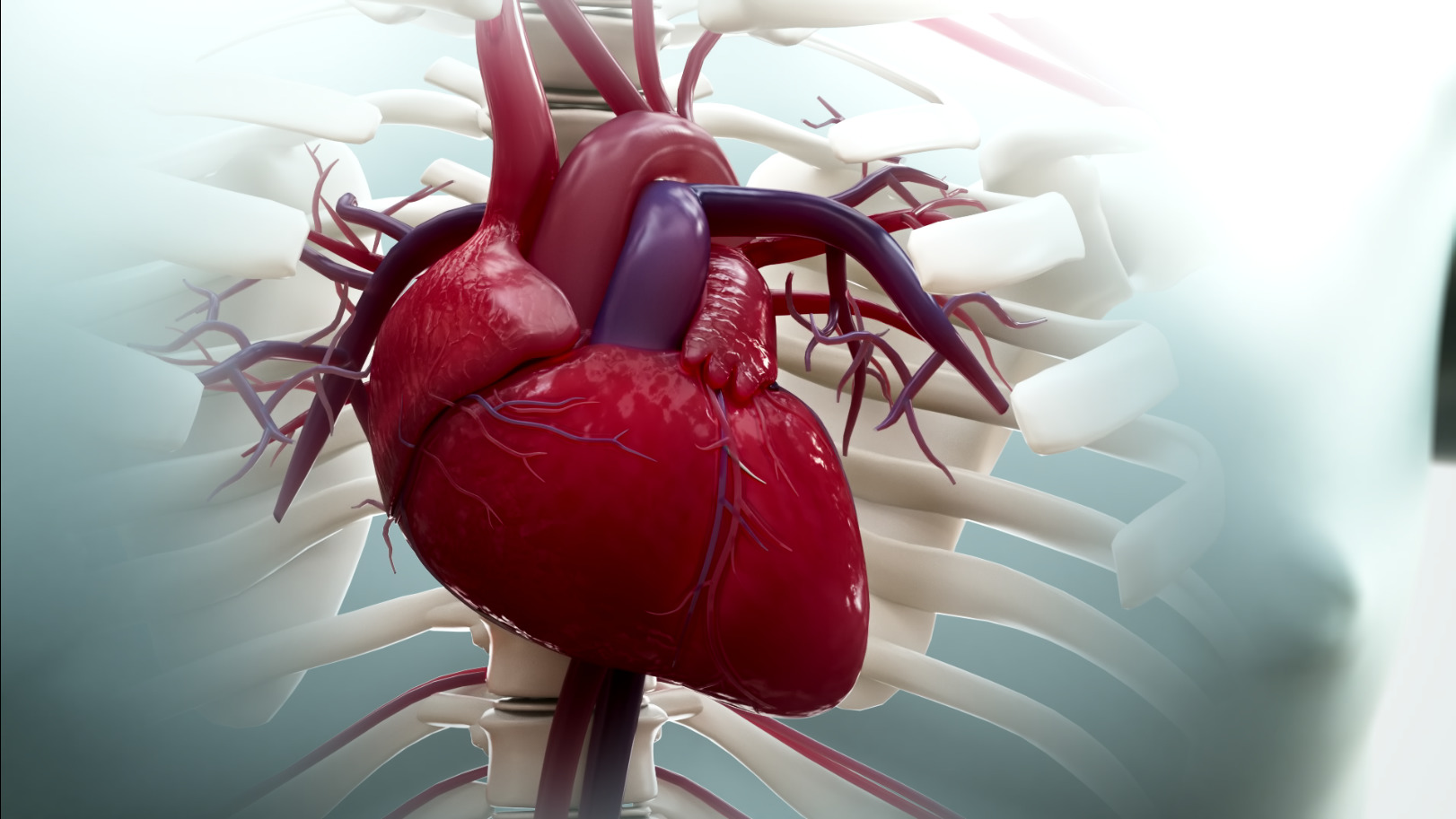
Definition & mechanisms
- Tricuspid regurgitation (TR) occurs when the tricuspid valve does not close properly, causing a reversal of blood flow through the valve
- TR can be of primary or secondary origin:
- Primary (organic) TR: Pathology of the tricuspid valve complex, may be of rheumatic, degenerative, congenital, infectious, traumatic, or iatrogenic origin
- Secondary (functional) TR: Related to right ventricular dilatation and/or dysfunction, annular dilatation, and leaflet tethering, usually secondary to left-sided valvular heart disease, atrial fibrillation or pulmonary hypertension
Signs & symptoms
- Often clinically silent and symptoms usually relate to concomitant left-sided valvular heart disease
- General fatigue and reduced exercise capacity
- Upper abdominal pain
- Peripheral lower limb edema
- Systolic jugular distension
- Pulsatile hepatomegaly
- Ascites, liver failure, and cachexia may be observed in end-stage disease
- Electrocardiogram frequently shows right bundle branch block and atrial fibrillation reflects disease evolution.
Severity assessment
| Parameters | Mild | Moderate | Severe | |
|---|---|---|---|---|
| Qualitative | TV morphology | Normal/abnormal | Normal/abnormal | Abnormal/flail/large coaptation defect |
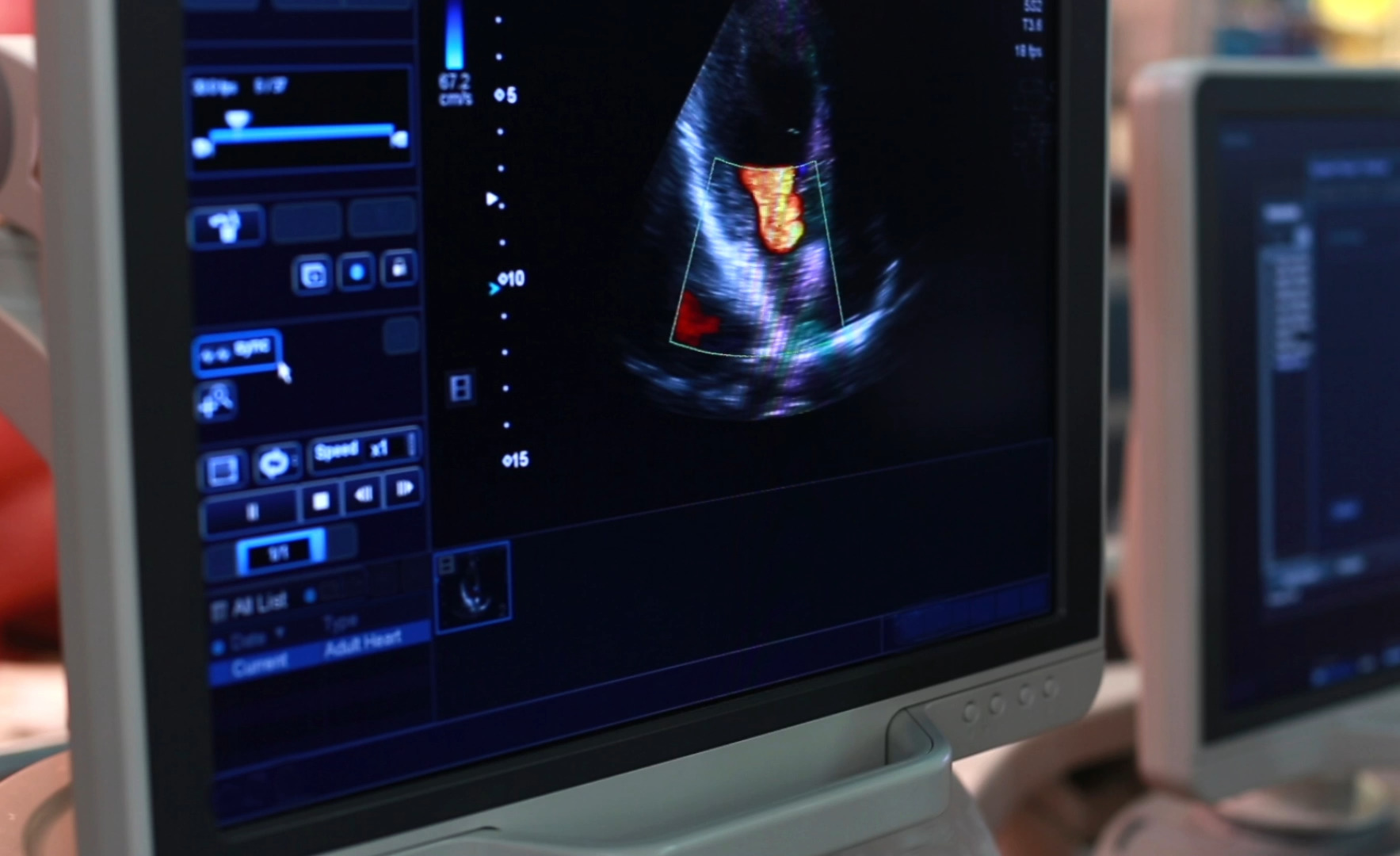
| Color flow TR jet | Small, central | Intermediate | Very large central jet or eccentric wall impinging jet | |
| CW signal of TR jet | Faint/parabolic | Dense/parabolic | Dense/triangular with early peaking (peak <2 m/s in massive TR) | |
| Semi-quantitative | VC width (mm) | Not defined | <7 | >7 |
| PISA radius (mm) | ≤5 | 6–9 | >9 | |
| Hepatic vein flow | Systolic dominance | Systolic blunting | Systolic flow reversal | |
| Tricuspid inflow | Normal | Normal | E-wave dominant (≥1 m/s) | |
| Quantitative | EROA (mm2) | Not defined | Not defined | ≥40 |
| R Vol (ml) | Not defined | Not defined | ≥45 |
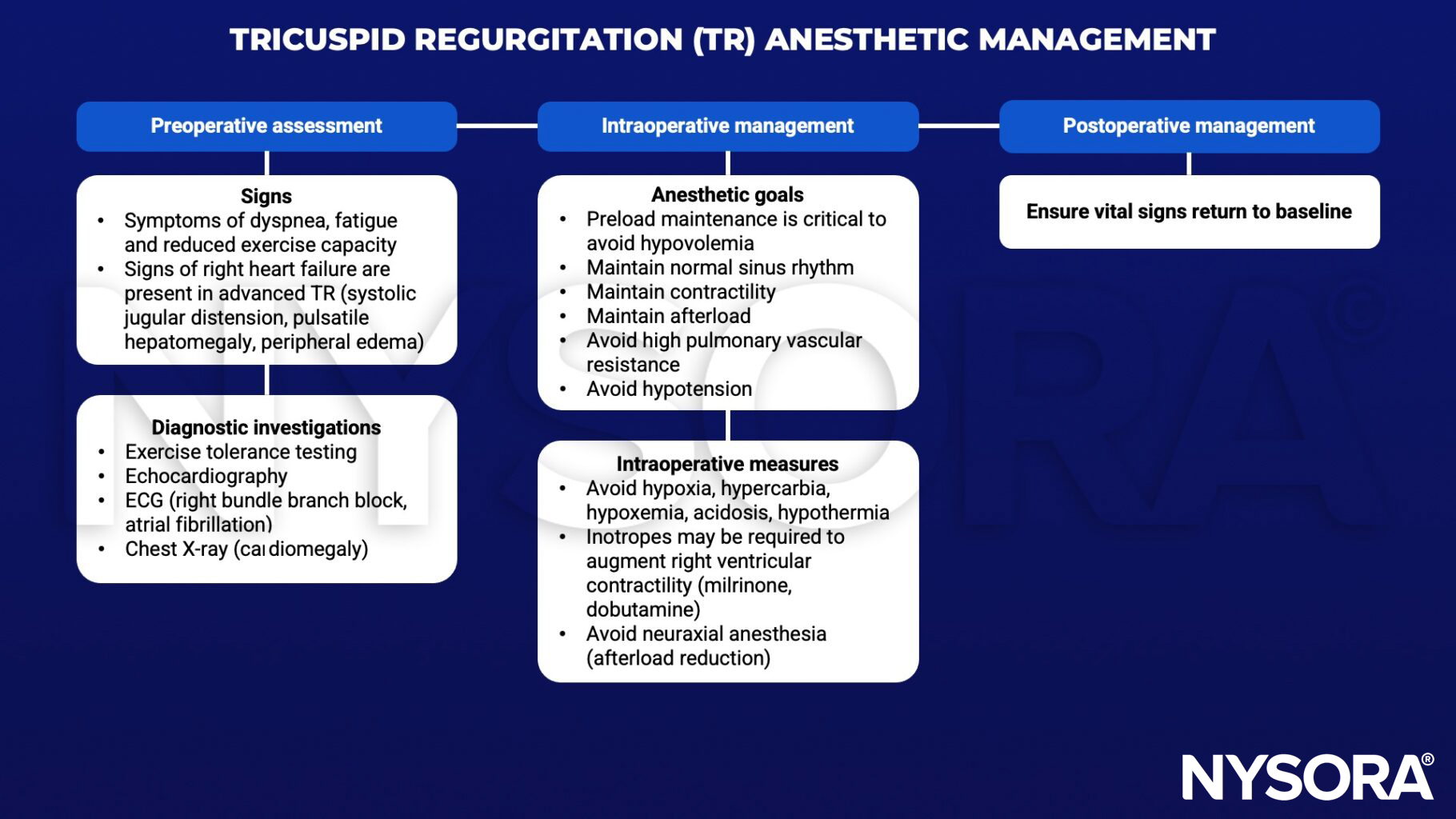
Management
Keep in mind
Tricuspid regurgitation is most commonly secondary to other morbidities, which might require further attention.
Suggested reading
- Antunes MJ, Rodríguez-Palomares J, Prendergast B, De Bonis M, Rosenhek R, Al-Attar N, et al. Management of tricuspid valve regurgitation: Position statement of the European Society of Cardiology Working Groups of Cardiovascular Surgery and Valvular Heart Disease. European Journal of Cardio-Thoracic Surgery. 2017;52(6):1022-30.