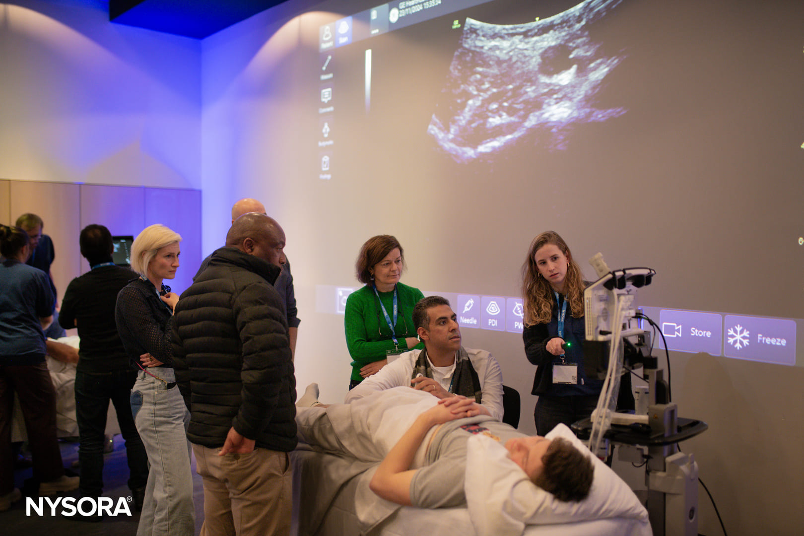
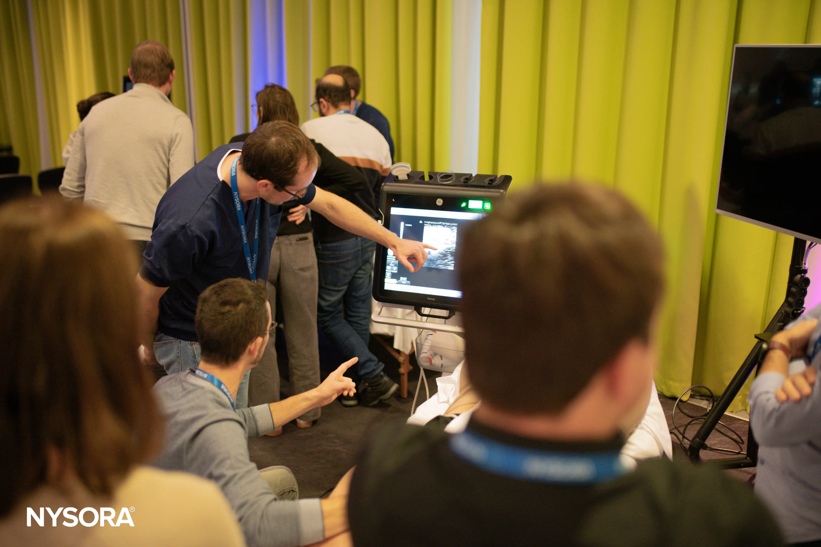
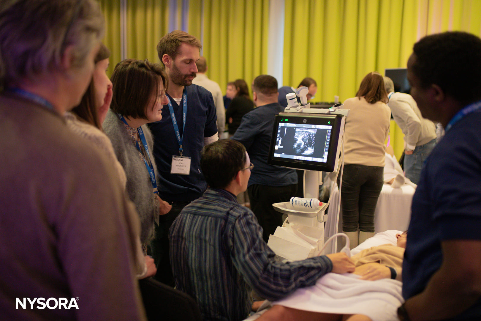
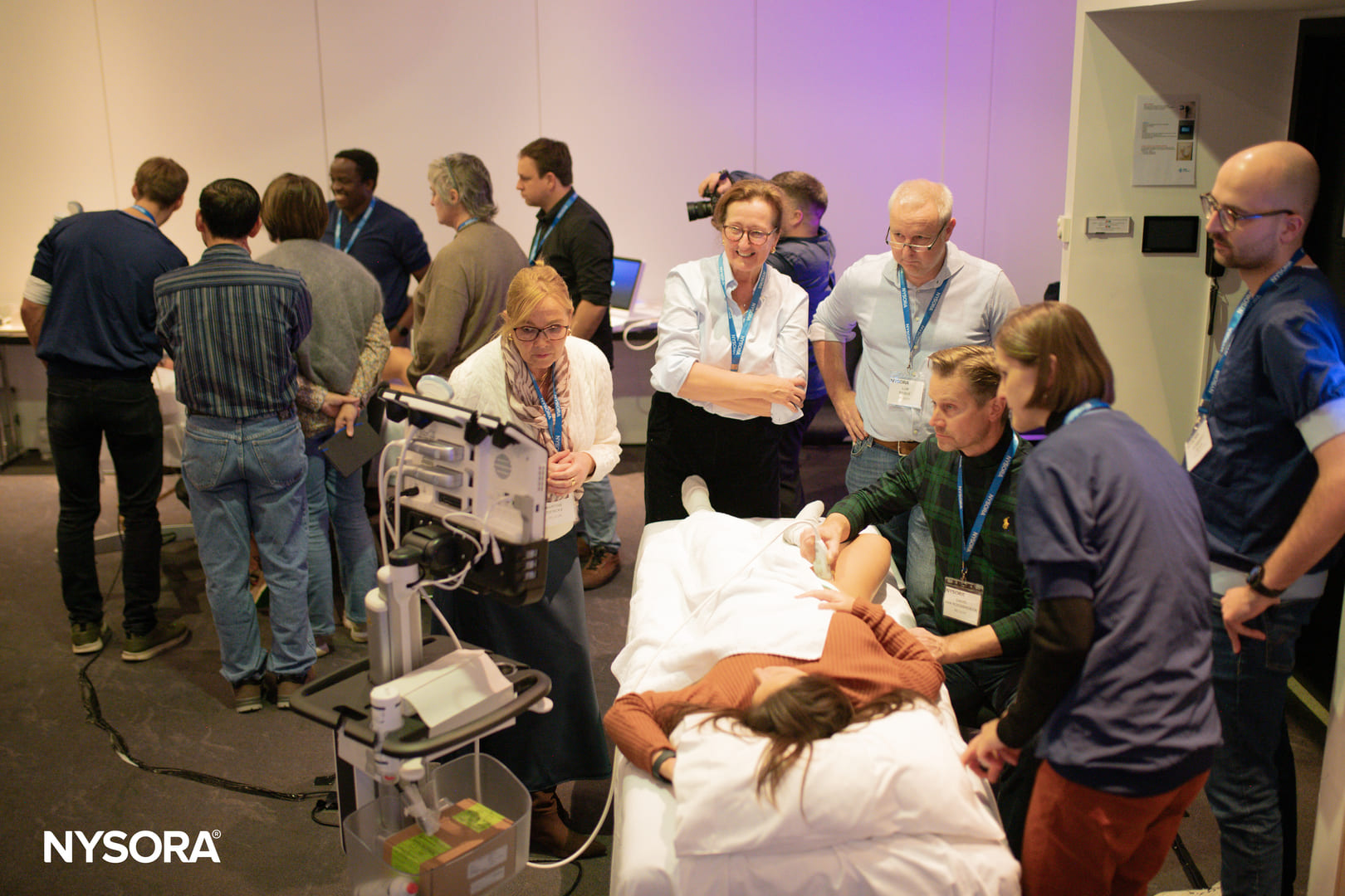
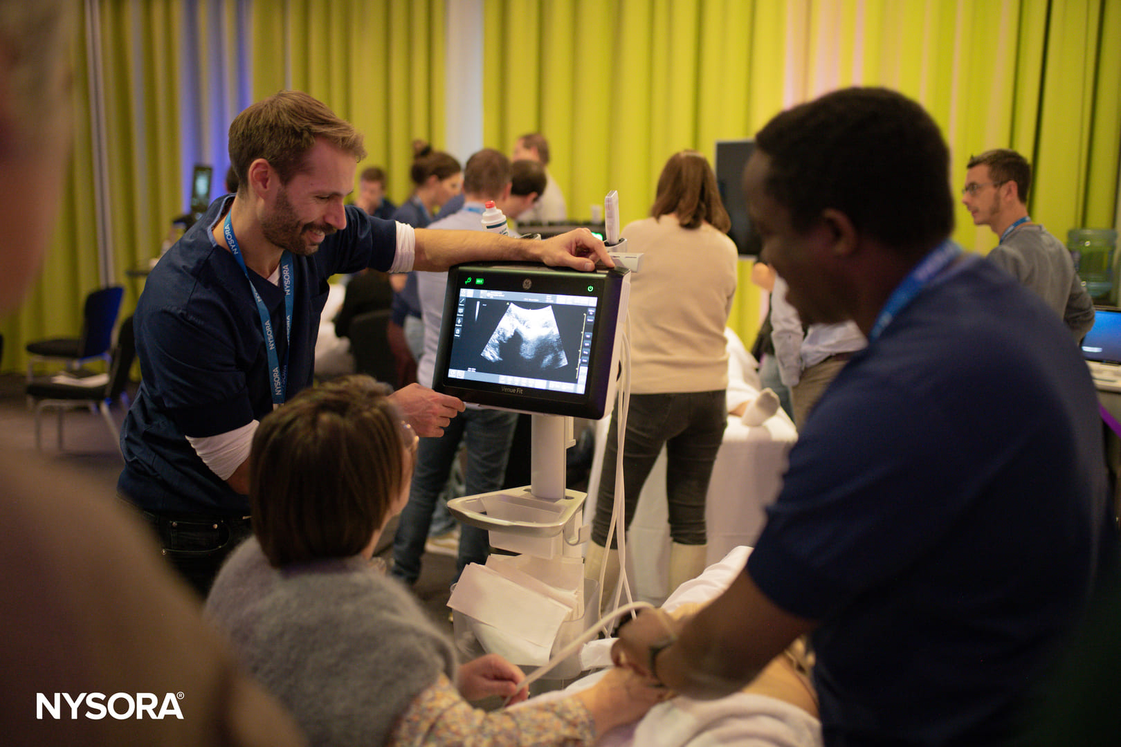
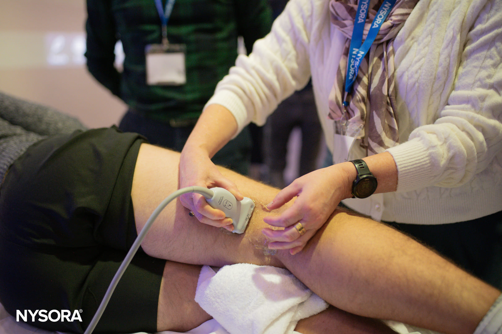
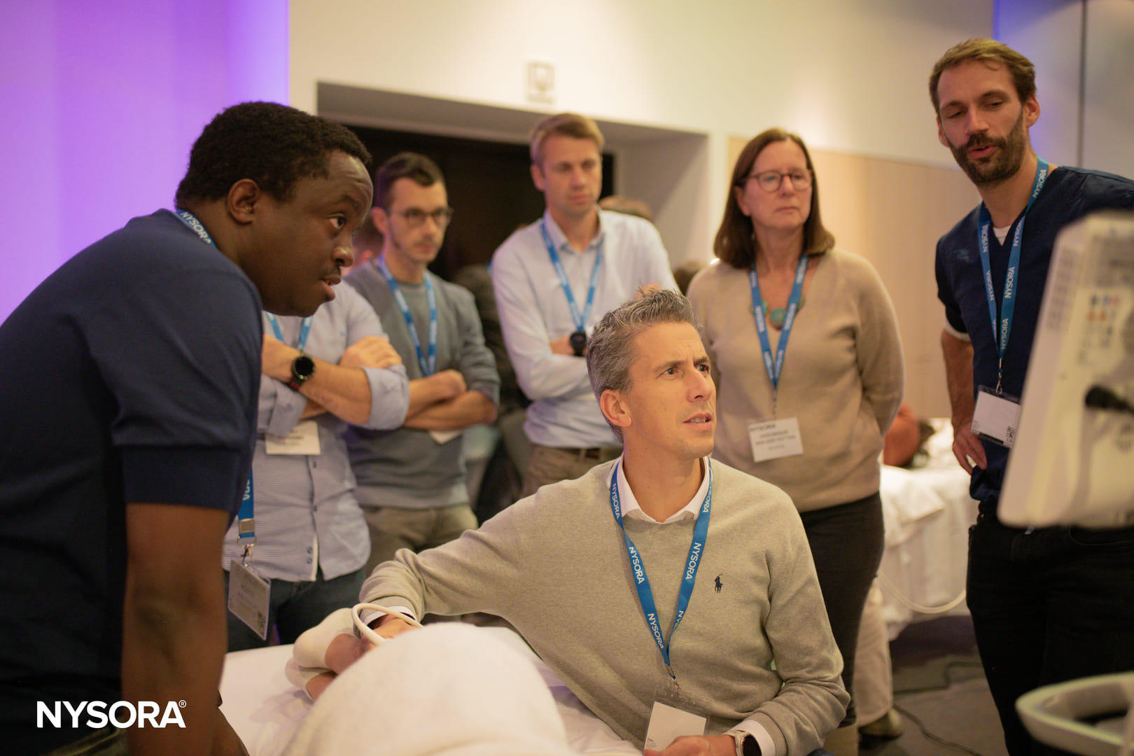
Join NYSORA’s Boutique Workshops for hands-on, practical training in small, focused groups - held across key locations in Europe and the USA.
3
Countries
10K+
Attendees
20+
Speakers
NYSORA Workshop Programs and Information Management
NYSORA's workshop series offers focused, hands-on learning to sharpen clinical skills in regional anesthesia and pain management, guided by expert instructors.
Experience hands-on, personalized training in small-group workshops across Europe, combining NYSORA’s decades of educational expertise with practical sessions, and advanced learning tools in regional anesthesia, POCUS, pain and nerve block techniques.
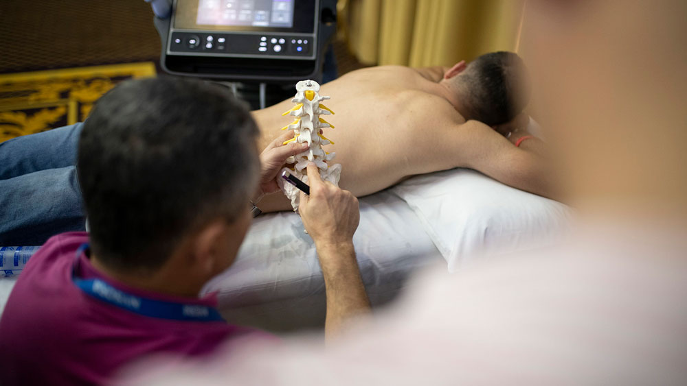
Neuraxial Ultrasound Scanning: Landmarks And Why It Matters
Four Critical Ultrasound Findings:
Midline Identification
Spinal Level Determination
Estimated Depth To Epidural/Intrathecal Space
Best Interspace For Access
Morning Session (09:30 - 09:45)
Coffee Break & Simulation Training
Morning Session (09:45 - 10:30)
LECTURE
Perioperative Management Of Nerve Blocks For Success: Premeds/Intraop/Multimodal
When Not To Perform Nerve Blocks
Prevention And Management Of Local Anesthetic Systemic Toxicity (LAST)
Morning Session (10:30 - 12:00)
MODULE 6: Lower Extremity — Hip & Knee Pathways
Femoral Nerve Block
Fascia Iliaca Block: Infrainguinal Vs Suprainguinal
PENG Block: When, Why, And How
When And Why: Femoral Vs Fascia Iliaca Vs PENG Blocks For Hip
Morning Session (12:00 - 12:30)
Lunch Break & Simulation Training
Afternoon Session (12:30 - 13:15)
Lower Extremity: Knee, Foot & Ankle
Femoral Triangle Vs Adductor Canal Block: The Truth Is In The Middle
IPACK And Genicular Nerve Concepts For Knee Analgesia
Ankle Block:
Tibial
Deep And Superficial Peroneal
Saphenous
Sural Nerves
Afternoon Session (13:15 - 14:30)
MODULE 7 REVIEW: More Scanning Top-Down
Afternoon Session (14:30 - 14:45)
Final Review & Q&A
What We Have Learned
Key Take-Home Messages
Must-Have Equipment, Documentation, And Sterility Principles
How To Safely Implement And Continue Developing These Techniques In Your Practice
Learning objectives
Interpret various sonographic artifacts and outline a plan for troubleshooting these using various transducer maneuvers and ultrasound machine setting adjustments;
Demonstrate the correct sonographic technique for identification of the brachial plexus and individual nerves of the lower extremity, and associated vascular and musculoskeletal structures;
Demonstrate the correct sonographic technique for performance of truncal blocks including transversus abdominis plane block, rectus sheath block, and paravertebral and neuraxial block;
Develop a framework for the establishment of a regional anesthesia service using strategies to increase surgical, anesthetic and hospital buy-in;
Discuss the rational choice of local anesthetics and adjuvants for various peripheral nerve blocks.
Faculty
Dr. Kris Abbas
Assistant professor
McMaster University
Department of Anesthesia
St. Joseph’s Healthcare
50 Charlton St. E.
Hamilton, ON. L8N 4A6
Canada
Dr. Kim Wong
BSc.P.T., M.D., FRCPC
Anesthesiologist, Acute Pain Service Physician Lead -St Joseph’s Healthcare Hamilton. Assistant Professor McMaster University
Justine Denomme
MD, FRCPC
Assistant Professor, Department of Anesthesiology, McMaster University, St Joseph’s Healthcare, ON, Canada
Neuraxial Ultrasound Scanning: Landmarks And Why It Matters
Four Critical Ultrasound Findings:
Midline Identification
Spinal Level Determination
Estimated Depth To Epidural/Intrathecal Space
Best Interspace For Access
Morning Session (09:30 - 09:45)
Coffee Break & Simulation Training
Morning Session (09:45 - 10:30)
LECTURE
Perioperative Management Of Nerve Blocks For Success: Premeds/Intraop/Multimodal
When Not To Perform Nerve Blocks
Prevention And Management Of Local Anesthetic Systemic Toxicity (LAST)
Morning Session (10:30 - 12:00)
MODULE 6: Lower Extremity — Hip & Knee Pathways
Femoral Nerve Block
Fascia Iliaca Block: Infrainguinal Vs Suprainguinal
PENG Block: When, Why, And How
When And Why: Femoral Vs Fascia Iliaca Vs PENG Blocks For Hip
Morning Session (12:00 - 12:30)
Lunch Break & Simulation Training
Afternoon Session (12:45 - 13:15)
Lower Extremity: Knee, Foot & Ankle
Femoral Triangle Vs Adductor Canal Block: The Truth Is In The Middle
IPACK And Genicular Nerve Concepts For Knee Analgesia
Ankle Block:
Tibial
Deep And Superficial Peroneal
Saphenous
Sural Nerves
Afternoon Session (13:15 - 14:30)
MODULE 7 REVIEW: More Scanning Top-Down
Afternoon Session (14:30 - 14:45)
Final Review & Q&A
What We Have Learned
Key Take-Home Messages
Must-Have Equipment, Documentation, And Sterility Principles
How To Safely Implement And Continue Developing These Techniques In Your Practice
Learning objectives
Interpret various sonographic artifacts and outline a plan for troubleshooting these using various transducer maneuvers and ultrasound machine setting adjustments;
Demonstrate the correct sonographic technique for identification of the brachial plexus and individual nerves of the lower extremity, and associated vascular and musculoskeletal structures;
Demonstrate the correct sonographic technique for performance of truncal blocks including transversus abdominis plane block, rectus sheath block, and paravertebral and neuraxial block;
Develop a framework for the establishment of a regional anesthesia service using strategies to increase surgical, anesthetic and hospital buy-in;
Discuss the rational choice of local anesthetics and adjuvants for various peripheral nerve blocks.
Faculty
Coming soon.
Workshops
Ultrasound in Interventional Pain Medicine Comprehensive Workshop (Plus CIPS Exam preparation Instructions)
Hip Joint, Greater Trochanter Complex, Iliopsoas Bursa and Tendon, IT Band, Lateral Femoral Cutaneous Nerve, Articular Branches of Femoral, Obturator and Accessory Obturator Nerve
Develop a step wise approach to various pain management procedures using ultrasound guidance
Understand curriculum based procedures as per the CIPS examination curriculum
Learn how to prepare and to pass the CIPS examination
Learn how to perform in clinical case scenarios, approach and assessment
Interpret various sonographic artifacts and outline a plan for troubleshooting these using various transducer manoeuvres and ultrasound machine setting adjustments
Faculty
Sadiq Bhayani
FRCA, MD, MBBS
Staff Physician, Pain Medicine at Cleveland Clinic Abu Dhabi, UAE
Rahul Bhansali
MD
Anaesthetist and Pain Management Consultant at Cesterfield Royal Hospital NHS Foundation Trust
United Kingdom
Queenayda Kroon
MD
Anesthesiologist at AZ Zeno, Belgium
Jose Pereira
MD
Physical Medicine and Rehabilitation Physician at Centre National de Rééducation Fonctionnelle et de Réadaptation – Rehazenter Luxembourg
Kevin Lathouwers
MD
Anesthesiologist at Ziekenhuis Oost-Limburg Hospital Belgium
Neuraxial Ultrasound Scanning: Landmarks And Why It Matters
Four Critical Ultrasound Findings:
Midline Identification
Spinal Level Determination
Estimated Depth To Epidural/Intrathecal Space
Best Interspace For Access
Coffee Break & Simulation Training
LECTURE
Perioperative Management Of Nerve Blocks For Success: Premeds/Intraop/Multimodal
When Not To Perform Nerve Blocks
Prevention And Management Of Local Anesthetic Systemic Toxicity (LAST)
MODULE 6: Lower Extremity — Hip & Knee Pathways
Femoral Nerve Block
Fascia Iliaca Block: Infrainguinal Vs Suprainguinal
PENG Block: When, Why, And How
When And Why: Femoral Vs Fascia Iliaca Vs PENG Blocks For Hip
Lunch Break & Simulation Training
Afternoon Session (12:30 - 14:45)
Lower Extremity: Knee, Foot & Ankle
Femoral Triangle Vs Adductor Canal Block: The Truth Is In The Middle
IPACK And Genicular Nerve Concepts For Knee Analgesia
Ankle Block:
Tibial
Deep And Superficial Peroneal
Saphenous
Sural Nerves
MODULE 7 REVIEW: More Scanning Top-Down
Final Review & Q&A
What We Have Learned
Key Take-Home Messages
Must-Have Equipment, Documentation, And Sterility Principles
How To Safely Implement And Continue Developing These Techniques In Your Practice
Learning objectives
Clinical Goals:
To solidify three-dimensional anatomical understanding of clinically relevant regional anesthesia targets, using high-fidelity cadaver dissection integrated with ultrasound imaging, ensuring participants can reliably translate sono-anatomy into real patient care.
To improve the safety, precision, and consistency of needling techniques for both common and advanced regional anesthesia blocks by practicing on cadaveric models that accurately replicate tissue planes, depth relationships, and anatomical variability.
To bridge expert knowledge and bedside decision-making, enabling participants to move beyond “how to perform a block” toward understanding why, when, and when not to perform specific techniques in complex perioperative scenarios.
To enhance procedural confidence and efficiency in daily clinical practice, through deliberate hands-on repetition, expert faculty feedback, and focused small-group instruction—directly targeting common sources of uncertainty and error.
To strengthen clinical judgment in regional anesthesia, including block selection, risk assessment, and complication avoidance, with the ultimate goal of improving patient outcomes and perioperative care quality.
Faculty
Admir Hadzic
MD, PhD.
Director – NYSORA, Anesthesiologist, Professor of Anesthesiology, Department of Anesthesiology, ZOL, Genk, Belgium
Catherine Vandepitte
MD, PhD.
Anesthesiologist at Ziekenhuis Oost-Limburg (ZOL), Genk, Belgium
Imré Van Herreweghe
MD
Anesthesiologist at Hospital Oost-Limburg
Genk, Belgium
Peter Merjavy
MD, EDRA Board Member
Consultant Anaesthetist at Craigavon Area Hospital, Southern HSC Trust
Craigavon, Northern Ireland, United Kingdom
Steve Coppens
MD, PhD
University Hospitals Leuven, Belgium
Danny Feike Hoogma
MD, PhD
University Hospitals Leuven, Belgium
Matthias Desmet
MD
AZ Groeninge, Belgium
Sari Casaer
MD
Ziekenhuis aan de Stroom, Belgium
Morné Wolmarans
MD
Norfolk & Norwich University Hospitals, UK
Hanne-Rose Honis
MD
Institute of Clinical and Functional Anatomy Innsbruck, Austria
Neuraxial Ultrasound Scanning: Landmarks And Why It Matters
Four Critical Ultrasound Findings:
Midline Identification
Spinal Level Determination
Estimated Depth To Epidural/Intrathecal Space
Best Interspace For Access
Morning Session (09:30 - 09:45)
Coffee Break & Simulation Training
Morning Session (09:45 - 10:30)
LECTURE
Perioperative Management Of Nerve Blocks For Success: Premeds/Intraop/Multimodal
When Not To Perform Nerve Blocks
Prevention And Management Of Local Anesthetic Systemic Toxicity (LAST)
Morning Session (10:30 - 12:00)
MODULE 6: Lower Extremity — Hip & Knee Pathways
Femoral Nerve Block
Fascia Iliaca Block: Infrainguinal Vs Suprainguinal
PENG Block: When, Why, And How
When And Why: Femoral Vs Fascia Iliaca Vs PENG Blocks For Hip
Morning Session (12:00 - 12:30)
Lunch Break & Simulation Training
Afternoon Session (12:30 - 13:15)
Lower Extremity: Knee, Foot & Ankle
Femoral Triangle Vs Adductor Canal Block: The Truth Is In The Middle
IPACK And Genicular Nerve Concepts For Knee Analgesia
Ankle Block:
Tibial
Deep And Superficial Peroneal
Saphenous
Sural Nerves
Afternoon Session (13:15 - 14:30)
MODULE 7 REVIEW: More Scanning Top-Down
Afternoon Session (14:30 - 14:45)
Final Review & Q&A
What We Have Learned
Key Take-Home Messages
Must-Have Equipment, Documentation, And Sterility Principles
How To Safely Implement And Continue Developing These Techniques In Your Practice
Learning objectives
Interpret various sonographic artifacts and outline a plan for troubleshooting these using various transducer maneuvers and ultrasound machine setting adjustments;
Demonstrate the correct sonographic technique for identification of the brachial plexus and individual nerves of the lower extremity, and associated vascular and musculoskeletal structures;
Demonstrate the correct sonographic technique for performance of truncal blocks including transversus abdominis plane block, rectus sheath block, and paravertebral and neuraxial block;
Develop a framework for the establishment of a regional anesthesia service using strategies to increase surgical, anesthetic and hospital buy-in;
Discuss the rational choice of local anesthetics and adjuvants for various peripheral nerve blocks.
Neuraxial Ultrasound Scanning: Landmarks And Why It Matters
Four Critical Ultrasound Findings:
Midline Identification
Spinal Level Determination
Estimated Depth To Epidural/Intrathecal Space
Best Interspace For Access
Morning Session (09:30 - 09:45)
Coffee Break & Simulation Training
Morning Session (09:45 - 10:30)
LECTURE
Perioperative Management Of Nerve Blocks For Success: Premeds/Intraop/Multimodal
When Not To Perform Nerve
Prevention And Management Of Local Anesthetic Systemic Toxicity (LAST)
Morning Session (10:30 - 12:00)
MODULE 6: Lower Extremity — Hip & Knee Pathways
Femoral Nerve Block
Fascia Iliaca Block: Infrainguinal Vs Suprainguinal
PENG Block: When, Why, And How
When And Why: Femoral Vs Fascia Iliaca Vs PENG Blocks For Hip
Morning Session (12:00 - 12:30)
Lunch Break & Simulation Training
Afternoon Session (12:30 - 13:15)
Lower Extremity: Knee, Foot & Ankle
Femoral Triangle Vs Adductor Canal Block: The Truth Is In The Middle
IPACK And Genicular Nerve Concepts For Knee Analgesia
Ankle Block:
Tibial
Deep And Superficial Peroneal
Saphenous
Sural Nerves
Afternoon Session (13:15 - 14:30)
MODULE 7 REVIEW: More Scanning Top-Down
Afternoon Session (14:30 - 14:45)
Final Review & Q&A
What We Have Learned
Key Take-Home Messages
Must-Have Equipment, Documentation, And Sterility Principles
How To Safely Implement And Continue Developing These Techniques In Your Practice
Learning objectives
Interpret various sonographic artifacts and outline a plan for troubleshooting these using various transducer maneuvers and ultrasound machine setting adjustments;
Demonstrate the correct sonographic technique for identification of the brachial plexus and individual nerves of the lower extremity, and associated vascular and musculoskeletal structures;
Demonstrate the correct sonographic technique for performance of truncal blocks including transversus abdominis plane block, rectus sheath block, and paravertebral and neuraxial block;
Develop a framework for the establishment of a regional anesthesia service using strategies to increase surgical, anesthetic and hospital buy-in;
Discuss the rational choice of local anesthetics and adjuvants for various peripheral nerve blocks.
Neuraxial Ultrasound Scanning: Landmarks And Why It Matters
Four Critical Ultrasound Findings:
Midline Identification
Spinal Level Determination
Estimated Depth To Epidural/Intrathecal Space
Best Interspace For Access
Morning Session (09:30 - 09:45)
Coffee Break & Simulation Training
Morning Session (09:45 - 10:30)
LECTURE
Perioperative Management Of Nerve Blocks For Success: Premeds/Intraop/Multimodal
When Not To Perform Nerve Blocks
Prevention And Management Of Local Anesthetic Systemic Toxicity (LAST)
Morning Session (10:30 - 12:00)
MODULE 6: Lower Extremity — Hip & Knee Pathways
Femoral Nerve Block
Fascia Iliaca Block: Infrainguinal Vs Suprainguinal
PENG Block: When, Why, And How
When And Why: Femoral Vs Fascia Iliaca Vs PENG Blocks For Hip
Morning Session (12:00 - 12:30)
Lunch Break & Simulation Training
Afternoon Session (12:30 - 13:15)
Lower Extremity: Knee, Foot & Ankle
Femoral Triangle Vs Adductor Canal Block: The Truth Is In The Middle
IPACK And Genicular Nerve Concepts For Knee Analgesia
Ankle Block:
Tibial
Deep And Superficial Peroneal
Saphenous
Sural Nerves
Afternoon Session (13:15 - 14:30)
MODULE 7 REVIEW: More Scanning Top-Down
Afternoon Session (14:30 - 14:45)
Final Review & Q&A
What We Have Learned
Key Take-Home Messages
Must-Have Equipment, Documentation, And Sterility Principles
How To Safely Implement And Continue Developing These Techniques In Your Practice
Learning objectives
Interpret various sonographic artifacts and outline a plan for troubleshooting these using various transducer maneuvers and ultrasound machine setting adjustments;
Demonstrate the correct sonographic technique for identification of the brachial plexus and individual nerves of the lower extremity, and associated vascular and musculoskeletal structures;
Demonstrate the correct sonographic technique for performance of truncal blocks including transversus abdominis plane block, rectus sheath block, and paravertebral and neuraxial block;
Develop a framework for the establishment of a regional anesthesia service using strategies to increase surgical, anesthetic and hospital buy-in;
Discuss the rational choice of local anesthetics and adjuvants for various peripheral nerve blocks.
Neuraxial Ultrasound Scanning: Landmarks And Why It Matters
Four Critical Ultrasound Findings:
Midline Identification
Spinal Level Determination
Estimated Depth To Epidural/Intrathecal Space
Best Interspace For Access
Morning Session (09:30 - 09:45)
Coffee Break & Simulation Training
Morning Session (09:45 - 10:30)
LECTURE
Perioperative Management Of Nerve Blocks For Success: Premeds/Intraop/Multimodal
When Not To Perform Nerve Blocks
Prevention And Management Of Local Anesthetic Systemic Toxicity (LAST)
Morning Session (10:30 - 12:00)
MODULE 6: Lower Extremity — Hip & Knee Pathways
Femoral Nerve Block
Fascia Iliaca Block: Infrainguinal Vs Suprainguinal
PENG Block: When, Why, And How
When And Why: Femoral Vs Fascia Iliaca Vs PENG Blocks For Hip
Morning Session (12:00 - 12:30)
Lunch Break & Simulation Training
Afternoon Session (12:30 - 13:15)
Lower Extremity: Knee, Foot & Ankle
Femoral Triangle Vs Adductor Canal Block: The Truth Is In The Middle
IPACK And Genicular Nerve Concepts For Knee Analgesia
Ankle Block:
Tibial
Deep And Superficial Peroneal
Saphenous
Sural Nerves
Afternoon Session (13:15 - 14:30)
MODULE 7 REVIEW: More Scanning Top-Down
Afternoon Session (14:30 - 14:45)
Final Review & Q&A
What We Have Learned
Key Take-Home Messages
Must-Have Equipment, Documentation, And Sterility Principles
How To Safely Implement And Continue Developing These Techniques In Your Practice
Learning objectives
Interpret various sonographic artifacts and outline a plan for troubleshooting these using various transducer maneuvers and ultrasound machine setting adjustments;
Demonstrate the correct sonographic technique for identification of the brachial plexus and individual nerves of the lower extremity, and associated vascular and musculoskeletal structures;
Demonstrate the correct sonographic technique for performance of truncal blocks including transversus abdominis plane block, rectus sheath block, and paravertebral and neuraxial block;
Develop a framework for the establishment of a regional anesthesia service using strategies to increase surgical, anesthetic and hospital buy-in;
Discuss the rational choice of local anesthetics and adjuvants for various peripheral nerve blocks.
Neuraxial Ultrasound Scanning: Landmarks And Why It Matters
Four Critical Ultrasound Findings:
Midline Identification
Spinal Level Determination
Estimated Depth To Epidural/Intrathecal Space
Best Interspace For Access
Morning Session (09:30 - 09:45)
Coffee Break & Simulation Training
Morning Session (09:45 - 10:30)
Perioperative Management Of Nerve Blocks For Success: Premeds/Intraop/Multimodal
When Not To Perform Nerve Blocks
Prevention And Management Of Local Anesthetic Systemic Toxicity (LAST)
Morning Session (10:30 - 12:00)
MODULE 6: Lower Extremity — Hip & Knee Pathways
Femoral Nerve Block
Fascia Iliaca Block: Infrainguinal Vs Suprainguinal
PENG Block: When, Why, And How
When And Why: Femoral Vs Fascia Iliaca Vs PENG Blocks For Hip
Morning Session (12:00 - 12:45)
Lunch Break & Simulation Training
Afternoon Session (12:45 - 13:15)
Lower Extremity: Knee, Foot & Ankle
Femoral Triangle Vs Adductor Canal Block: The Truth Is In The Middle
IPACK And Genicular Nerve Concepts For Knee Analgesia
Ankle Block:
Tibial
Deep And Superficial Peroneal
Saphenous
Sural Nerves
Afternoon Session (13:15 - 14:30)
MODULE 7 REVIEW: More Scanning Top-Down
Afternoon Session (14:30 - 14:45)
Final Review & Q&A
What We Have Learned
Key Take-Home Messages
Must-Have Equipment, Documentation, And Sterility Principles
How To Safely Implement And Continue Developing These Techniques In Your Practice
Learning objectives
Interpret various sonographic artifacts and outline a plan for troubleshooting these using various transducer maneuvers and ultrasound machine setting adjustments;
Demonstrate the correct sonographic technique for identification of the brachial plexus and individual nerves of the lower extremity, and associated vascular and musculoskeletal structures;
Demonstrate the correct sonographic technique for performance of truncal blocks including transversus abdominis plane block, rectus sheath block, and paravertebral and neuraxial block;
Develop a framework for the establishment of a regional anesthesia service using strategies to increase surgical, anesthetic and hospital buy-in;
Discuss the rational choice of local anesthetics and adjuvants for various peripheral nerve blocks.
Neuraxial Ultrasound Scanning: Landmarks And Why It Matters
Four Critical Ultrasound Findings:
Midline Identification
Spinal Level Determination
Estimated Depth To Epidural/Intrathecal Space
Best Interspace For Access
Morning Session (09:30 - 09:45)
Coffee Break & Simulation Training
Morning Session (09:45 - 10:30)
LECTURE
Perioperative Management Of Nerve Blocks For Success: Premeds/Intraop/Multimodal
When Not To Perform Nerve Blocks
Prevention And Management Of Local Anesthetic Systemic Toxicity (LAST)
Morning Session (10:30 - 12:00)
MODULE 6: Lower Extremity — Hip & Knee Pathways
Femoral Nerve Block
Fascia Iliaca Block: Infrainguinal Vs Suprainguinal
PENG Block: When, Why, And How
When And Why: Femoral Vs Fascia Iliaca Vs PENG Blocks For Hip
Morning Session (12:00 - 12:30)
Lunch Break & Simulation Training
Afternoon Session (12:30 - 13:15)
Lower Extremity: Knee, Foot & Ankle
Femoral Triangle Vs Adductor Canal Block: The Truth Is In The Middle
IPACK And Genicular Nerve Concepts For Knee Analgesia
Ankle Block:
Tibial
Deep And Superficial Peroneal
Saphenous
Sural Nerves
Afternoon Session (13:15 - 14:30)
MODULE 7 REVIEW: More Scanning Top-Down
Afternoon Session (14:30 - 14:45)
Final Review & Q&A
What We Have Learned
Key Take-Home Messages
Must-Have Equipment, Documentation, And Sterility Principles
How To Safely Implement And Continue Developing These Techniques In Your Practice
Learning objectives
Interpret various sonographic artifacts and outline a plan for troubleshooting these using various transducer maneuvers and ultrasound machine setting adjustments;
Demonstrate the correct sonographic technique for identification of the brachial plexus and individual nerves of the lower extremity, and associated vascular and musculoskeletal structures;
Demonstrate the correct sonographic technique for performance of truncal blocks including transversus abdominis plane block, rectus sheath block, and paravertebral and neuraxial block;
Develop a framework for the establishment of a regional anesthesia service using strategies to increase surgical, anesthetic and hospital buy-in;
Discuss the rational choice of local anesthetics and adjuvants for various peripheral nerve blocks.
Neuraxial Ultrasound Scanning: Landmarks And Why It Matters
Four Critical Ultrasound Findings:
Midline Identification
Spinal Level Determination
Estimated Depth To Epidural/Intrathecal Space
Best Interspace For Access
Morning Session (09:30 - 09:45)
Coffee Break & Simulation Training
Morning Session (09:45 - 10:30)
LECTURE
Perioperative Management Of Nerve Blocks For Success: Premeds/Intraop/Multimodal
When Not To Perform Nerve Blocks
Prevention And Management Of Local Anesthetic Systemic Toxicity (LAST)
Morning Session (10:30 - 12:00)
MODULE 6: Lower Extremity — Hip & Knee Pathways
Femoral Nerve Block
Fascia Iliaca Block: Infrainguinal Vs Suprainguinal
PENG Block: When, Why, And How
When And Why: Femoral Vs Fascia Iliaca Vs PENG Blocks For Hip
Morning Session (12:00 - 12:30)
Lunch Break & Simulation Training
Afternoon Session (12:30 - 13:15)
Lower Extremity: Knee, Foot & Ankle
Femoral Triangle Vs Adductor Canal Block: The Truth Is In The Middle
IPACK And Genicular Nerve Concepts For Knee Analgesia
Ankle Block:
Tibial
Deep And Superficial Peroneal
Saphenous
Sural Nerves
Afternoon Session (13:15 - 14:30)
MODULE 7 REVIEW: More Scanning Top-Down
Afternoon Session (14:30 - 14:45)
Final Review & Q&A
What We Have Learned
Key Take-Home Messages
Must-Have Equipment, Documentation, And Sterility Principles
How To Safely Implement And Continue Developing These Techniques In Your Practice
Learning objectives
Interpret various sonographic artifacts and outline a plan for troubleshooting these using various transducer maneuvers and ultrasound machine setting adjustments;
Demonstrate the correct sonographic technique for identification of the brachial plexus and individual nerves of the lower extremity, and associated vascular and musculoskeletal structures;
Demonstrate the correct sonographic technique for performance of truncal blocks including transversus abdominis plane block, rectus sheath block, and paravertebral and neuraxial block;
Develop a framework for the establishment of a regional anesthesia service using strategies to increase surgical, anesthetic and hospital buy-in;
Discuss the rational choice of local anesthetics and adjuvants for various peripheral nerve blocks.
Faculty
Dr. Kris Abbas
Assistant professor
McMaster University
Department of Anesthesia
St. Joseph’s Healthcare
50 Charlton St. E.
Hamilton, ON. L8N 4A6
Canada
Neuraxial Ultrasound Scanning: Landmarks And Why It Matters
Four Critical Ultrasound Findings:
Midline Identification
Spinal Level Determination
Estimated Depth To Epidural/Intrathecal Space
Best Interspace For Access
Morning Session (09:30 - 09:45)
Coffee Break & Simulation Training
Morning Session (09:45 - 10:30)
LECTURE
Perioperative Management Of Nerve Blocks For Success: Premeds/Intraop/Multimodal
When Not To Perform Nerve Blocks
Prevention And Management Of Local Anesthetic Systemic Toxicity (LAST)
Morning Session (10:30 - 12:00)
MODULE 6: Lower Extremity — Hip & Knee Pathways
Femoral Nerve Block
Fascia Iliaca Block: Infrainguinal Vs Suprainguinal
PENG Block: When, Why, And How
When And Why: Femoral Vs Fascia Iliaca Vs PENG Blocks For Hip
Morning Session (12:00 - 12:30)
Lunch Break & Simulation Training
Afternoon Session (12:30 - 13:15)
Lower Extremity: Knee, Foot & Ankle
Femoral Triangle Vs Adductor Canal Block: The Truth Is In The Middle
IPACK And Genicular Nerve Concepts For Knee Analgesia
Ankle Block:
Tibial
Deep And Superficial Peroneal
Saphenous
Sural Nerves
Afternoon Session (13:15 - 14:30)
MODULE 7 REVIEW: More Scanning Top-Down
Afternoon Session (14:30 - 14:45)
Final Review & Q&A
What We Have Learned
Key Take-Home Messages
Must-Have Equipment, Documentation, And Sterility Principles
How To Safely Implement And Continue Developing These Techniques In Your Practice
Learning objectives
Interpret various sonographic artifacts and outline a plan for troubleshooting these using various transducer maneuvers and ultrasound machine setting adjustments;
Demonstrate the correct sonographic technique for identification of the brachial plexus and individual nerves of the lower extremity, and associated vascular and musculoskeletal structures;
Demonstrate the correct sonographic technique for performance of truncal blocks including transversus abdominis plane block, rectus sheath block, and paravertebral and neuraxial block;
Develop a framework for the establishment of a regional anesthesia service using strategies to increase surgical, anesthetic and hospital buy-in;
Discuss the rational choice of local anesthetics and adjuvants for various peripheral nerve blocks.
Neuraxial Ultrasound Scanning: Landmarks And Why It Matters
Four Critical Ultrasound Findings:
Midline Identification
Spinal Level Determination
Estimated Depth To Epidural/Intrathecal Space
Best Interspace For Access
Morning Session (09:30 - 09:45)
Coffee Break & Simulation Training
Morning Session (09:45 - 10:30)
LECTURE
Perioperative Management Of Nerve Blocks For Success: Premeds/Intraop/Multimodal
When Not To Perform Nerve Blocks
Prevention And Management Of Local Anesthetic Systemic Toxicity (LAST)
Morning Session (10:30 - 12:00)
MODULE 6: Lower Extremity — Hip & Knee Pathways
Femoral Nerve Block
Fascia Iliaca Block: Infrainguinal Vs Suprainguinal
PENG Block: When, Why, And How
When And Why: Femoral Vs Fascia Iliaca Vs PENG Blocks For Hip
Morning Session (12:00 - 12:30)
Lunch Break & Simulation Training
Afternoon Session (12:45 - 13:15)
Lower Extremity: Knee, Foot & Ankle
Femoral Triangle Vs Adductor Canal Block: The Truth Is In The Middle
IPACK And Genicular Nerve Concepts For Knee Analgesia
Ankle Block:
Tibial
Deep And Superficial Peroneal
Saphenous
Sural Nerves
Afternoon Session (13:15 - 14:30)
MODULE 7 REVIEW: More Scanning Top-Down
Afternoon Session (14:30 - 14:45)
Final Review & Q&A
What We Have Learned
Key Take-Home Messages
Must-Have Equipment, Documentation, And Sterility Principles
How To Safely Implement And Continue Developing These Techniques In Your Practice
Learning objectives
Interpret various sonographic artifacts and outline a plan for troubleshooting these using various transducer maneuvers and ultrasound machine setting adjustments;
Demonstrate the correct sonographic technique for identification of the brachial plexus and individual nerves of the lower extremity, and associated vascular and musculoskeletal structures;
Demonstrate the correct sonographic technique for performance of truncal blocks including transversus abdominis plane block, rectus sheath block, and paravertebral and neuraxial block;
Develop a framework for the establishment of a regional anesthesia service using strategies to increase surgical, anesthetic and hospital buy-in;
Discuss the rational choice of local anesthetics and adjuvants for various peripheral nerve blocks.
Neuraxial Ultrasound Scanning: Landmarks And Why It Matters
Four Critical Ultrasound Findings:
Midline Identification
Spinal Level Determination
Estimated Depth To Epidural/Intrathecal Space
Best Interspace For Access
Morning Session (09:30 - 09:45)
Coffee Break & Simulation Training
Morning Session (09:45 - 10:30)
LECTURE
Perioperative Management Of Nerve Blocks For Success: Premeds/Intraop/Multimodal
When Not To Perform Nerve Blocks
Prevention And Management Of Local Anesthetic Systemic Toxicity (LAST)
Morning Session (10:30 - 12:00)
MODULE 6: Lower Extremity — Hip & Knee Pathways
Femoral Nerve Block
Fascia Iliaca Block: Infrainguinal Vs Suprainguinal
PENG Block: When, Why, And How
When And Why: Femoral Vs Fascia Iliaca Vs PENG Blocks For Hip
Morning Session (12:00 - 12:30)
Lunch Break & Simulation Training
Afternoon Session (12:30 - 13:15)
Lower Extremity: Knee, Foot & Ankle
Femoral Triangle Vs Adductor Canal Block: The Truth Is In The Middle
IPACK And Genicular Nerve Concepts For Knee Analgesia
Ankle Block:
Tibial
Deep And Superficial Peroneal
Saphenous
Sural Nerves
Afternoon Session (13:15 - 14:30)
MODULE 7 REVIEW: More Scanning Top-Down
Afternoon Session (14:30 - 14:45)
Final Review & Q&A
What We Have Learned
Key Take-Home Messages
Must-Have Equipment, Documentation, And Sterility Principles
How To Safely Implement And Continue Developing These Techniques In Your Practice
Learning objectives
Interpret various sonographic artifacts and outline a plan for troubleshooting these using various transducer maneuvers and ultrasound machine setting adjustments;
Demonstrate the correct sonographic technique for identification of the brachial plexus and individual nerves of the lower extremity, and associated vascular and musculoskeletal structures;
Demonstrate the correct sonographic technique for performance of truncal blocks including transversus abdominis plane block, rectus sheath block, and paravertebral and neuraxial block;
Develop a framework for the establishment of a regional anesthesia service using strategies to increase surgical, anesthetic and hospital buy-in;
Discuss the rational choice of local anesthetics and adjuvants for various peripheral nerve blocks.
Neuraxial Ultrasound Scanning: Landmarks And Why It Matters
Four Critical Ultrasound Findings:
Midline Identification
Spinal Level Determination
Estimated Depth To Epidural/Intrathecal Space
Best Interspace For Access
Morning Session (09:30 - 09:45)
Coffee Break & Simulation Training
Morning Session (09:45 - 10:30)
LECTURE
Perioperative Management Of Nerve Blocks For Success: Premeds/Intraop/Multimodal
When Not To Perform Nerve
Prevention And Management Of Local Anesthetic Systemic Toxicity (LAST)
Morning Session (10:30 - 12:00)
MODULE 6: Lower Extremity — Hip & Knee Pathways
Femoral Nerve Block
Fascia Iliaca Block: Infrainguinal Vs Suprainguinal
PENG Block: When, Why, And How
When And Why: Femoral Vs Fascia Iliaca Vs PENG Blocks For Hip
Morning Session (12:00 - 12:30)
Lunch Break & Simulation Training
Afternoon Session (12:30 - 13:15)
Lower Extremity: Knee, Foot & Ankle
Femoral Triangle Vs Adductor Canal Block: The Truth Is In The Middle
IPACK And Genicular Nerve Concepts For Knee Analgesia
Ankle Block:
Tibial
Deep And Superficial Peroneal
Saphenous
Sural Nerves
Afternoon Session (13:15 - 14:30)
MODULE 7 REVIEW: More Scanning Top-Down
Afternoon Session (14:30 - 14:45)
Final Review & Q&A
What We Have Learned
Key Take-Home Messages
Must-Have Equipment, Documentation, And Sterility Principles
How To Safely Implement And Continue Developing These Techniques In Your Practice
Learning objectives
Interpret various sonographic artifacts and outline a plan for troubleshooting these using various transducer maneuvers and ultrasound machine setting adjustments;
Demonstrate the correct sonographic technique for identification of the brachial plexus and individual nerves of the lower extremity, and associated vascular and musculoskeletal structures;
Demonstrate the correct sonographic technique for performance of truncal blocks including transversus abdominis plane block, rectus sheath block, and paravertebral and neuraxial block;
Develop a framework for the establishment of a regional anesthesia service using strategies to increase surgical, anesthetic and hospital buy-in;
Discuss the rational choice of local anesthetics and adjuvants for various peripheral nerve blocks.
Neuraxial Ultrasound Scanning: Landmarks And Why It Matters
Four Critical Ultrasound Findings:
Midline Identification
Spinal Level Determination
Estimated Depth To Epidural/Intrathecal Space
Best Interspace For Access
Morning Session (09:30 - 09:45)
Coffee Break & Simulation Training
Morning Session (09:45 - 10:30)
LECTURE
Perioperative Management Of Nerve Blocks For Success: Premeds/Intraop/Multimodal
When Not To Perform Nerve Blocks
Prevention And Management Of Local Anesthetic Systemic Toxicity (LAST)
Morning Session (10:30 - 12:00)
MODULE 6: Lower Extremity — Hip & Knee Pathways
Femoral Nerve Block
Fascia Iliaca Block: Infrainguinal Vs Suprainguinal
PENG Block: When, Why, And How
When And Why: Femoral Vs Fascia Iliaca Vs PENG Blocks For Hip
Morning Session (12:00 - 12:30)
Lunch Break & Simulation Training
Afternoon Session (12:30 - 13:15)
Lower Extremity: Knee, Foot & Ankle
Femoral Triangle Vs Adductor Canal Block: The Truth Is In The Middle
IPACK And Genicular Nerve Concepts For Knee Analgesia
Ankle Block:
Tibial
Deep And Superficial Peroneal
Saphenous
Sural Nerves
Afternoon Session (13:15 - 14:30)
MODULE 7 REVIEW: More Scanning Top-Down
Afternoon Session (14:30 - 14:45)
Final Review & Q&A
What We Have Learned
Key Take-Home Messages
Must-Have Equipment, Documentation, And Sterility Principles
How To Safely Implement And Continue Developing These Techniques In Your Practice
Learning objectives
Interpret various sonographic artifacts and outline a plan for troubleshooting these using various transducer maneuvers and ultrasound machine setting adjustments;
Demonstrate the correct sonographic technique for identification of the brachial plexus and individual nerves of the lower extremity, and associated vascular and musculoskeletal structures;
Demonstrate the correct sonographic technique for performance of truncal blocks including transversus abdominis plane block, rectus sheath block, and paravertebral and neuraxial block;
Develop a framework for the establishment of a regional anesthesia service using strategies to increase surgical, anesthetic and hospital buy-in;
Discuss the rational choice of local anesthetics and adjuvants for various peripheral nerve blocks.
Neuraxial Ultrasound Scanning: Landmarks And Why It Matters
Four Critical Ultrasound Findings:
Midline Identification
Spinal Level Determination
Estimated Depth To Epidural/Intrathecal Space
Best Interspace For Access
Morning Session (09:30 - 09:45)
Coffee Break & Simulation Training
Morning Session (09:45 - 10:30)
LECTURE
Perioperative Management Of Nerve Blocks For Success: Premeds/Intraop/Multimodal
When Not To Perform Nerve Blocks
Prevention And Management Of Local Anesthetic Systemic Toxicity (LAST)
Morning Session (10:30 - 12:00)
MODULE 6: Lower Extremity — Hip & Knee Pathways
Femoral Nerve Block
Fascia Iliaca Block: Infrainguinal Vs Suprainguinal
PENG Block: When, Why, And How
When And Why: Femoral Vs Fascia Iliaca Vs PENG Blocks For Hip
Morning Session (12:00 - 12:30)
Lunch Break & Simulation Training
Afternoon Session (12:30 - 13:15)
Lower Extremity: Knee, Foot & Ankle
Femoral Triangle Vs Adductor Canal Block: The Truth Is In The Middle
IPACK And Genicular Nerve Concepts For Knee Analgesia
Ankle Block:
Tibial
Deep And Superficial Peroneal
Saphenous
Sural Nerves
Afternoon Session (13:15 - 14:30)
MODULE 7 REVIEW: More Scanning Top-Down
Afternoon Session (14:30 - 14:45)
Final Review & Q&A
What We Have Learned
Key Take-Home Messages
Must-Have Equipment, Documentation, And Sterility Principles
How To Safely Implement And Continue Developing These Techniques In Your Practice
Learning objectives
Interpret various sonographic artifacts and outline a plan for troubleshooting these using various transducer maneuvers and ultrasound machine setting adjustments;
Demonstrate the correct sonographic technique for identification of the brachial plexus and individual nerves of the lower extremity, and associated vascular and musculoskeletal structures;
Demonstrate the correct sonographic technique for performance of truncal blocks including transversus abdominis plane block, rectus sheath block, and paravertebral and neuraxial block;
Develop a framework for the establishment of a regional anesthesia service using strategies to increase surgical, anesthetic and hospital buy-in;
Discuss the rational choice of local anesthetics and adjuvants for various peripheral nerve blocks.
Neuraxial Ultrasound Scanning: Landmarks And Why It Matters
Four Critical Ultrasound Findings:
Midline Identification
Spinal Level Determination
Estimated Depth To Epidural/Intrathecal Space
Best Interspace For Access
Morning Session (09:30 - 09:45)
Coffee Break & Simulation Training
Morning Session (09:45 - 10:30)
LECTURE
Perioperative Management Of Nerve Blocks For Success: Premeds/Intraop/Multimodal
When Not To Perform Nerve Blocks
Prevention And Management Of Local Anesthetic Systemic Toxicity (LAST)
Morning Session (10:30 - 12:00)
MODULE 6: Lower Extremity — Hip & Knee Pathways
Femoral Nerve Block
Fascia Iliaca Block: Infrainguinal Vs Suprainguinal
PENG Block: When, Why, And How
When And Why: Femoral Vs Fascia Iliaca Vs PENG Blocks For Hip
Morning Session (12:00 - 12:30)
Lunch Break & Simulation Training
Afternoon Session (12:30 - 13:15)
Lower Extremity: Knee, Foot & Ankle
Femoral Triangle Vs Adductor Canal Block: The Truth Is In The Middle
IPACK And Genicular Nerve Concepts For Knee Analgesia
Ankle Block:
Tibial
Deep And Superficial Peroneal
Saphenous
Sural Nerves
Afternoon Session (13:15 - 14:30)
MODULE 7 REVIEW: More Scanning Top-Down
Afternoon Session (14:30 - 14:45)
Final Review & Q&A
What We Have Learned
Key Take-Home Messages
Must-Have Equipment, Documentation, And Sterility Principles
How To Safely Implement And Continue Developing These Techniques In Your Practice
Learning objectives
Interpret various sonographic artifacts and outline a plan for troubleshooting these using various transducer maneuvers and ultrasound machine setting adjustments;
Demonstrate the correct sonographic technique for identification of the brachial plexus and individual nerves of the lower extremity, and associated vascular and musculoskeletal structures;
Demonstrate the correct sonographic technique for performance of truncal blocks including transversus abdominis plane block, rectus sheath block, and paravertebral and neuraxial block;
Develop a framework for the establishment of a regional anesthesia service using strategies to increase surgical, anesthetic and hospital buy-in;
Discuss the rational choice of local anesthetics and adjuvants for various peripheral nerve blocks.
Neuraxial Ultrasound Scanning: Landmarks And Why It Matters
Four Critical Ultrasound Findings:
Midline Identification
Spinal Level Determination
Estimated Depth To Epidural/Intrathecal Space
Best Interspace For Access
Morning Session (09:30 - 09:45)
Coffee Break & Simulation Training
Morning Session (09:45 - 10:30)
Perioperative Management Of Nerve Blocks For Success: Premeds/Intraop/Multimodal
When Not To Perform Nerve Blocks
Prevention And Management Of Local Anesthetic Systemic Toxicity (LAST)
Morning Session (10:30 - 12:00)
MODULE 6: Lower Extremity — Hip & Knee Pathways
Femoral Nerve Block
Fascia Iliaca Block: Infrainguinal Vs Suprainguinal
PENG Block: When, Why, And How
When And Why: Femoral Vs Fascia Iliaca Vs PENG Blocks For Hip
Morning Session (12:00 - 12:45)
Lunch Break & Simulation Training
Afternoon Session (12:45 - 13:15)
Lower Extremity: Knee, Foot & Ankle
Femoral Triangle Vs Adductor Canal Block: The Truth Is In The Middle
IPACK And Genicular Nerve Concepts For Knee Analgesia
Ankle Block:
Tibial
Deep And Superficial Peroneal
Saphenous
Sural Nerves
Afternoon Session (13:15 - 14:30)
MODULE 7 REVIEW: More Scanning Top-Down
Afternoon Session (14:30 - 14:45)
Final Review & Q&A
What We Have Learned
Key Take-Home Messages
Must-Have Equipment, Documentation, And Sterility Principles
How To Safely Implement And Continue Developing These Techniques In Your Practice
Learning objectives
Interpret various sonographic artifacts and outline a plan for troubleshooting these using various transducer maneuvers and ultrasound machine setting adjustments;
Demonstrate the correct sonographic technique for identification of the brachial plexus and individual nerves of the lower extremity, and associated vascular and musculoskeletal structures;
Demonstrate the correct sonographic technique for performance of truncal blocks including transversus abdominis plane block, rectus sheath block, and paravertebral and neuraxial block;
Develop a framework for the establishment of a regional anesthesia service using strategies to increase surgical, anesthetic and hospital buy-in;
Discuss the rational choice of local anesthetics and adjuvants for various peripheral nerve blocks.
Neuraxial Ultrasound Scanning: Landmarks And Why It Matters
Four Critical Ultrasound Findings:
Midline Identification
Spinal Level Determination
Estimated Depth To Epidural/Intrathecal Space
Best Interspace For Access
Morning Session (09:30 - 09:45)
Coffee Break & Simulation Training
Morning Session (09:45 - 10:30)
LECTURE
Perioperative Management Of Nerve Blocks For Success: Premeds/Intraop/Multimodal
When Not To Perform Nerve Blocks
Prevention And Management Of Local Anesthetic Systemic Toxicity (LAST)
Morning Session (10:30 - 12:00)
MODULE 6: Lower Extremity — Hip & Knee Pathways
Femoral Nerve Block
Fascia Iliaca Block: Infrainguinal Vs Suprainguinal
PENG Block: When, Why, And How
When And Why: Femoral Vs Fascia Iliaca Vs PENG Blocks For Hip
Morning Session (12:00 - 12:30)
Lunch Break & Simulation Training
Afternoon Session (12:30 - 13:15)
Lower Extremity: Knee, Foot & Ankle
Femoral Triangle Vs Adductor Canal Block: The Truth Is In The Middle
IPACK And Genicular Nerve Concepts For Knee Analgesia
Ankle Block:
Tibial
Deep And Superficial Peroneal
Saphenous
Sural Nerves
Afternoon Session (13:15 - 14:30)
MODULE 7 REVIEW: More Scanning Top-Down
Afternoon Session (14:30 - 14:45)
Final Review & Q&A
What We Have Learned
Key Take-Home Messages
Must-Have Equipment, Documentation, And Sterility Principles
How To Safely Implement And Continue Developing These Techniques In Your Practice
Learning objectives
Interpret various sonographic artifacts and outline a plan for troubleshooting these using various transducer maneuvers and ultrasound machine setting adjustments;
Demonstrate the correct sonographic technique for identification of the brachial plexus and individual nerves of the lower extremity, and associated vascular and musculoskeletal structures;
Demonstrate the correct sonographic technique for performance of truncal blocks including transversus abdominis plane block, rectus sheath block, and paravertebral and neuraxial block;
Develop a framework for the establishment of a regional anesthesia service using strategies to increase surgical, anesthetic and hospital buy-in;
Discuss the rational choice of local anesthetics and adjuvants for various peripheral nerve blocks.
Neuraxial Ultrasound Scanning: Landmarks And Why It Matters
Four Critical Ultrasound Findings:
Midline Identification
Spinal Level Determination
Estimated Depth To Epidural/Intrathecal Space
Best Interspace For Access
Morning Session (09:30 - 09:45)
Coffee Break & Simulation Training
Morning Session (09:45 - 10:30)
LECTURE
Perioperative Management Of Nerve Blocks For Success: Premeds/Intraop/Multimodal
When Not To Perform Nerve Blocks
Prevention And Management Of Local Anesthetic Systemic Toxicity (LAST)
Morning Session (10:30 - 12:00)
MODULE 6: Lower Extremity — Hip & Knee Pathways
Femoral Nerve Block
Fascia Iliaca Block: Infrainguinal Vs Suprainguinal
PENG Block: When, Why, And How
When And Why: Femoral Vs Fascia Iliaca Vs PENG Blocks For Hip
Morning Session (12:00 - 12:30)
Lunch Break & Simulation Training
Afternoon Session (12:30 - 13:15)
Lower Extremity: Knee, Foot & Ankle
Femoral Triangle Vs Adductor Canal Block: The Truth Is In The Middle
IPACK And Genicular Nerve Concepts For Knee Analgesia
Ankle Block:
Tibial
Deep And Superficial Peroneal
Saphenous
Sural Nerves
Afternoon Session (13:15 - 14:30)
MODULE 7 REVIEW: More Scanning Top-Down
Afternoon Session (14:30 - 14:45)
Final Review & Q&A
What We Have Learned
Key Take-Home Messages
Must-Have Equipment, Documentation, And Sterility Principles
How To Safely Implement And Continue Developing These Techniques In Your Practice
Learning objectives
Interpret various sonographic artifacts and outline a plan for troubleshooting these using various transducer maneuvers and ultrasound machine setting adjustments;
Demonstrate the correct sonographic technique for identification of the brachial plexus and individual nerves of the lower extremity, and associated vascular and musculoskeletal structures;
Demonstrate the correct sonographic technique for performance of truncal blocks including transversus abdominis plane block, rectus sheath block, and paravertebral and neuraxial block;
Develop a framework for the establishment of a regional anesthesia service using strategies to increase surgical, anesthetic and hospital buy-in;
Discuss the rational choice of local anesthetics and adjuvants for various peripheral nerve blocks.
Neuraxial Ultrasound Scanning: Landmarks And Why It Matters
Four Critical Ultrasound Findings:
Midline Identification
Spinal Level Determination
Estimated Depth To Epidural/Intrathecal Space
Best Interspace For Access
Morning Session (09:30 - 09:45)
Coffee Break & Simulation Training
Morning Session (09:45 - 10:30)
LECTURE
Perioperative Management Of Nerve Blocks For Success: Premeds/Intraop/Multimodal
When Not To Perform Nerve Blocks
Prevention And Management Of Local Anesthetic Systemic Toxicity (LAST)
Morning Session (10:30 - 12:00)
MODULE 6: Lower Extremity — Hip & Knee Pathways
Femoral Nerve Block
Fascia Iliaca Block: Infrainguinal Vs Suprainguinal
PENG Block: When, Why, And How
When And Why: Femoral Vs Fascia Iliaca Vs PENG Blocks For Hip
Morning Session (12:00 - 12:30)
Lunch Break & Simulation Training
Afternoon Session (12:30 - 13:15)
Lower Extremity: Knee, Foot & Ankle
Femoral Triangle Vs Adductor Canal Block: The Truth Is In The Middle
IPACK And Genicular Nerve Concepts For Knee Analgesia
Ankle Block:
Tibial
Deep And Superficial Peroneal
Saphenous
Sural Nerves
Afternoon Session (13:15 - 14:30)
MODULE 7 REVIEW: More Scanning Top-Down
Afternoon Session (14:30 - 14:45)
Final Review & Q&A
What We Have Learned
Key Take-Home Messages
Must-Have Equipment, Documentation, And Sterility Principles
How To Safely Implement And Continue Developing These Techniques In Your Practice
Learning objectives
Interpret various sonographic artifacts and outline a plan for troubleshooting these using various transducer maneuvers and ultrasound machine setting adjustments;
Demonstrate the correct sonographic technique for identification of the brachial plexus and individual nerves of the lower extremity, and associated vascular and musculoskeletal structures;
Demonstrate the correct sonographic technique for performance of truncal blocks including transversus abdominis plane block, rectus sheath block, and paravertebral and neuraxial block;
Develop a framework for the establishment of a regional anesthesia service using strategies to increase surgical, anesthetic and hospital buy-in;
Discuss the rational choice of local anesthetics and adjuvants for various peripheral nerve blocks.
Faculty
Coming soon.
2026
Workshops
Ultrasound in Interventional Pain Medicine Comprehensive Workshop (Plus CIPS Exam preparation Instructions)
Hip Joint, Greater Trochanter Complex, Iliopsoas Bursa and Tendon, IT Band, Lateral Femoral Cutaneous Nerve, Articular Branches of Femoral, Obturator and Accessory Obturator Nerve
Develop a step wise approach to various pain management procedures using ultrasound guidance
Understand curriculum based procedures as per the CIPS examination curriculum
Learn how to prepare and to pass the CIPS examination
Learn how to perform in clinical case scenarios, approach and assessment
Interpret various sonographic artifacts and outline a plan for troubleshooting these using various transducer manoeuvres and ultrasound machine setting adjustments
Faculty
Sadiq Bhayani
FRCA, MD, MBBS
Staff Physician, Pain Medicine at Cleveland Clinic Abu Dhabi, UAE
Rahul Bhansali
MD
Anaesthetist and Pain Management Consultant at Cesterfield Royal Hospital NHS Foundation Trust
United Kingdom
Queenayda Kroon
MD
Anesthesiologist at AZ Zeno, Belgium
Jose Pereira
MD
Physical Medicine and Rehabilitation Physician at Centre National de Rééducation Fonctionnelle et de Réadaptation – Rehazenter Luxembourg
Kevin Lathouwers
MD
Anesthesiologist at Ziekenhuis Oost-Limburg Hospital Belgium
Neuraxial Ultrasound Scanning: Landmarks And Why It Matters
Four Critical Ultrasound Findings:
Midline Identification
Spinal Level Determination
Estimated Depth To Epidural/Intrathecal Space
Best Interspace For Access
Coffee Break & Simulation Training
LECTURE
Perioperative Management Of Nerve Blocks For Success: Premeds/Intraop/Multimodal
When Not To Perform Nerve Blocks
Prevention And Management Of Local Anesthetic Systemic Toxicity (LAST)
MODULE 6: Lower Extremity — Hip & Knee Pathways
Femoral Nerve Block
Fascia Iliaca Block: Infrainguinal Vs Suprainguinal
PENG Block: When, Why, And How
When And Why: Femoral Vs Fascia Iliaca Vs PENG Blocks For Hip
Lunch Break & Simulation Training
Afternoon Session (12:30 - 14:45)
Lower Extremity: Knee, Foot & Ankle
Femoral Triangle Vs Adductor Canal Block: The Truth Is In The Middle
IPACK And Genicular Nerve Concepts For Knee Analgesia
Ankle Block:
Tibial
Deep And Superficial Peroneal
Saphenous
Sural Nerves
MODULE 7 REVIEW: More Scanning Top-Down
Final Review & Q&A
What We Have Learned
Key Take-Home Messages
Must-Have Equipment, Documentation, And Sterility Principles
How To Safely Implement And Continue Developing These Techniques In Your Practice
Learning objectives
Clinical Goals:
To solidify three-dimensional anatomical understanding of clinically relevant regional anesthesia targets, using high-fidelity cadaver dissection integrated with ultrasound imaging, ensuring participants can reliably translate sono-anatomy into real patient care.
To improve the safety, precision, and consistency of needling techniques for both common and advanced regional anesthesia blocks by practicing on cadaveric models that accurately replicate tissue planes, depth relationships, and anatomical variability.
To bridge expert knowledge and bedside decision-making, enabling participants to move beyond “how to perform a block” toward understanding why, when, and when not to perform specific techniques in complex perioperative scenarios.
To enhance procedural confidence and efficiency in daily clinical practice, through deliberate hands-on repetition, expert faculty feedback, and focused small-group instruction—directly targeting common sources of uncertainty and error.
To strengthen clinical judgment in regional anesthesia, including block selection, risk assessment, and complication avoidance, with the ultimate goal of improving patient outcomes and perioperative care quality.
Faculty
Admir Hadzic
MD, PhD.
Director – NYSORA, Anesthesiologist, Professor of Anesthesiology, Department of Anesthesiology, ZOL, Genk, Belgium
Catherine Vandepitte
MD, PhD.
Anesthesiologist at Ziekenhuis Oost-Limburg (ZOL), Genk, Belgium
Imré Van Herreweghe
MD
Anesthesiologist at Hospital Oost-Limburg
Genk, Belgium
Peter Merjavy
MD, EDRA Board Member
Consultant Anaesthetist at Craigavon Area Hospital, Southern HSC Trust
Craigavon, Northern Ireland, United Kingdom
Steve Coppens
MD, PhD
University Hospitals Leuven, Belgium
Danny Feike Hoogma
MD, PhD
University Hospitals Leuven, Belgium
Matthias Desmet
MD
AZ Groeninge, Belgium
Sari Casaer
MD
Ziekenhuis aan de Stroom, Belgium
Morné Wolmarans
MD
Norfolk & Norwich University Hospitals, UK
Hanne-Rose Honis
MD
Institute of Clinical and Functional Anatomy Innsbruck, Austria
Neuraxial Ultrasound Scanning: Landmarks And Why It Matters
Four Critical Ultrasound Findings:
Midline Identification
Spinal Level Determination
Estimated Depth To Epidural/Intrathecal Space
Best Interspace For Access
Morning Session (09:30 - 09:45)
Coffee Break & Simulation Training
Morning Session (09:45 - 10:30)
LECTURE
Perioperative Management Of Nerve Blocks For Success: Premeds/Intraop/Multimodal
When Not To Perform Nerve Blocks
Prevention And Management Of Local Anesthetic Systemic Toxicity (LAST)
Morning Session (10:30 - 12:00)
MODULE 6: Lower Extremity — Hip & Knee Pathways
Femoral Nerve Block
Fascia Iliaca Block: Infrainguinal Vs Suprainguinal
PENG Block: When, Why, And How
When And Why: Femoral Vs Fascia Iliaca Vs PENG Blocks For Hip
Morning Session (12:00 - 12:30)
Lunch Break & Simulation Training
Afternoon Session (12:30 - 13:15)
Lower Extremity: Knee, Foot & Ankle
Femoral Triangle Vs Adductor Canal Block: The Truth Is In The Middle
IPACK And Genicular Nerve Concepts For Knee Analgesia
Ankle Block:
Tibial
Deep And Superficial Peroneal
Saphenous
Sural Nerves
Afternoon Session (13:15 - 14:30)
MODULE 7 REVIEW: More Scanning Top-Down
Afternoon Session (14:30 - 14:45)
Final Review & Q&A
What We Have Learned
Key Take-Home Messages
Must-Have Equipment, Documentation, And Sterility Principles
How To Safely Implement And Continue Developing These Techniques In Your Practice
Learning objectives
Interpret various sonographic artifacts and outline a plan for troubleshooting these using various transducer maneuvers and ultrasound machine setting adjustments;
Demonstrate the correct sonographic technique for identification of the brachial plexus and individual nerves of the lower extremity, and associated vascular and musculoskeletal structures;
Demonstrate the correct sonographic technique for performance of truncal blocks including transversus abdominis plane block, rectus sheath block, and paravertebral and neuraxial block;
Develop a framework for the establishment of a regional anesthesia service using strategies to increase surgical, anesthetic and hospital buy-in;
Discuss the rational choice of local anesthetics and adjuvants for various peripheral nerve blocks.
Neuraxial Ultrasound Scanning: Landmarks And Why It Matters
Four Critical Ultrasound Findings:
Midline Identification
Spinal Level Determination
Estimated Depth To Epidural/Intrathecal Space
Best Interspace For Access
Morning Session (09:30 - 09:45)
Coffee Break & Simulation Training
Morning Session (09:45 - 10:30)
LECTURE
Perioperative Management Of Nerve Blocks For Success: Premeds/Intraop/Multimodal
When Not To Perform Nerve Blocks
Prevention And Management Of Local Anesthetic Systemic Toxicity (LAST)
Morning Session (10:30 - 12:00)
MODULE 6: Lower Extremity — Hip & Knee Pathways
Femoral Nerve Block
Fascia Iliaca Block: Infrainguinal Vs Suprainguinal
PENG Block: When, Why, And How
When And Why: Femoral Vs Fascia Iliaca Vs PENG Blocks For Hip
Morning Session (12:00 - 12:30)
Lunch Break & Simulation Training
Afternoon Session (12:30 - 13:15)
Lower Extremity: Knee, Foot & Ankle
Femoral Triangle Vs Adductor Canal Block: The Truth Is In The Middle
IPACK And Genicular Nerve Concepts For Knee Analgesia
Ankle Block:
Tibial
Deep And Superficial Peroneal
Saphenous
Sural Nerves
Afternoon Session (13:15 - 14:30)
MODULE 7 REVIEW: More Scanning Top-Down
Afternoon Session (14:30 - 14:45)
Final Review & Q&A
What We Have Learned
Key Take-Home Messages
Must-Have Equipment, Documentation, And Sterility Principles
How To Safely Implement And Continue Developing These Techniques In Your Practice
Learning objectives
Interpret various sonographic artifacts and outline a plan for troubleshooting these using various transducer maneuvers and ultrasound machine setting adjustments;
Demonstrate the correct sonographic technique for identification of the brachial plexus and individual nerves of the lower extremity, and associated vascular and musculoskeletal structures;
Demonstrate the correct sonographic technique for performance of truncal blocks including transversus abdominis plane block, rectus sheath block, and paravertebral and neuraxial block;
Develop a framework for the establishment of a regional anesthesia service using strategies to increase surgical, anesthetic and hospital buy-in;
Discuss the rational choice of local anesthetics and adjuvants for various peripheral nerve blocks.
Faculty
Dr. Kris Abbas
Assistant professor
McMaster University
Department of Anesthesia
St. Joseph’s Healthcare
50 Charlton St. E.
Hamilton, ON. L8N 4A6
Canada
Dr. Kim Wong
BSc.P.T., M.D., FRCPC
Anesthesiologist, Acute Pain Service Physician Lead -St Joseph’s Healthcare Hamilton. Assistant Professor McMaster University
Justine Denomme
MD, FRCPC
Assistant Professor, Department of Anesthesiology, McMaster University, St Joseph’s Healthcare, ON, Canada
Neuraxial Ultrasound Scanning: Landmarks And Why It Matters
Four Critical Ultrasound Findings:
Midline Identification
Spinal Level Determination
Estimated Depth To Epidural/Intrathecal Space
Best Interspace For Access
Morning Session (09:30 - 09:45)
Coffee Break & Simulation Training
Morning Session (09:45 - 10:30)
LECTURE
Perioperative Management Of Nerve Blocks For Success: Premeds/Intraop/Multimodal
When Not To Perform Nerve Blocks
Prevention And Management Of Local Anesthetic Systemic Toxicity (LAST)
Morning Session (10:30 - 12:00)
MODULE 6: Lower Extremity — Hip & Knee Pathways
Femoral Nerve Block
Fascia Iliaca Block: Infrainguinal Vs Suprainguinal
PENG Block: When, Why, And How
When And Why: Femoral Vs Fascia Iliaca Vs PENG Blocks For Hip
Morning Session (12:00 - 12:30)
Lunch Break & Simulation Training
Afternoon Session (12:30 - 13:15)
Lower Extremity: Knee, Foot & Ankle
Femoral Triangle Vs Adductor Canal Block: The Truth Is In The Middle
IPACK And Genicular Nerve Concepts For Knee Analgesia
Ankle Block:
Tibial
Deep And Superficial Peroneal
Saphenous
Sural Nerves
Afternoon Session (13:15 - 14:30)
MODULE 7 REVIEW: More Scanning Top-Down
Afternoon Session (14:30 - 14:45)
Final Review & Q&A
What We Have Learned
Key Take-Home Messages
Must-Have Equipment, Documentation, And Sterility Principles
How To Safely Implement And Continue Developing These Techniques In Your Practice
Learning objectives
Interpret various sonographic artifacts and outline a plan for troubleshooting these using various transducer maneuvers and ultrasound machine setting adjustments;
Demonstrate the correct sonographic technique for identification of the brachial plexus and individual nerves of the lower extremity, and associated vascular and musculoskeletal structures;
Demonstrate the correct sonographic technique for performance of truncal blocks including transversus abdominis plane block, rectus sheath block, and paravertebral and neuraxial block;
Develop a framework for the establishment of a regional anesthesia service using strategies to increase surgical, anesthetic and hospital buy-in;
Discuss the rational choice of local anesthetics and adjuvants for various peripheral nerve blocks.
Faculty
Dr. Kris Abbas
Assistant professor
McMaster University
Department of Anesthesia
St. Joseph’s Healthcare
50 Charlton St. E.
Hamilton, ON. L8N 4A6
Canada
Dr. Kim Wong
BSc.P.T., M.D., FRCPC
Anesthesiologist, Acute Pain Service Physician Lead -St Joseph’s Healthcare Hamilton. Assistant Professor McMaster University
Justine Denomme
MD, FRCPC
Assistant Professor, Department of Anesthesiology, McMaster University, St Joseph’s Healthcare, ON, Canada
Neuraxial Ultrasound Scanning: Landmarks And Why It Matters
Four Critical Ultrasound Findings:
Midline Identification
Spinal Level Determination
Estimated Depth To Epidural/Intrathecal Space
Best Interspace For Access
Morning Session (09:30 - 09:45)
Coffee Break & Simulation Training
Morning Session (09:45 - 10:30)
LECTURE
Perioperative Management Of Nerve Blocks For Success: Premeds/Intraop/Multimodal
When Not To Perform Nerve Blocks
Prevention And Management Of Local Anesthetic Systemic Toxicity (LAST)
Morning Session (10:30 - 12:00)
MODULE 6: Lower Extremity — Hip & Knee Pathways
Femoral Nerve Block
Fascia Iliaca Block: Infrainguinal Vs Suprainguinal
PENG Block: When, Why, And How
When And Why: Femoral Vs Fascia Iliaca Vs PENG Blocks For Hip
Morning Session (12:00 - 12:30)
Lunch Break & Simulation Training
Afternoon Session (12:45 - 13:15)
Lower Extremity: Knee, Foot & Ankle
Femoral Triangle Vs Adductor Canal Block: The Truth Is In The Middle
IPACK And Genicular Nerve Concepts For Knee Analgesia
Ankle Block:
Tibial
Deep And Superficial Peroneal
Saphenous
Sural Nerves
Afternoon Session (13:15 - 14:30)
MODULE 7 REVIEW: More Scanning Top-Down
Afternoon Session (14:30 - 14:45)
Final Review & Q&A
What We Have Learned
Key Take-Home Messages
Must-Have Equipment, Documentation, And Sterility Principles
How To Safely Implement And Continue Developing These Techniques In Your Practice
Learning objectives
Interpret various sonographic artifacts and outline a plan for troubleshooting these using various transducer maneuvers and ultrasound machine setting adjustments;
Demonstrate the correct sonographic technique for identification of the brachial plexus and individual nerves of the lower extremity, and associated vascular and musculoskeletal structures;
Demonstrate the correct sonographic technique for performance of truncal blocks including transversus abdominis plane block, rectus sheath block, and paravertebral and neuraxial block;
Develop a framework for the establishment of a regional anesthesia service using strategies to increase surgical, anesthetic and hospital buy-in;
Discuss the rational choice of local anesthetics and adjuvants for various peripheral nerve blocks.
Faculty
Coming soon.
Workshops
Ultrasound in Interventional Pain Medicine Comprehensive Workshop (Plus CIPS Exam preparation Instructions)
Hip Joint, Greater Trochanter Complex, Iliopsoas Bursa and Tendon, IT Band, Lateral Femoral Cutaneous Nerve, Articular Branches of Femoral, Obturator and Accessory Obturator Nerve
Develop a step wise approach to various pain management procedures using ultrasound guidance
Understand curriculum based procedures as per the CIPS examination curriculum
Learn how to prepare and to pass the CIPS examination
Learn how to perform in clinical case scenarios, approach and assessment
Interpret various sonographic artifacts and outline a plan for troubleshooting these using various transducer manoeuvres and ultrasound machine setting adjustments
Faculty
Sadiq Bhayani
FRCA, MD, MBBS
Staff Physician, Pain Medicine at Cleveland Clinic Abu Dhabi, UAE
Rahul Bhansali
MD
Anaesthetist and Pain Management Consultant at Cesterfield Royal Hospital NHS Foundation Trust
United Kingdom
Queenayda Kroon
MD
Anesthesiologist at AZ Zeno, Belgium
Jose Pereira
MD
Physical Medicine and Rehabilitation Physician at Centre National de Rééducation Fonctionnelle et de Réadaptation – Rehazenter Luxembourg
Kevin Lathouwers
MD
Anesthesiologist at Ziekenhuis Oost-Limburg Hospital Belgium
Neuraxial Ultrasound Scanning: Landmarks And Why It Matters
Four Critical Ultrasound Findings:
Midline Identification
Spinal Level Determination
Estimated Depth To Epidural/Intrathecal Space
Best Interspace For Access
Coffee Break & Simulation Training
LECTURE
Perioperative Management Of Nerve Blocks For Success: Premeds/Intraop/Multimodal
When Not To Perform Nerve Blocks
Prevention And Management Of Local Anesthetic Systemic Toxicity (LAST)
MODULE 6: Lower Extremity — Hip & Knee Pathways
Femoral Nerve Block
Fascia Iliaca Block: Infrainguinal Vs Suprainguinal
PENG Block: When, Why, And How
When And Why: Femoral Vs Fascia Iliaca Vs PENG Blocks For Hip
Lunch Break & Simulation Training
Afternoon Session (12:30 - 14:45)
Lower Extremity: Knee, Foot & Ankle
Femoral Triangle Vs Adductor Canal Block: The Truth Is In The Middle
IPACK And Genicular Nerve Concepts For Knee Analgesia
Ankle Block:
Tibial
Deep And Superficial Peroneal
Saphenous
Sural Nerves
MODULE 7 REVIEW: More Scanning Top-Down
Final Review & Q&A
What We Have Learned
Key Take-Home Messages
Must-Have Equipment, Documentation, And Sterility Principles
How To Safely Implement And Continue Developing These Techniques In Your Practice
Learning objectives
Clinical Goals:
To solidify three-dimensional anatomical understanding of clinically relevant regional anesthesia targets, using high-fidelity cadaver dissection integrated with ultrasound imaging, ensuring participants can reliably translate sono-anatomy into real patient care.
To improve the safety, precision, and consistency of needling techniques for both common and advanced regional anesthesia blocks by practicing on cadaveric models that accurately replicate tissue planes, depth relationships, and anatomical variability.
To bridge expert knowledge and bedside decision-making, enabling participants to move beyond “how to perform a block” toward understanding why, when, and when not to perform specific techniques in complex perioperative scenarios.
To enhance procedural confidence and efficiency in daily clinical practice, through deliberate hands-on repetition, expert faculty feedback, and focused small-group instruction—directly targeting common sources of uncertainty and error.
To strengthen clinical judgment in regional anesthesia, including block selection, risk assessment, and complication avoidance, with the ultimate goal of improving patient outcomes and perioperative care quality.
Faculty
Admir Hadzic
MD, PhD.
Director – NYSORA, Anesthesiologist, Professor of Anesthesiology, Department of Anesthesiology, ZOL, Genk, Belgium
Catherine Vandepitte
MD, PhD.
Anesthesiologist at Ziekenhuis Oost-Limburg (ZOL), Genk, Belgium
Imré Van Herreweghe
MD
Anesthesiologist at Hospital Oost-Limburg
Genk, Belgium
Peter Merjavy
MD, EDRA Board Member
Consultant Anaesthetist at Craigavon Area Hospital, Southern HSC Trust
Craigavon, Northern Ireland, United Kingdom
Steve Coppens
MD, PhD
University Hospitals Leuven, Belgium
Danny Feike Hoogma
MD, PhD
University Hospitals Leuven, Belgium
Matthias Desmet
MD
AZ Groeninge, Belgium
Sari Casaer
MD
Ziekenhuis aan de Stroom, Belgium
Morné Wolmarans
MD
Norfolk & Norwich University Hospitals, UK
Hanne-Rose Honis
MD
Institute of Clinical and Functional Anatomy Innsbruck, Austria
Neuraxial Ultrasound Scanning: Landmarks And Why It Matters
Four Critical Ultrasound Findings:
Midline Identification
Spinal Level Determination
Estimated Depth To Epidural/Intrathecal Space
Best Interspace For Access
Morning Session (09:30 - 09:45)
Coffee Break & Simulation Training
Morning Session (09:45 - 10:30)
LECTURE
Perioperative Management Of Nerve Blocks For Success: Premeds/Intraop/Multimodal
When Not To Perform Nerve Blocks
Prevention And Management Of Local Anesthetic Systemic Toxicity (LAST)
Morning Session (10:30 - 12:00)
MODULE 6: Lower Extremity — Hip & Knee Pathways
Femoral Nerve Block
Fascia Iliaca Block: Infrainguinal Vs Suprainguinal
PENG Block: When, Why, And How
When And Why: Femoral Vs Fascia Iliaca Vs PENG Blocks For Hip
Morning Session (12:00 - 12:30)
Lunch Break & Simulation Training
Afternoon Session (12:30 - 13:15)
Lower Extremity: Knee, Foot & Ankle
Femoral Triangle Vs Adductor Canal Block: The Truth Is In The Middle
IPACK And Genicular Nerve Concepts For Knee Analgesia
Ankle Block:
Tibial
Deep And Superficial Peroneal
Saphenous
Sural Nerves
Afternoon Session (13:15 - 14:30)
MODULE 7 REVIEW: More Scanning Top-Down
Afternoon Session (14:30 - 14:45)
Final Review & Q&A
What We Have Learned
Key Take-Home Messages
Must-Have Equipment, Documentation, And Sterility Principles
How To Safely Implement And Continue Developing These Techniques In Your Practice
Learning objectives
Interpret various sonographic artifacts and outline a plan for troubleshooting these using various transducer maneuvers and ultrasound machine setting adjustments;
Demonstrate the correct sonographic technique for identification of the brachial plexus and individual nerves of the lower extremity, and associated vascular and musculoskeletal structures;
Demonstrate the correct sonographic technique for performance of truncal blocks including transversus abdominis plane block, rectus sheath block, and paravertebral and neuraxial block;
Develop a framework for the establishment of a regional anesthesia service using strategies to increase surgical, anesthetic and hospital buy-in;
Discuss the rational choice of local anesthetics and adjuvants for various peripheral nerve blocks.
Neuraxial Ultrasound Scanning: Landmarks And Why It Matters
Four Critical Ultrasound Findings:
Midline Identification
Spinal Level Determination
Estimated Depth To Epidural/Intrathecal Space
Best Interspace For Access
Morning Session (09:30 - 09:45)
Coffee Break & Simulation Training
Morning Session (09:45 - 10:30)
LECTURE
Perioperative Management Of Nerve Blocks For Success: Premeds/Intraop/Multimodal
When Not To Perform Nerve
Prevention And Management Of Local Anesthetic Systemic Toxicity (LAST)
Morning Session (10:30 - 12:00)
MODULE 6: Lower Extremity — Hip & Knee Pathways
Femoral Nerve Block
Fascia Iliaca Block: Infrainguinal Vs Suprainguinal
PENG Block: When, Why, And How
When And Why: Femoral Vs Fascia Iliaca Vs PENG Blocks For Hip
Morning Session (12:00 - 12:30)
Lunch Break & Simulation Training
Afternoon Session (12:30 - 13:15)
Lower Extremity: Knee, Foot & Ankle
Femoral Triangle Vs Adductor Canal Block: The Truth Is In The Middle
IPACK And Genicular Nerve Concepts For Knee Analgesia
Ankle Block:
Tibial
Deep And Superficial Peroneal
Saphenous
Sural Nerves
Afternoon Session (13:15 - 14:30)
MODULE 7 REVIEW: More Scanning Top-Down
Afternoon Session (14:30 - 14:45)
Final Review & Q&A
What We Have Learned
Key Take-Home Messages
Must-Have Equipment, Documentation, And Sterility Principles
How To Safely Implement And Continue Developing These Techniques In Your Practice
Learning objectives
Interpret various sonographic artifacts and outline a plan for troubleshooting these using various transducer maneuvers and ultrasound machine setting adjustments;
Demonstrate the correct sonographic technique for identification of the brachial plexus and individual nerves of the lower extremity, and associated vascular and musculoskeletal structures;
Demonstrate the correct sonographic technique for performance of truncal blocks including transversus abdominis plane block, rectus sheath block, and paravertebral and neuraxial block;
Develop a framework for the establishment of a regional anesthesia service using strategies to increase surgical, anesthetic and hospital buy-in;
Discuss the rational choice of local anesthetics and adjuvants for various peripheral nerve blocks.
Neuraxial Ultrasound Scanning: Landmarks And Why It Matters
Four Critical Ultrasound Findings:
Midline Identification
Spinal Level Determination
Estimated Depth To Epidural/Intrathecal Space
Best Interspace For Access
Morning Session (09:30 - 09:45)
Coffee Break & Simulation Training
Morning Session (09:45 - 10:30)
LECTURE
Perioperative Management Of Nerve Blocks For Success: Premeds/Intraop/Multimodal
When Not To Perform Nerve Blocks
Prevention And Management Of Local Anesthetic Systemic Toxicity (LAST)
Morning Session (10:30 - 12:00)
MODULE 6: Lower Extremity — Hip & Knee Pathways
Femoral Nerve Block
Fascia Iliaca Block: Infrainguinal Vs Suprainguinal
PENG Block: When, Why, And How
When And Why: Femoral Vs Fascia Iliaca Vs PENG Blocks For Hip
Morning Session (12:00 - 12:30)
Lunch Break & Simulation Training
Afternoon Session (12:30 - 13:15)
Lower Extremity: Knee, Foot & Ankle
Femoral Triangle Vs Adductor Canal Block: The Truth Is In The Middle
IPACK And Genicular Nerve Concepts For Knee Analgesia
Ankle Block:
Tibial
Deep And Superficial Peroneal
Saphenous
Sural Nerves
Afternoon Session (13:15 - 14:30)
MODULE 7 REVIEW: More Scanning Top-Down
Afternoon Session (14:30 - 14:45)
Final Review & Q&A
What We Have Learned
Key Take-Home Messages
Must-Have Equipment, Documentation, And Sterility Principles
How To Safely Implement And Continue Developing These Techniques In Your Practice
Learning objectives
Interpret various sonographic artifacts and outline a plan for troubleshooting these using various transducer maneuvers and ultrasound machine setting adjustments;
Demonstrate the correct sonographic technique for identification of the brachial plexus and individual nerves of the lower extremity, and associated vascular and musculoskeletal structures;
Demonstrate the correct sonographic technique for performance of truncal blocks including transversus abdominis plane block, rectus sheath block, and paravertebral and neuraxial block;
Develop a framework for the establishment of a regional anesthesia service using strategies to increase surgical, anesthetic and hospital buy-in;
Discuss the rational choice of local anesthetics and adjuvants for various peripheral nerve blocks.
Neuraxial Ultrasound Scanning: Landmarks And Why It Matters
Four Critical Ultrasound Findings:
Midline Identification
Spinal Level Determination
Estimated Depth To Epidural/Intrathecal Space
Best Interspace For Access
Morning Session (09:30 - 09:45)
Coffee Break & Simulation Training
Morning Session (09:45 - 10:30)
LECTURE
Perioperative Management Of Nerve Blocks For Success: Premeds/Intraop/Multimodal
When Not To Perform Nerve Blocks
Prevention And Management Of Local Anesthetic Systemic Toxicity (LAST)
Morning Session (10:30 - 12:00)
MODULE 6: Lower Extremity — Hip & Knee Pathways
Femoral Nerve Block
Fascia Iliaca Block: Infrainguinal Vs Suprainguinal
PENG Block: When, Why, And How
When And Why: Femoral Vs Fascia Iliaca Vs PENG Blocks For Hip
Morning Session (12:00 - 12:30)
Lunch Break & Simulation Training
Afternoon Session (12:30 - 13:15)
Lower Extremity: Knee, Foot & Ankle
Femoral Triangle Vs Adductor Canal Block: The Truth Is In The Middle
IPACK And Genicular Nerve Concepts For Knee Analgesia
Ankle Block:
Tibial
Deep And Superficial Peroneal
Saphenous
Sural Nerves
Afternoon Session (13:15 - 14:30)
MODULE 7 REVIEW: More Scanning Top-Down
Afternoon Session (14:30 - 14:45)
Final Review & Q&A
What We Have Learned
Key Take-Home Messages
Must-Have Equipment, Documentation, And Sterility Principles
How To Safely Implement And Continue Developing These Techniques In Your Practice
Learning objectives
Interpret various sonographic artifacts and outline a plan for troubleshooting these using various transducer maneuvers and ultrasound machine setting adjustments;
Demonstrate the correct sonographic technique for identification of the brachial plexus and individual nerves of the lower extremity, and associated vascular and musculoskeletal structures;
Demonstrate the correct sonographic technique for performance of truncal blocks including transversus abdominis plane block, rectus sheath block, and paravertebral and neuraxial block;
Develop a framework for the establishment of a regional anesthesia service using strategies to increase surgical, anesthetic and hospital buy-in;
Discuss the rational choice of local anesthetics and adjuvants for various peripheral nerve blocks.
Neuraxial Ultrasound Scanning: Landmarks And Why It Matters
Four Critical Ultrasound Findings:
Midline Identification
Spinal Level Determination
Estimated Depth To Epidural/Intrathecal Space
Best Interspace For Access
Morning Session (09:30 - 09:45)
Coffee Break & Simulation Training
Morning Session (09:45 - 10:30)
LECTURE
Perioperative Management Of Nerve Blocks For Success: Premeds/Intraop/Multimodal
When Not To Perform Nerve Blocks
Prevention And Management Of Local Anesthetic Systemic Toxicity (LAST)
Morning Session (10:30 - 12:00)
MODULE 6: Lower Extremity — Hip & Knee Pathways
Femoral Nerve Block
Fascia Iliaca Block: Infrainguinal Vs Suprainguinal
PENG Block: When, Why, And How
When And Why: Femoral Vs Fascia Iliaca Vs PENG Blocks For Hip
Morning Session (12:00 - 12:30)
Lunch Break & Simulation Training
Afternoon Session (12:30 - 13:15)
Lower Extremity: Knee, Foot & Ankle
Femoral Triangle Vs Adductor Canal Block: The Truth Is In The Middle
IPACK And Genicular Nerve Concepts For Knee Analgesia
Ankle Block:
Tibial
Deep And Superficial Peroneal
Saphenous
Sural Nerves
Afternoon Session (13:15 - 14:30)
MODULE 7 REVIEW: More Scanning Top-Down
Afternoon Session (14:30 - 14:45)
Final Review & Q&A
What We Have Learned
Key Take-Home Messages
Must-Have Equipment, Documentation, And Sterility Principles
How To Safely Implement And Continue Developing These Techniques In Your Practice
Learning objectives
Interpret various sonographic artifacts and outline a plan for troubleshooting these using various transducer maneuvers and ultrasound machine setting adjustments;
Demonstrate the correct sonographic technique for identification of the brachial plexus and individual nerves of the lower extremity, and associated vascular and musculoskeletal structures;
Demonstrate the correct sonographic technique for performance of truncal blocks including transversus abdominis plane block, rectus sheath block, and paravertebral and neuraxial block;
Develop a framework for the establishment of a regional anesthesia service using strategies to increase surgical, anesthetic and hospital buy-in;
Discuss the rational choice of local anesthetics and adjuvants for various peripheral nerve blocks.
Neuraxial Ultrasound Scanning: Landmarks And Why It Matters
Four Critical Ultrasound Findings:
Midline Identification
Spinal Level Determination
Estimated Depth To Epidural/Intrathecal Space
Best Interspace For Access
Morning Session (09:30 - 09:45)
Coffee Break & Simulation Training
Morning Session (09:45 - 10:30)
Perioperative Management Of Nerve Blocks For Success: Premeds/Intraop/Multimodal
When Not To Perform Nerve Blocks
Prevention And Management Of Local Anesthetic Systemic Toxicity (LAST)
Morning Session (10:30 - 12:00)
MODULE 6: Lower Extremity — Hip & Knee Pathways
Femoral Nerve Block
Fascia Iliaca Block: Infrainguinal Vs Suprainguinal
PENG Block: When, Why, And How
When And Why: Femoral Vs Fascia Iliaca Vs PENG Blocks For Hip
Morning Session (12:00 - 12:45)
Lunch Break & Simulation Training
Afternoon Session (12:45 - 13:15)
Lower Extremity: Knee, Foot & Ankle
Femoral Triangle Vs Adductor Canal Block: The Truth Is In The Middle
IPACK And Genicular Nerve Concepts For Knee Analgesia
Ankle Block:
Tibial
Deep And Superficial Peroneal
Saphenous
Sural Nerves
Afternoon Session (13:15 - 14:30)
MODULE 7 REVIEW: More Scanning Top-Down
Afternoon Session (14:30 - 14:45)
Final Review & Q&A
What We Have Learned
Key Take-Home Messages
Must-Have Equipment, Documentation, And Sterility Principles
How To Safely Implement And Continue Developing These Techniques In Your Practice
Learning objectives
Interpret various sonographic artifacts and outline a plan for troubleshooting these using various transducer maneuvers and ultrasound machine setting adjustments;
Demonstrate the correct sonographic technique for identification of the brachial plexus and individual nerves of the lower extremity, and associated vascular and musculoskeletal structures;
Demonstrate the correct sonographic technique for performance of truncal blocks including transversus abdominis plane block, rectus sheath block, and paravertebral and neuraxial block;
Develop a framework for the establishment of a regional anesthesia service using strategies to increase surgical, anesthetic and hospital buy-in;
Discuss the rational choice of local anesthetics and adjuvants for various peripheral nerve blocks.
Neuraxial Ultrasound Scanning: Landmarks And Why It Matters
Four Critical Ultrasound Findings:
Midline Identification
Spinal Level Determination
Estimated Depth To Epidural/Intrathecal Space
Best Interspace For Access
Morning Session (09:30 - 09:45)
Coffee Break & Simulation Training
Morning Session (09:45 - 10:30)
LECTURE
Perioperative Management Of Nerve Blocks For Success: Premeds/Intraop/Multimodal
When Not To Perform Nerve Blocks
Prevention And Management Of Local Anesthetic Systemic Toxicity (LAST)
Morning Session (10:30 - 12:00)
MODULE 6: Lower Extremity — Hip & Knee Pathways
Femoral Nerve Block
Fascia Iliaca Block: Infrainguinal Vs Suprainguinal
PENG Block: When, Why, And How
When And Why: Femoral Vs Fascia Iliaca Vs PENG Blocks For Hip
Morning Session (12:00 - 12:30)
Lunch Break & Simulation Training
Afternoon Session (12:30 - 13:15)
Lower Extremity: Knee, Foot & Ankle
Femoral Triangle Vs Adductor Canal Block: The Truth Is In The Middle
IPACK And Genicular Nerve Concepts For Knee Analgesia
Ankle Block:
Tibial
Deep And Superficial Peroneal
Saphenous
Sural Nerves
Afternoon Session (13:15 - 14:30)
MODULE 7 REVIEW: More Scanning Top-Down
Afternoon Session (14:30 - 14:45)
Final Review & Q&A
What We Have Learned
Key Take-Home Messages
Must-Have Equipment, Documentation, And Sterility Principles
How To Safely Implement And Continue Developing These Techniques In Your Practice
Learning objectives
Interpret various sonographic artifacts and outline a plan for troubleshooting these using various transducer maneuvers and ultrasound machine setting adjustments;
Demonstrate the correct sonographic technique for identification of the brachial plexus and individual nerves of the lower extremity, and associated vascular and musculoskeletal structures;
Demonstrate the correct sonographic technique for performance of truncal blocks including transversus abdominis plane block, rectus sheath block, and paravertebral and neuraxial block;
Develop a framework for the establishment of a regional anesthesia service using strategies to increase surgical, anesthetic and hospital buy-in;
Discuss the rational choice of local anesthetics and adjuvants for various peripheral nerve blocks.
Neuraxial Ultrasound Scanning: Landmarks And Why It Matters
Four Critical Ultrasound Findings:
Midline Identification
Spinal Level Determination
Estimated Depth To Epidural/Intrathecal Space
Best Interspace For Access
Morning Session (09:30 - 09:45)
Coffee Break & Simulation Training
Morning Session (09:45 - 10:30)
LECTURE
Perioperative Management Of Nerve Blocks For Success: Premeds/Intraop/Multimodal
When Not To Perform Nerve Blocks
Prevention And Management Of Local Anesthetic Systemic Toxicity (LAST)
Morning Session (10:30 - 12:00)
MODULE 6: Lower Extremity — Hip & Knee Pathways
Femoral Nerve Block
Fascia Iliaca Block: Infrainguinal Vs Suprainguinal
PENG Block: When, Why, And How
When And Why: Femoral Vs Fascia Iliaca Vs PENG Blocks For Hip
Morning Session (12:00 - 12:30)
Lunch Break & Simulation Training
Afternoon Session (12:30 - 13:15)
Lower Extremity: Knee, Foot & Ankle
Femoral Triangle Vs Adductor Canal Block: The Truth Is In The Middle
IPACK And Genicular Nerve Concepts For Knee Analgesia
Ankle Block:
Tibial
Deep And Superficial Peroneal
Saphenous
Sural Nerves
Afternoon Session (13:15 - 14:30)
MODULE 7 REVIEW: More Scanning Top-Down
Afternoon Session (14:30 - 14:45)
Final Review & Q&A
What We Have Learned
Key Take-Home Messages
Must-Have Equipment, Documentation, And Sterility Principles
How To Safely Implement And Continue Developing These Techniques In Your Practice
Learning objectives
Interpret various sonographic artifacts and outline a plan for troubleshooting these using various transducer maneuvers and ultrasound machine setting adjustments;
Demonstrate the correct sonographic technique for identification of the brachial plexus and individual nerves of the lower extremity, and associated vascular and musculoskeletal structures;
Demonstrate the correct sonographic technique for performance of truncal blocks including transversus abdominis plane block, rectus sheath block, and paravertebral and neuraxial block;
Develop a framework for the establishment of a regional anesthesia service using strategies to increase surgical, anesthetic and hospital buy-in;
Discuss the rational choice of local anesthetics and adjuvants for various peripheral nerve blocks.
Neuraxial Ultrasound Scanning: Landmarks And Why It Matters
Four Critical Ultrasound Findings:
Midline Identification
Spinal Level Determination
Estimated Depth To Epidural/Intrathecal Space
Best Interspace For Access
Morning Session (09:30 - 09:45)
Coffee Break & Simulation Training
Morning Session (09:45 - 10:30)
LECTURE
Perioperative Management Of Nerve Blocks For Success: Premeds/Intraop/Multimodal
When Not To Perform Nerve Blocks
Prevention And Management Of Local Anesthetic Systemic Toxicity (LAST)
Morning Session (10:30 - 12:00)
MODULE 6: Lower Extremity — Hip & Knee Pathways
Femoral Nerve Block
Fascia Iliaca Block: Infrainguinal Vs Suprainguinal
PENG Block: When, Why, And How
When And Why: Femoral Vs Fascia Iliaca Vs PENG Blocks For Hip
Morning Session (12:00 - 12:30)
Lunch Break & Simulation Training
Afternoon Session (12:30 - 13:15)
Lower Extremity: Knee, Foot & Ankle
Femoral Triangle Vs Adductor Canal Block: The Truth Is In The Middle
IPACK And Genicular Nerve Concepts For Knee Analgesia
Ankle Block:
Tibial
Deep And Superficial Peroneal
Saphenous
Sural Nerves
Afternoon Session (13:15 - 14:30)
MODULE 7 REVIEW: More Scanning Top-Down
Afternoon Session (14:30 - 14:45)
Final Review & Q&A
What We Have Learned
Key Take-Home Messages
Must-Have Equipment, Documentation, And Sterility Principles
How To Safely Implement And Continue Developing These Techniques In Your Practice
Learning objectives
Interpret various sonographic artifacts and outline a plan for troubleshooting these using various transducer maneuvers and ultrasound machine setting adjustments;
Demonstrate the correct sonographic technique for identification of the brachial plexus and individual nerves of the lower extremity, and associated vascular and musculoskeletal structures;
Demonstrate the correct sonographic technique for performance of truncal blocks including transversus abdominis plane block, rectus sheath block, and paravertebral and neuraxial block;
Develop a framework for the establishment of a regional anesthesia service using strategies to increase surgical, anesthetic and hospital buy-in;
Discuss the rational choice of local anesthetics and adjuvants for various peripheral nerve blocks.
Faculty
Dr. Kris Abbas
Assistant professor
McMaster University
Department of Anesthesia
St. Joseph’s Healthcare
50 Charlton St. E.
Hamilton, ON. L8N 4A6
Canada
Neuraxial Ultrasound Scanning: Landmarks And Why It Matters
Four Critical Ultrasound Findings:
Midline Identification
Spinal Level Determination
Estimated Depth To Epidural/Intrathecal Space
Best Interspace For Access
Morning Session (09:30 - 09:45)
Coffee Break & Simulation Training
Morning Session (09:45 - 10:30)
LECTURE
Perioperative Management Of Nerve Blocks For Success: Premeds/Intraop/Multimodal
When Not To Perform Nerve Blocks
Prevention And Management Of Local Anesthetic Systemic Toxicity (LAST)
Morning Session (10:30 - 12:00)
MODULE 6: Lower Extremity — Hip & Knee Pathways
Femoral Nerve Block
Fascia Iliaca Block: Infrainguinal Vs Suprainguinal
PENG Block: When, Why, And How
When And Why: Femoral Vs Fascia Iliaca Vs PENG Blocks For Hip
Morning Session (12:00 - 12:30)
Lunch Break & Simulation Training
Afternoon Session (12:30 - 13:15)
Lower Extremity: Knee, Foot & Ankle
Femoral Triangle Vs Adductor Canal Block: The Truth Is In The Middle
IPACK And Genicular Nerve Concepts For Knee Analgesia
Ankle Block:
Tibial
Deep And Superficial Peroneal
Saphenous
Sural Nerves
Afternoon Session (13:15 - 14:30)
MODULE 7 REVIEW: More Scanning Top-Down
Afternoon Session (14:30 - 14:45)
Final Review & Q&A
What We Have Learned
Key Take-Home Messages
Must-Have Equipment, Documentation, And Sterility Principles
How To Safely Implement And Continue Developing These Techniques In Your Practice
Learning objectives
Interpret various sonographic artifacts and outline a plan for troubleshooting these using various transducer maneuvers and ultrasound machine setting adjustments;
Demonstrate the correct sonographic technique for identification of the brachial plexus and individual nerves of the lower extremity, and associated vascular and musculoskeletal structures;
Demonstrate the correct sonographic technique for performance of truncal blocks including transversus abdominis plane block, rectus sheath block, and paravertebral and neuraxial block;
Develop a framework for the establishment of a regional anesthesia service using strategies to increase surgical, anesthetic and hospital buy-in;
Discuss the rational choice of local anesthetics and adjuvants for various peripheral nerve blocks.
Neuraxial Ultrasound Scanning: Landmarks And Why It Matters
Four Critical Ultrasound Findings:
Midline Identification
Spinal Level Determination
Estimated Depth To Epidural/Intrathecal Space
Best Interspace For Access
Morning Session (09:30 - 09:45)
Coffee Break & Simulation Training
Morning Session (09:45 - 10:30)
LECTURE
Perioperative Management Of Nerve Blocks For Success: Premeds/Intraop/Multimodal
When Not To Perform Nerve Blocks
Prevention And Management Of Local Anesthetic Systemic Toxicity (LAST)
Morning Session (10:30 - 12:00)
MODULE 6: Lower Extremity — Hip & Knee Pathways
Femoral Nerve Block
Fascia Iliaca Block: Infrainguinal Vs Suprainguinal
PENG Block: When, Why, And How
When And Why: Femoral Vs Fascia Iliaca Vs PENG Blocks For Hip
Morning Session (12:00 - 12:30)
Lunch Break & Simulation Training
Afternoon Session (12:45 - 13:15)
Lower Extremity: Knee, Foot & Ankle
Femoral Triangle Vs Adductor Canal Block: The Truth Is In The Middle
IPACK And Genicular Nerve Concepts For Knee Analgesia
Ankle Block:
Tibial
Deep And Superficial Peroneal
Saphenous
Sural Nerves
Afternoon Session (13:15 - 14:30)
MODULE 7 REVIEW: More Scanning Top-Down
Afternoon Session (14:30 - 14:45)
Final Review & Q&A
What We Have Learned
Key Take-Home Messages
Must-Have Equipment, Documentation, And Sterility Principles
How To Safely Implement And Continue Developing These Techniques In Your Practice
Learning objectives
Interpret various sonographic artifacts and outline a plan for troubleshooting these using various transducer maneuvers and ultrasound machine setting adjustments;
Demonstrate the correct sonographic technique for identification of the brachial plexus and individual nerves of the lower extremity, and associated vascular and musculoskeletal structures;
Demonstrate the correct sonographic technique for performance of truncal blocks including transversus abdominis plane block, rectus sheath block, and paravertebral and neuraxial block;
Develop a framework for the establishment of a regional anesthesia service using strategies to increase surgical, anesthetic and hospital buy-in;
Discuss the rational choice of local anesthetics and adjuvants for various peripheral nerve blocks.
Neuraxial Ultrasound Scanning: Landmarks And Why It Matters
Four Critical Ultrasound Findings:
Midline Identification
Spinal Level Determination
Estimated Depth To Epidural/Intrathecal Space
Best Interspace For Access
Morning Session (09:30 - 09:45)
Coffee Break & Simulation Training
Morning Session (09:45 - 10:30)
LECTURE
Perioperative Management Of Nerve Blocks For Success: Premeds/Intraop/Multimodal
When Not To Perform Nerve Blocks
Prevention And Management Of Local Anesthetic Systemic Toxicity (LAST)
Morning Session (10:30 - 12:00)
MODULE 6: Lower Extremity — Hip & Knee Pathways
Femoral Nerve Block
Fascia Iliaca Block: Infrainguinal Vs Suprainguinal
PENG Block: When, Why, And How
When And Why: Femoral Vs Fascia Iliaca Vs PENG Blocks For Hip
Morning Session (12:00 - 12:30)
Lunch Break & Simulation Training
Afternoon Session (12:30 - 13:15)
Lower Extremity: Knee, Foot & Ankle
Femoral Triangle Vs Adductor Canal Block: The Truth Is In The Middle
IPACK And Genicular Nerve Concepts For Knee Analgesia
Ankle Block:
Tibial
Deep And Superficial Peroneal
Saphenous
Sural Nerves
Afternoon Session (13:15 - 14:30)
MODULE 7 REVIEW: More Scanning Top-Down
Afternoon Session (14:30 - 14:45)
Final Review & Q&A
What We Have Learned
Key Take-Home Messages
Must-Have Equipment, Documentation, And Sterility Principles
How To Safely Implement And Continue Developing These Techniques In Your Practice
Learning objectives
Interpret various sonographic artifacts and outline a plan for troubleshooting these using various transducer maneuvers and ultrasound machine setting adjustments;
Demonstrate the correct sonographic technique for identification of the brachial plexus and individual nerves of the lower extremity, and associated vascular and musculoskeletal structures;
Demonstrate the correct sonographic technique for performance of truncal blocks including transversus abdominis plane block, rectus sheath block, and paravertebral and neuraxial block;
Develop a framework for the establishment of a regional anesthesia service using strategies to increase surgical, anesthetic and hospital buy-in;
Discuss the rational choice of local anesthetics and adjuvants for various peripheral nerve blocks.
Neuraxial Ultrasound Scanning: Landmarks And Why It Matters
Four Critical Ultrasound Findings:
Midline Identification
Spinal Level Determination
Estimated Depth To Epidural/Intrathecal Space
Best Interspace For Access
Morning Session (09:30 - 09:45)
Coffee Break & Simulation Training
Morning Session (09:45 - 10:30)
LECTURE
Perioperative Management Of Nerve Blocks For Success: Premeds/Intraop/Multimodal
When Not To Perform Nerve
Prevention And Management Of Local Anesthetic Systemic Toxicity (LAST)
Morning Session (10:30 - 12:00)
MODULE 6: Lower Extremity — Hip & Knee Pathways
Femoral Nerve Block
Fascia Iliaca Block: Infrainguinal Vs Suprainguinal
PENG Block: When, Why, And How
When And Why: Femoral Vs Fascia Iliaca Vs PENG Blocks For Hip
Morning Session (12:00 - 12:30)
Lunch Break & Simulation Training
Afternoon Session (12:30 - 13:15)
Lower Extremity: Knee, Foot & Ankle
Femoral Triangle Vs Adductor Canal Block: The Truth Is In The Middle
IPACK And Genicular Nerve Concepts For Knee Analgesia
Ankle Block:
Tibial
Deep And Superficial Peroneal
Saphenous
Sural Nerves
Afternoon Session (13:15 - 14:30)
MODULE 7 REVIEW: More Scanning Top-Down
Afternoon Session (14:30 - 14:45)
Final Review & Q&A
What We Have Learned
Key Take-Home Messages
Must-Have Equipment, Documentation, And Sterility Principles
How To Safely Implement And Continue Developing These Techniques In Your Practice
Learning objectives
Interpret various sonographic artifacts and outline a plan for troubleshooting these using various transducer maneuvers and ultrasound machine setting adjustments;
Demonstrate the correct sonographic technique for identification of the brachial plexus and individual nerves of the lower extremity, and associated vascular and musculoskeletal structures;
Demonstrate the correct sonographic technique for performance of truncal blocks including transversus abdominis plane block, rectus sheath block, and paravertebral and neuraxial block;
Develop a framework for the establishment of a regional anesthesia service using strategies to increase surgical, anesthetic and hospital buy-in;
Discuss the rational choice of local anesthetics and adjuvants for various peripheral nerve blocks.
Neuraxial Ultrasound Scanning: Landmarks And Why It Matters
Four Critical Ultrasound Findings:
Midline Identification
Spinal Level Determination
Estimated Depth To Epidural/Intrathecal Space
Best Interspace For Access
Morning Session (09:30 - 09:45)
Coffee Break & Simulation Training
Morning Session (09:45 - 10:30)
LECTURE
Perioperative Management Of Nerve Blocks For Success: Premeds/Intraop/Multimodal
When Not To Perform Nerve Blocks
Prevention And Management Of Local Anesthetic Systemic Toxicity (LAST)
Morning Session (10:30 - 12:00)
MODULE 6: Lower Extremity — Hip & Knee Pathways
Femoral Nerve Block
Fascia Iliaca Block: Infrainguinal Vs Suprainguinal
PENG Block: When, Why, And How
When And Why: Femoral Vs Fascia Iliaca Vs PENG Blocks For Hip
Morning Session (12:00 - 12:30)
Lunch Break & Simulation Training
Afternoon Session (12:30 - 13:15)
Lower Extremity: Knee, Foot & Ankle
Femoral Triangle Vs Adductor Canal Block: The Truth Is In The Middle
IPACK And Genicular Nerve Concepts For Knee Analgesia
Ankle Block:
Tibial
Deep And Superficial Peroneal
Saphenous
Sural Nerves
Afternoon Session (13:15 - 14:30)
MODULE 7 REVIEW: More Scanning Top-Down
Afternoon Session (14:30 - 14:45)
Final Review & Q&A
What We Have Learned
Key Take-Home Messages
Must-Have Equipment, Documentation, And Sterility Principles
How To Safely Implement And Continue Developing These Techniques In Your Practice
Learning objectives
Interpret various sonographic artifacts and outline a plan for troubleshooting these using various transducer maneuvers and ultrasound machine setting adjustments;
Demonstrate the correct sonographic technique for identification of the brachial plexus and individual nerves of the lower extremity, and associated vascular and musculoskeletal structures;
Demonstrate the correct sonographic technique for performance of truncal blocks including transversus abdominis plane block, rectus sheath block, and paravertebral and neuraxial block;
Develop a framework for the establishment of a regional anesthesia service using strategies to increase surgical, anesthetic and hospital buy-in;
Discuss the rational choice of local anesthetics and adjuvants for various peripheral nerve blocks.
Neuraxial Ultrasound Scanning: Landmarks And Why It Matters
Four Critical Ultrasound Findings:
Midline Identification
Spinal Level Determination
Estimated Depth To Epidural/Intrathecal Space
Best Interspace For Access
Morning Session (09:30 - 09:45)
Coffee Break & Simulation Training
Morning Session (09:45 - 10:30)
LECTURE
Perioperative Management Of Nerve Blocks For Success: Premeds/Intraop/Multimodal
When Not To Perform Nerve Blocks
Prevention And Management Of Local Anesthetic Systemic Toxicity (LAST)
Morning Session (10:30 - 12:00)
MODULE 6: Lower Extremity — Hip & Knee Pathways
Femoral Nerve Block
Fascia Iliaca Block: Infrainguinal Vs Suprainguinal
PENG Block: When, Why, And How
When And Why: Femoral Vs Fascia Iliaca Vs PENG Blocks For Hip
Morning Session (12:00 - 12:30)
Lunch Break & Simulation Training
Afternoon Session (12:30 - 13:15)
Lower Extremity: Knee, Foot & Ankle
Femoral Triangle Vs Adductor Canal Block: The Truth Is In The Middle
IPACK And Genicular Nerve Concepts For Knee Analgesia
Ankle Block:
Tibial
Deep And Superficial Peroneal
Saphenous
Sural Nerves
Afternoon Session (13:15 - 14:30)
MODULE 7 REVIEW: More Scanning Top-Down
Afternoon Session (14:30 - 14:45)
Final Review & Q&A
What We Have Learned
Key Take-Home Messages
Must-Have Equipment, Documentation, And Sterility Principles
How To Safely Implement And Continue Developing These Techniques In Your Practice
Learning objectives
Interpret various sonographic artifacts and outline a plan for troubleshooting these using various transducer maneuvers and ultrasound machine setting adjustments;
Demonstrate the correct sonographic technique for identification of the brachial plexus and individual nerves of the lower extremity, and associated vascular and musculoskeletal structures;
Demonstrate the correct sonographic technique for performance of truncal blocks including transversus abdominis plane block, rectus sheath block, and paravertebral and neuraxial block;
Develop a framework for the establishment of a regional anesthesia service using strategies to increase surgical, anesthetic and hospital buy-in;
Discuss the rational choice of local anesthetics and adjuvants for various peripheral nerve blocks.
Neuraxial Ultrasound Scanning: Landmarks And Why It Matters
Four Critical Ultrasound Findings:
Midline Identification
Spinal Level Determination
Estimated Depth To Epidural/Intrathecal Space
Best Interspace For Access
Morning Session (09:30 - 09:45)
Coffee Break & Simulation Training
Morning Session (09:45 - 10:30)
LECTURE
Perioperative Management Of Nerve Blocks For Success: Premeds/Intraop/Multimodal
When Not To Perform Nerve Blocks
Prevention And Management Of Local Anesthetic Systemic Toxicity (LAST)
Morning Session (10:30 - 12:00)
MODULE 6: Lower Extremity — Hip & Knee Pathways
Femoral Nerve Block
Fascia Iliaca Block: Infrainguinal Vs Suprainguinal
PENG Block: When, Why, And How
When And Why: Femoral Vs Fascia Iliaca Vs PENG Blocks For Hip
Morning Session (12:00 - 12:30)
Lunch Break & Simulation Training
Afternoon Session (12:30 - 13:15)
Lower Extremity: Knee, Foot & Ankle
Femoral Triangle Vs Adductor Canal Block: The Truth Is In The Middle
IPACK And Genicular Nerve Concepts For Knee Analgesia
Ankle Block:
Tibial
Deep And Superficial Peroneal
Saphenous
Sural Nerves
Afternoon Session (13:15 - 14:30)
MODULE 7 REVIEW: More Scanning Top-Down
Afternoon Session (14:30 - 14:45)
Final Review & Q&A
What We Have Learned
Key Take-Home Messages
Must-Have Equipment, Documentation, And Sterility Principles
How To Safely Implement And Continue Developing These Techniques In Your Practice
Learning objectives
Interpret various sonographic artifacts and outline a plan for troubleshooting these using various transducer maneuvers and ultrasound machine setting adjustments;
Demonstrate the correct sonographic technique for identification of the brachial plexus and individual nerves of the lower extremity, and associated vascular and musculoskeletal structures;
Demonstrate the correct sonographic technique for performance of truncal blocks including transversus abdominis plane block, rectus sheath block, and paravertebral and neuraxial block;
Develop a framework for the establishment of a regional anesthesia service using strategies to increase surgical, anesthetic and hospital buy-in;
Discuss the rational choice of local anesthetics and adjuvants for various peripheral nerve blocks.
Neuraxial Ultrasound Scanning: Landmarks And Why It Matters
Four Critical Ultrasound Findings:
Midline Identification
Spinal Level Determination
Estimated Depth To Epidural/Intrathecal Space
Best Interspace For Access
Morning Session (09:30 - 09:45)
Coffee Break & Simulation Training
Morning Session (09:45 - 10:30)
Perioperative Management Of Nerve Blocks For Success: Premeds/Intraop/Multimodal
When Not To Perform Nerve Blocks
Prevention And Management Of Local Anesthetic Systemic Toxicity (LAST)
Morning Session (10:30 - 12:00)
MODULE 6: Lower Extremity — Hip & Knee Pathways
Femoral Nerve Block
Fascia Iliaca Block: Infrainguinal Vs Suprainguinal
PENG Block: When, Why, And How
When And Why: Femoral Vs Fascia Iliaca Vs PENG Blocks For Hip
Morning Session (12:00 - 12:45)
Lunch Break & Simulation Training
Afternoon Session (12:45 - 13:15)
Lower Extremity: Knee, Foot & Ankle
Femoral Triangle Vs Adductor Canal Block: The Truth Is In The Middle
IPACK And Genicular Nerve Concepts For Knee Analgesia
Ankle Block:
Tibial
Deep And Superficial Peroneal
Saphenous
Sural Nerves
Afternoon Session (13:15 - 14:30)
MODULE 7 REVIEW: More Scanning Top-Down
Afternoon Session (14:30 - 14:45)
Final Review & Q&A
What We Have Learned
Key Take-Home Messages
Must-Have Equipment, Documentation, And Sterility Principles
How To Safely Implement And Continue Developing These Techniques In Your Practice
Learning objectives
Interpret various sonographic artifacts and outline a plan for troubleshooting these using various transducer maneuvers and ultrasound machine setting adjustments;
Demonstrate the correct sonographic technique for identification of the brachial plexus and individual nerves of the lower extremity, and associated vascular and musculoskeletal structures;
Demonstrate the correct sonographic technique for performance of truncal blocks including transversus abdominis plane block, rectus sheath block, and paravertebral and neuraxial block;
Develop a framework for the establishment of a regional anesthesia service using strategies to increase surgical, anesthetic and hospital buy-in;
Discuss the rational choice of local anesthetics and adjuvants for various peripheral nerve blocks.
Neuraxial Ultrasound Scanning: Landmarks And Why It Matters
Four Critical Ultrasound Findings:
Midline Identification
Spinal Level Determination
Estimated Depth To Epidural/Intrathecal Space
Best Interspace For Access
Morning Session (09:30 - 09:45)
Coffee Break & Simulation Training
Morning Session (09:45 - 10:30)
LECTURE
Perioperative Management Of Nerve Blocks For Success: Premeds/Intraop/Multimodal
When Not To Perform Nerve Blocks
Prevention And Management Of Local Anesthetic Systemic Toxicity (LAST)
Morning Session (10:30 - 12:00)
MODULE 6: Lower Extremity — Hip & Knee Pathways
Femoral Nerve Block
Fascia Iliaca Block: Infrainguinal Vs Suprainguinal
PENG Block: When, Why, And How
When And Why: Femoral Vs Fascia Iliaca Vs PENG Blocks For Hip
Morning Session (12:00 - 12:30)
Lunch Break & Simulation Training
Afternoon Session (12:30 - 13:15)
Lower Extremity: Knee, Foot & Ankle
Femoral Triangle Vs Adductor Canal Block: The Truth Is In The Middle
IPACK And Genicular Nerve Concepts For Knee Analgesia
Ankle Block:
Tibial
Deep And Superficial Peroneal
Saphenous
Sural Nerves
Afternoon Session (13:15 - 14:30)
MODULE 7 REVIEW: More Scanning Top-Down
Afternoon Session (14:30 - 14:45)
Final Review & Q&A
What We Have Learned
Key Take-Home Messages
Must-Have Equipment, Documentation, And Sterility Principles
How To Safely Implement And Continue Developing These Techniques In Your Practice
Learning objectives
Interpret various sonographic artifacts and outline a plan for troubleshooting these using various transducer maneuvers and ultrasound machine setting adjustments;
Demonstrate the correct sonographic technique for identification of the brachial plexus and individual nerves of the lower extremity, and associated vascular and musculoskeletal structures;
Demonstrate the correct sonographic technique for performance of truncal blocks including transversus abdominis plane block, rectus sheath block, and paravertebral and neuraxial block;
Develop a framework for the establishment of a regional anesthesia service using strategies to increase surgical, anesthetic and hospital buy-in;
Discuss the rational choice of local anesthetics and adjuvants for various peripheral nerve blocks.
Neuraxial Ultrasound Scanning: Landmarks And Why It Matters
Four Critical Ultrasound Findings:
Midline Identification
Spinal Level Determination
Estimated Depth To Epidural/Intrathecal Space
Best Interspace For Access
Morning Session (09:30 - 09:45)
Coffee Break & Simulation Training
Morning Session (09:45 - 10:30)
LECTURE
Perioperative Management Of Nerve Blocks For Success: Premeds/Intraop/Multimodal
When Not To Perform Nerve Blocks
Prevention And Management Of Local Anesthetic Systemic Toxicity (LAST)
Morning Session (10:30 - 12:00)
MODULE 6: Lower Extremity — Hip & Knee Pathways
Femoral Nerve Block
Fascia Iliaca Block: Infrainguinal Vs Suprainguinal
PENG Block: When, Why, And How
When And Why: Femoral Vs Fascia Iliaca Vs PENG Blocks For Hip
Morning Session (12:00 - 12:30)
Lunch Break & Simulation Training
Afternoon Session (12:30 - 13:15)
Lower Extremity: Knee, Foot & Ankle
Femoral Triangle Vs Adductor Canal Block: The Truth Is In The Middle
IPACK And Genicular Nerve Concepts For Knee Analgesia
Ankle Block:
Tibial
Deep And Superficial Peroneal
Saphenous
Sural Nerves
Afternoon Session (13:15 - 14:30)
MODULE 7 REVIEW: More Scanning Top-Down
Afternoon Session (14:30 - 14:45)
Final Review & Q&A
What We Have Learned
Key Take-Home Messages
Must-Have Equipment, Documentation, And Sterility Principles
How To Safely Implement And Continue Developing These Techniques In Your Practice
Learning objectives
Interpret various sonographic artifacts and outline a plan for troubleshooting these using various transducer maneuvers and ultrasound machine setting adjustments;
Demonstrate the correct sonographic technique for identification of the brachial plexus and individual nerves of the lower extremity, and associated vascular and musculoskeletal structures;
Demonstrate the correct sonographic technique for performance of truncal blocks including transversus abdominis plane block, rectus sheath block, and paravertebral and neuraxial block;
Develop a framework for the establishment of a regional anesthesia service using strategies to increase surgical, anesthetic and hospital buy-in;
Discuss the rational choice of local anesthetics and adjuvants for various peripheral nerve blocks.
Neuraxial Ultrasound Scanning: Landmarks And Why It Matters
Four Critical Ultrasound Findings:
Midline Identification
Spinal Level Determination
Estimated Depth To Epidural/Intrathecal Space
Best Interspace For Access
Morning Session (09:30 - 09:45)
Coffee Break & Simulation Training
Morning Session (09:45 - 10:30)
LECTURE
Perioperative Management Of Nerve Blocks For Success: Premeds/Intraop/Multimodal
When Not To Perform Nerve Blocks
Prevention And Management Of Local Anesthetic Systemic Toxicity (LAST)
Morning Session (10:30 - 12:00)
MODULE 6: Lower Extremity — Hip & Knee Pathways
Femoral Nerve Block
Fascia Iliaca Block: Infrainguinal Vs Suprainguinal
PENG Block: When, Why, And How
When And Why: Femoral Vs Fascia Iliaca Vs PENG Blocks For Hip
Morning Session (12:00 - 12:30)
Lunch Break & Simulation Training
Afternoon Session (12:30 - 13:15)
Lower Extremity: Knee, Foot & Ankle
Femoral Triangle Vs Adductor Canal Block: The Truth Is In The Middle
IPACK And Genicular Nerve Concepts For Knee Analgesia
Ankle Block:
Tibial
Deep And Superficial Peroneal
Saphenous
Sural Nerves
Afternoon Session (13:15 - 14:30)
MODULE 7 REVIEW: More Scanning Top-Down
Afternoon Session (14:30 - 14:45)
Final Review & Q&A
What We Have Learned
Key Take-Home Messages
Must-Have Equipment, Documentation, And Sterility Principles
How To Safely Implement And Continue Developing These Techniques In Your Practice
Learning objectives
Interpret various sonographic artifacts and outline a plan for troubleshooting these using various transducer maneuvers and ultrasound machine setting adjustments;
Demonstrate the correct sonographic technique for identification of the brachial plexus and individual nerves of the lower extremity, and associated vascular and musculoskeletal structures;
Demonstrate the correct sonographic technique for performance of truncal blocks including transversus abdominis plane block, rectus sheath block, and paravertebral and neuraxial block;
Develop a framework for the establishment of a regional anesthesia service using strategies to increase surgical, anesthetic and hospital buy-in;
Discuss the rational choice of local anesthetics and adjuvants for various peripheral nerve blocks.
Faculty
Coming soon.
2026
Workshops
Ultrasound in Interventional Pain Medicine Comprehensive Workshop (Plus CIPS Exam preparation Instructions)
Hip Joint, Greater Trochanter Complex, Iliopsoas Bursa and Tendon, IT Band, Lateral Femoral Cutaneous Nerve, Articular Branches of Femoral, Obturator and Accessory Obturator Nerve
Develop a step wise approach to various pain management procedures using ultrasound guidance
Understand curriculum based procedures as per the CIPS examination curriculum
Learn how to prepare and to pass the CIPS examination
Learn how to perform in clinical case scenarios, approach and assessment
Interpret various sonographic artifacts and outline a plan for troubleshooting these using various transducer manoeuvres and ultrasound machine setting adjustments
Faculty
Sadiq Bhayani
FRCA, MD, MBBS
Staff Physician, Pain Medicine at Cleveland Clinic Abu Dhabi, UAE
Rahul Bhansali
MD
Anaesthetist and Pain Management Consultant at Cesterfield Royal Hospital NHS Foundation Trust
United Kingdom
Queenayda Kroon
MD
Anesthesiologist at AZ Zeno, Belgium
Jose Pereira
MD
Physical Medicine and Rehabilitation Physician at Centre National de Rééducation Fonctionnelle et de Réadaptation – Rehazenter Luxembourg
Kevin Lathouwers
MD
Anesthesiologist at Ziekenhuis Oost-Limburg Hospital Belgium
Neuraxial Ultrasound Scanning: Landmarks And Why It Matters
Four Critical Ultrasound Findings:
Midline Identification
Spinal Level Determination
Estimated Depth To Epidural/Intrathecal Space
Best Interspace For Access
Coffee Break & Simulation Training
LECTURE
Perioperative Management Of Nerve Blocks For Success: Premeds/Intraop/Multimodal
When Not To Perform Nerve Blocks
Prevention And Management Of Local Anesthetic Systemic Toxicity (LAST)
MODULE 6: Lower Extremity — Hip & Knee Pathways
Femoral Nerve Block
Fascia Iliaca Block: Infrainguinal Vs Suprainguinal
PENG Block: When, Why, And How
When And Why: Femoral Vs Fascia Iliaca Vs PENG Blocks For Hip
Lunch Break & Simulation Training
Afternoon Session (12:30 - 14:45)
Lower Extremity: Knee, Foot & Ankle
Femoral Triangle Vs Adductor Canal Block: The Truth Is In The Middle
IPACK And Genicular Nerve Concepts For Knee Analgesia
Ankle Block:
Tibial
Deep And Superficial Peroneal
Saphenous
Sural Nerves
MODULE 7 REVIEW: More Scanning Top-Down
Final Review & Q&A
What We Have Learned
Key Take-Home Messages
Must-Have Equipment, Documentation, And Sterility Principles
How To Safely Implement And Continue Developing These Techniques In Your Practice
Learning objectives
Clinical Goals:
To solidify three-dimensional anatomical understanding of clinically relevant regional anesthesia targets, using high-fidelity cadaver dissection integrated with ultrasound imaging, ensuring participants can reliably translate sono-anatomy into real patient care.
To improve the safety, precision, and consistency of needling techniques for both common and advanced regional anesthesia blocks by practicing on cadaveric models that accurately replicate tissue planes, depth relationships, and anatomical variability.
To bridge expert knowledge and bedside decision-making, enabling participants to move beyond “how to perform a block” toward understanding why, when, and when not to perform specific techniques in complex perioperative scenarios.
To enhance procedural confidence and efficiency in daily clinical practice, through deliberate hands-on repetition, expert faculty feedback, and focused small-group instruction—directly targeting common sources of uncertainty and error.
To strengthen clinical judgment in regional anesthesia, including block selection, risk assessment, and complication avoidance, with the ultimate goal of improving patient outcomes and perioperative care quality.
Faculty
Admir Hadzic
MD, PhD.
Director – NYSORA, Anesthesiologist, Professor of Anesthesiology, Department of Anesthesiology, ZOL, Genk, Belgium
Catherine Vandepitte
MD, PhD.
Anesthesiologist at Ziekenhuis Oost-Limburg (ZOL), Genk, Belgium
Imré Van Herreweghe
MD
Anesthesiologist at Hospital Oost-Limburg
Genk, Belgium
Peter Merjavy
MD, EDRA Board Member
Consultant Anaesthetist at Craigavon Area Hospital, Southern HSC Trust
Craigavon, Northern Ireland, United Kingdom
Steve Coppens
MD, PhD
University Hospitals Leuven, Belgium
Danny Feike Hoogma
MD, PhD
University Hospitals Leuven, Belgium
Matthias Desmet
MD
AZ Groeninge, Belgium
Sari Casaer
MD
Ziekenhuis aan de Stroom, Belgium
Morné Wolmarans
MD
Norfolk & Norwich University Hospitals, UK
Hanne-Rose Honis
MD
Institute of Clinical and Functional Anatomy Innsbruck, Austria
Neuraxial Ultrasound Scanning: Landmarks And Why It Matters
Four Critical Ultrasound Findings:
Midline Identification
Spinal Level Determination
Estimated Depth To Epidural/Intrathecal Space
Best Interspace For Access
Morning Session (09:30 - 09:45)
Coffee Break & Simulation Training
Morning Session (09:45 - 10:30)
LECTURE
Perioperative Management Of Nerve Blocks For Success: Premeds/Intraop/Multimodal
When Not To Perform Nerve Blocks
Prevention And Management Of Local Anesthetic Systemic Toxicity (LAST)
Morning Session (10:30 - 12:00)
MODULE 6: Lower Extremity — Hip & Knee Pathways
Femoral Nerve Block
Fascia Iliaca Block: Infrainguinal Vs Suprainguinal
PENG Block: When, Why, And How
When And Why: Femoral Vs Fascia Iliaca Vs PENG Blocks For Hip
Morning Session (12:00 - 12:30)
Lunch Break & Simulation Training
Afternoon Session (12:30 - 13:15)
Lower Extremity: Knee, Foot & Ankle
Femoral Triangle Vs Adductor Canal Block: The Truth Is In The Middle
IPACK And Genicular Nerve Concepts For Knee Analgesia
Ankle Block:
Tibial
Deep And Superficial Peroneal
Saphenous
Sural Nerves
Afternoon Session (13:15 - 14:30)
MODULE 7 REVIEW: More Scanning Top-Down
Afternoon Session (14:30 - 14:45)
Final Review & Q&A
What We Have Learned
Key Take-Home Messages
Must-Have Equipment, Documentation, And Sterility Principles
How To Safely Implement And Continue Developing These Techniques In Your Practice
Learning objectives
Interpret various sonographic artifacts and outline a plan for troubleshooting these using various transducer maneuvers and ultrasound machine setting adjustments;
Demonstrate the correct sonographic technique for identification of the brachial plexus and individual nerves of the lower extremity, and associated vascular and musculoskeletal structures;
Demonstrate the correct sonographic technique for performance of truncal blocks including transversus abdominis plane block, rectus sheath block, and paravertebral and neuraxial block;
Develop a framework for the establishment of a regional anesthesia service using strategies to increase surgical, anesthetic and hospital buy-in;
Discuss the rational choice of local anesthetics and adjuvants for various peripheral nerve blocks.
Neuraxial Ultrasound Scanning: Landmarks And Why It Matters
Four Critical Ultrasound Findings:
Midline Identification
Spinal Level Determination
Estimated Depth To Epidural/Intrathecal Space
Best Interspace For Access
Morning Session (09:30 - 09:45)
Coffee Break & Simulation Training
Morning Session (09:45 - 10:30)
LECTURE
Perioperative Management Of Nerve Blocks For Success: Premeds/Intraop/Multimodal
When Not To Perform Nerve Blocks
Prevention And Management Of Local Anesthetic Systemic Toxicity (LAST)
Morning Session (10:30 - 12:00)
MODULE 6: Lower Extremity — Hip & Knee Pathways
Femoral Nerve Block
Fascia Iliaca Block: Infrainguinal Vs Suprainguinal
PENG Block: When, Why, And How
When And Why: Femoral Vs Fascia Iliaca Vs PENG Blocks For Hip
Morning Session (12:00 - 12:30)
Lunch Break & Simulation Training
Afternoon Session (12:30 - 13:15)
Lower Extremity: Knee, Foot & Ankle
Femoral Triangle Vs Adductor Canal Block: The Truth Is In The Middle
IPACK And Genicular Nerve Concepts For Knee Analgesia
Ankle Block:
Tibial
Deep And Superficial Peroneal
Saphenous
Sural Nerves
Afternoon Session (13:15 - 14:30)
MODULE 7 REVIEW: More Scanning Top-Down
Afternoon Session (14:30 - 14:45)
Final Review & Q&A
What We Have Learned
Key Take-Home Messages
Must-Have Equipment, Documentation, And Sterility Principles
How To Safely Implement And Continue Developing These Techniques In Your Practice
Learning objectives
Interpret various sonographic artifacts and outline a plan for troubleshooting these using various transducer maneuvers and ultrasound machine setting adjustments;
Demonstrate the correct sonographic technique for identification of the brachial plexus and individual nerves of the lower extremity, and associated vascular and musculoskeletal structures;
Demonstrate the correct sonographic technique for performance of truncal blocks including transversus abdominis plane block, rectus sheath block, and paravertebral and neuraxial block;
Develop a framework for the establishment of a regional anesthesia service using strategies to increase surgical, anesthetic and hospital buy-in;
Discuss the rational choice of local anesthetics and adjuvants for various peripheral nerve blocks.
Neuraxial Ultrasound Scanning: Landmarks And Why It Matters
Four Critical Ultrasound Findings:
Midline Identification
Spinal Level Determination
Estimated Depth To Epidural/Intrathecal Space
Best Interspace For Access
Morning Session (09:30 - 09:45)
Coffee Break & Simulation Training
Morning Session (09:45 - 10:30)
LECTURE
Perioperative Management Of Nerve Blocks For Success: Premeds/Intraop/Multimodal
When Not To Perform Nerve Blocks
Prevention And Management Of Local Anesthetic Systemic Toxicity (LAST)
Morning Session (10:30 - 12:00)
MODULE 6: Lower Extremity — Hip & Knee Pathways
Femoral Nerve Block
Fascia Iliaca Block: Infrainguinal Vs Suprainguinal
PENG Block: When, Why, And How
When And Why: Femoral Vs Fascia Iliaca Vs PENG Blocks For Hip
Morning Session (12:00 - 12:30)
Lunch Break & Simulation Training
Afternoon Session (12:30 - 13:15)
Lower Extremity: Knee, Foot & Ankle
Femoral Triangle Vs Adductor Canal Block: The Truth Is In The Middle
IPACK And Genicular Nerve Concepts For Knee Analgesia
Ankle Block:
Tibial
Deep And Superficial Peroneal
Saphenous
Sural Nerves
Afternoon Session (13:15 - 14:30)
MODULE 7 REVIEW: More Scanning Top-Down
Afternoon Session (14:30 - 14:45)
Final Review & Q&A
What We Have Learned
Key Take-Home Messages
Must-Have Equipment, Documentation, And Sterility Principles
How To Safely Implement And Continue Developing These Techniques In Your Practice
Learning objectives
Interpret various sonographic artifacts and outline a plan for troubleshooting these using various transducer maneuvers and ultrasound machine setting adjustments;
Demonstrate the correct sonographic technique for identification of the brachial plexus and individual nerves of the lower extremity, and associated vascular and musculoskeletal structures;
Demonstrate the correct sonographic technique for performance of truncal blocks including transversus abdominis plane block, rectus sheath block, and paravertebral and neuraxial block;
Develop a framework for the establishment of a regional anesthesia service using strategies to increase surgical, anesthetic and hospital buy-in;
Discuss the rational choice of local anesthetics and adjuvants for various peripheral nerve blocks.
Neuraxial Ultrasound Scanning: Landmarks And Why It Matters
Four Critical Ultrasound Findings:
Midline Identification
Spinal Level Determination
Estimated Depth To Epidural/Intrathecal Space
Best Interspace For Access
Morning Session (09:30 - 09:45)
Coffee Break & Simulation Training
Morning Session (09:45 - 10:30)
LECTURE
Perioperative Management Of Nerve Blocks For Success: Premeds/Intraop/Multimodal
When Not To Perform Nerve Blocks
Prevention And Management Of Local Anesthetic Systemic Toxicity (LAST)
Morning Session (10:30 - 12:00)
MODULE 6: Lower Extremity — Hip & Knee Pathways
Femoral Nerve Block
Fascia Iliaca Block: Infrainguinal Vs Suprainguinal
PENG Block: When, Why, And How
When And Why: Femoral Vs Fascia Iliaca Vs PENG Blocks For Hip
Morning Session (12:00 - 12:30)
Lunch Break & Simulation Training
Afternoon Session (12:30 - 13:15)
Lower Extremity: Knee, Foot & Ankle
Femoral Triangle Vs Adductor Canal Block: The Truth Is In The Middle
IPACK And Genicular Nerve Concepts For Knee Analgesia
Ankle Block:
Tibial
Deep And Superficial Peroneal
Saphenous
Sural Nerves
Afternoon Session (13:15 - 14:30)
MODULE 7 REVIEW: More Scanning Top-Down
Afternoon Session (14:30 - 14:45)
Final Review & Q&A
What We Have Learned
Key Take-Home Messages
Must-Have Equipment, Documentation, And Sterility Principles
How To Safely Implement And Continue Developing These Techniques In Your Practice
Learning objectives
Interpret various sonographic artifacts and outline a plan for troubleshooting these using various transducer maneuvers and ultrasound machine setting adjustments;
Demonstrate the correct sonographic technique for identification of the brachial plexus and individual nerves of the lower extremity, and associated vascular and musculoskeletal structures;
Demonstrate the correct sonographic technique for performance of truncal blocks including transversus abdominis plane block, rectus sheath block, and paravertebral and neuraxial block;
Develop a framework for the establishment of a regional anesthesia service using strategies to increase surgical, anesthetic and hospital buy-in;
Discuss the rational choice of local anesthetics and adjuvants for various peripheral nerve blocks.
Faculty
Dr. Kris Abbas
Assistant professor
McMaster University
Department of Anesthesia
St. Joseph’s Healthcare
50 Charlton St. E.
Hamilton, ON. L8N 4A6
Canada
Dr. Kim Wong
BSc.P.T., M.D., FRCPC
Anesthesiologist, Acute Pain Service Physician Lead -St Joseph’s Healthcare Hamilton. Assistant Professor McMaster University
Justine Denomme
MD, FRCPC
Assistant Professor, Department of Anesthesiology, McMaster University, St Joseph’s Healthcare, ON, Canada
Neuraxial Ultrasound Scanning: Landmarks And Why It Matters
Four Critical Ultrasound Findings:
Midline Identification
Spinal Level Determination
Estimated Depth To Epidural/Intrathecal Space
Best Interspace For Access
Morning Session (09:30 - 09:45)
Coffee Break & Simulation Training
Morning Session (09:45 - 10:30)
LECTURE
Perioperative Management Of Nerve Blocks For Success: Premeds/Intraop/Multimodal
When Not To Perform Nerve Blocks
Prevention And Management Of Local Anesthetic Systemic Toxicity (LAST)
Morning Session (10:30 - 12:00)
MODULE 6: Lower Extremity — Hip & Knee Pathways
Femoral Nerve Block
Fascia Iliaca Block: Infrainguinal Vs Suprainguinal
PENG Block: When, Why, And How
When And Why: Femoral Vs Fascia Iliaca Vs PENG Blocks For Hip
Morning Session (12:00 - 12:30)
Lunch Break & Simulation Training
Afternoon Session (12:30 - 13:15)
Lower Extremity: Knee, Foot & Ankle
Femoral Triangle Vs Adductor Canal Block: The Truth Is In The Middle
IPACK And Genicular Nerve Concepts For Knee Analgesia
Ankle Block:
Tibial
Deep And Superficial Peroneal
Saphenous
Sural Nerves
Afternoon Session (13:15 - 14:30)
MODULE 7 REVIEW: More Scanning Top-Down
Afternoon Session (14:30 - 14:45)
Final Review & Q&A
What We Have Learned
Key Take-Home Messages
Must-Have Equipment, Documentation, And Sterility Principles
How To Safely Implement And Continue Developing These Techniques In Your Practice
Learning objectives
Interpret various sonographic artifacts and outline a plan for troubleshooting these using various transducer maneuvers and ultrasound machine setting adjustments;
Demonstrate the correct sonographic technique for identification of the brachial plexus and individual nerves of the lower extremity, and associated vascular and musculoskeletal structures;
Demonstrate the correct sonographic technique for performance of truncal blocks including transversus abdominis plane block, rectus sheath block, and paravertebral and neuraxial block;
Develop a framework for the establishment of a regional anesthesia service using strategies to increase surgical, anesthetic and hospital buy-in;
Discuss the rational choice of local anesthetics and adjuvants for various peripheral nerve blocks.
Faculty
Dr. Kris Abbas
Assistant professor
McMaster University
Department of Anesthesia
St. Joseph’s Healthcare
50 Charlton St. E.
Hamilton, ON. L8N 4A6
Canada
Workshop Gallery
Browse our gallery to see highlights from past workshops.
FAQ
Common questions about NYSORA Workshops & Boot Camps
NYSORA Workshops are small-group, hands-on training programs focused on ultrasound-guided techniques in regional anesthesia, pain procedures, and POCUS. This section also includes Boot Camps, which are immersive clinical observation experiences in a real hospital setting.
Workshops combine brief teaching with extensive hands-on practice using ultrasound machines, live models, and simulators.
Boot Camps are hospital-based clinical immersions where participants observe real-world ultrasound-guided procedures and decision-making during routine patient care.
NYSORA workshops are highly practical. While short lectures provide clinical context, most of the program is dedicated to hands-on scanning, supervised practice, and real-time feedback from faculty.
Depending on the course, topics may include:
Ultrasound-guided regional anesthesia and nerve blocks
Ultrasound-guided pain interventions (acute and chronic pain)
POCUS scanning and clinical interpretation
Needle visualization, sonoanatomy, and standardized step-by-step approaches
Each workshop page lists the exact procedures and learning objectives.
Workshops are intentionally kept small to maximize ultrasound time and instructor interaction.
Boot Camps are extremely limited-capacity to maintain a close faculty-to-participant ratio in the clinical environment.
Exact group size is shown on each event page.
Workshops are led by experienced clinicians and educators with deep expertise in regional anesthesia, pain medicine, and ultrasound-guided techniques.
Faculty composition varies by course and is listed on each workshop or boot camp page.
No formal prerequisites are required for most workshops. Courses support a range of skill levels—from trainees building fundamentals to experienced clinicians refining ultrasound-guided techniques.
Any course-specific requirements are listed on the event page.
Inclusions vary by event, but typically include:
Access to the full educational program
Use of ultrasound equipment, models, and training materials (for workshops)
Course resources and supporting materials
Lunch and breaks (when specified)
In some cases, access to digital learning tools such as NYSORA 360 or a relevant manual/app subscription
Travel and accommodation are generally not included. Where available, event pages provide guidance on nearby hotels and transportation.
You typically do not need to bring special equipment—ultrasound machines and training materials are provided.
Bring comfortable attire, a notebook, and optionally a tablet or laptop for accessing digital resources.
Boot Camp–specific instructions (such as hospital dress code) are provided after registration.