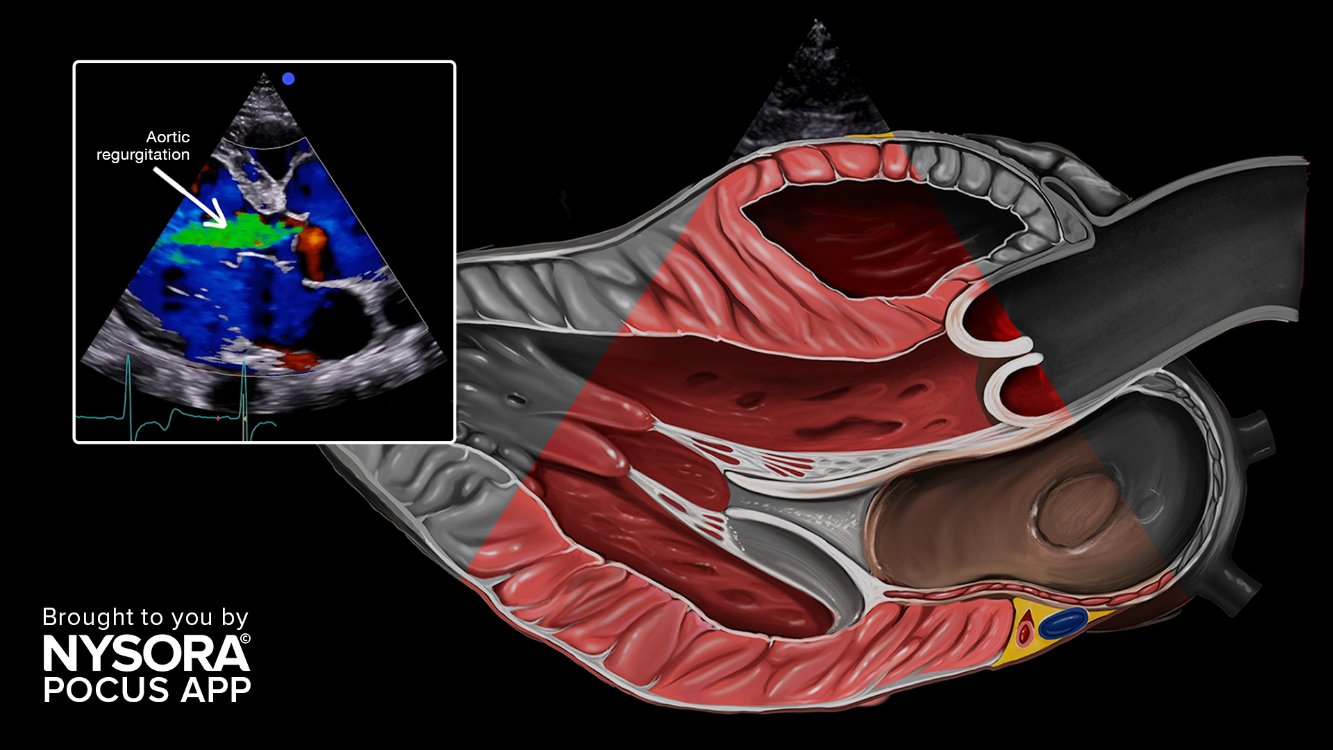

Ultrasound imaging is a widely used diagnostic tool that utilizes high-frequency sound waves to create images of internal organs and tissues. Ultrasound images are considered safe, non-invasive, and relatively inexpensive compared to other imaging modalities. However, like any medical imaging technique, ultrasound is prone to image artifacts, which can cause errors but also help in diagnosis and treatment planning.
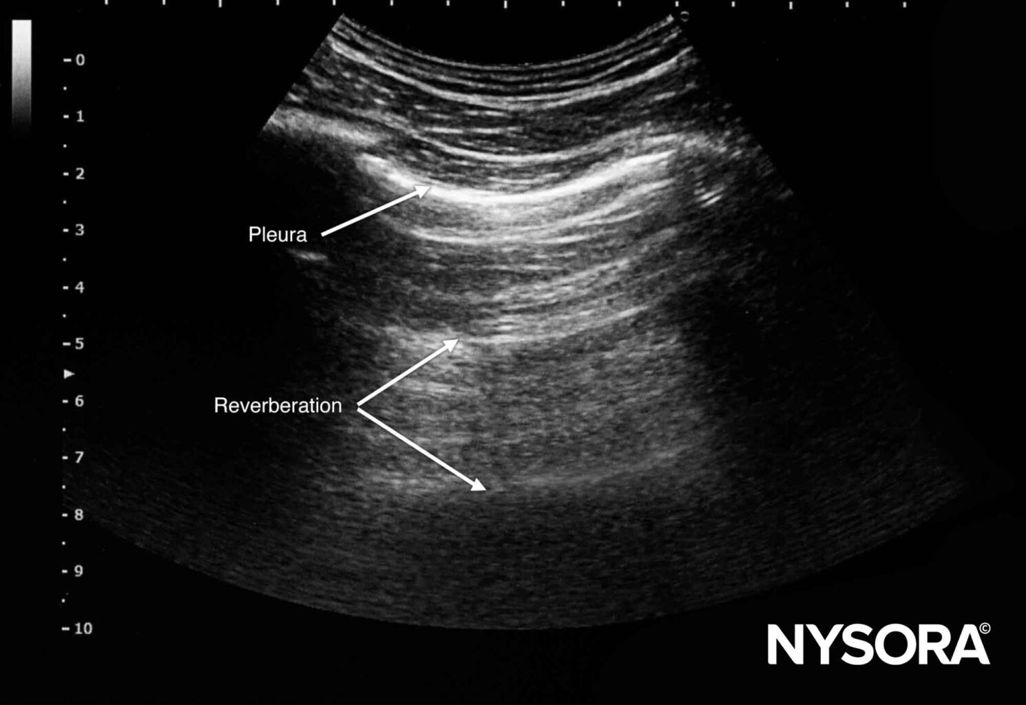
Reverberation artifacts
Reverberation artifacts occur when sound waves bounce back and forth between two surfaces, creating multiple echoes. These echoes appear as parallel lines on the image, giving the appearance of multiple objects or structures. Reverberation artifacts are common in areas where sound waves encounter multiple layers of tissue or air.
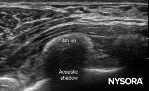
Shadowing artifacts
Shadowing artifacts occur when sound waves are blocked by a dense object, such as a bone or gas-filled organ, causing a loss of signal and a shadow to appear on the image. Shadowing artifacts can also occur when sound waves encounter an area of decreased sound transmission.
Edge artifacts
Edge artifacts occur when sound waves encounter an abrupt change in tissue density, such as the boundary between two organs. This can cause a bright line to appear on the image, which can obscure underlying structures.
Attenuation artifacts
Attenuation artifacts occur when sound waves lose energy as they travel through tissue, causing a loss of signal intensity. This can result in a hypoechoic or anechoic area on the image, making it difficult to visualize the underlying structure.
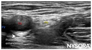
Enhancement artifacts
Structures with low acoustic impedance can artificially enhance the brightness below the structure, making it harder to visualize underlying structures (e.g., bladder, vessels).
Speckle artifacts
Speckle artifacts are caused by the interference of sound waves with each other, resulting in a granular or speckled appearance on the image. These artifacts can make it difficult to distinguish between small structures, such as blood vessels.
Doppler artifacts
Doppler artifacts occur when there is a high amount of motion in the area being imaged, such as blood flow. This can cause distortion of the Doppler waveform and make it difficult to accurately measure blood flow velocity.
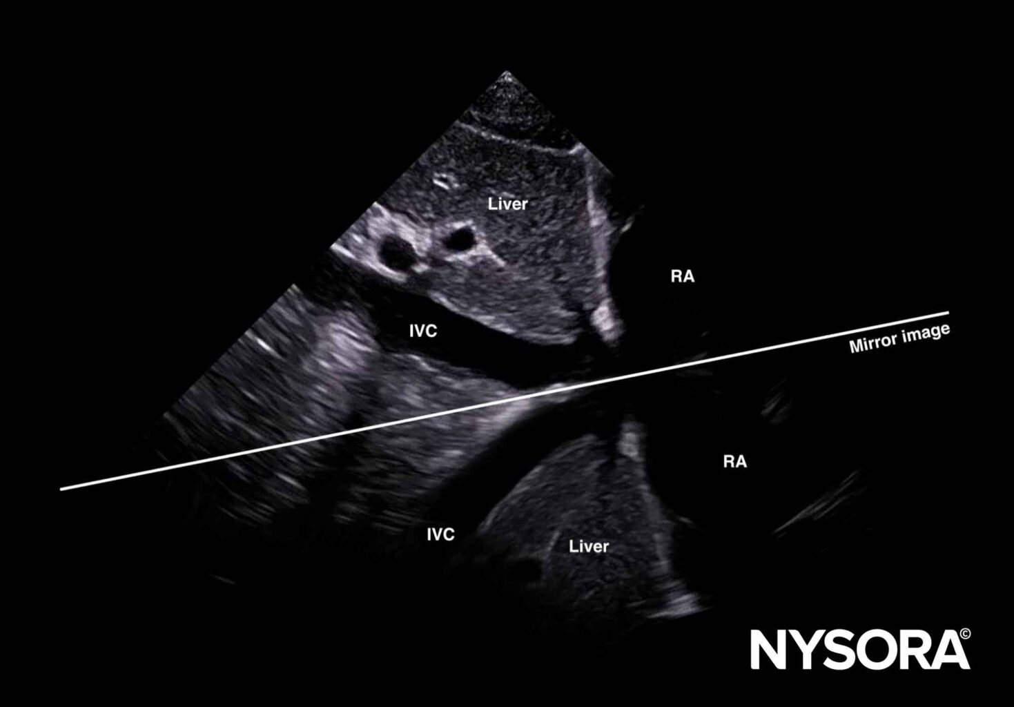
Mirror imaging
Ultrasound waves reflecting between structures can result in multiple reflections of the waves and mirroring of the structures between these layers. Mirrored structures can be found on the ultrasound image but not in the original structure.
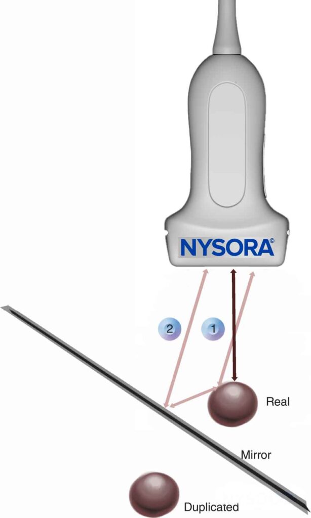
Mirror image: 1. Real or direct reflection of the ultrasound wave, 2. False or indirect reflection of the ultrasound wave.
Interesting fact
By moving the transducer, the mirrored structure will disappear but the original structure will remain.
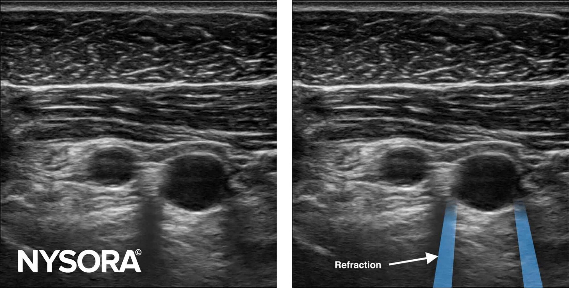
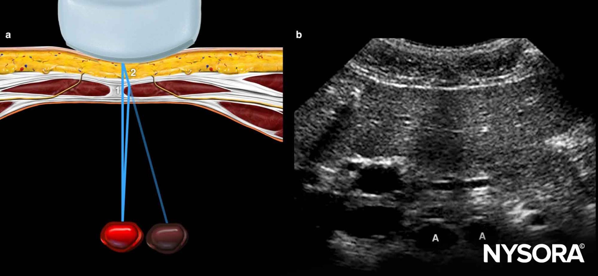
Refraction artifacts
Refraction is the appearance of the sound wave being bent when the ultrasound beam obliquely crosses an interface with tissues with different densities and, thus, different propagation speeds. It may result in an acoustic shadow at the border of the interface or a duplication artifact.
In conclusion, ultrasound image artifacts are a common occurrence in medical imaging and can have a significant impact on diagnosis and treatment planning. There are several ways to make use of ultrasound artifacts, including proper patient positioning, adjusting the gain and frequency settings, and selecting the appropriate transducer for the area being imaged. In addition, by understanding the causes and types of artifacts, healthcare professionals can ensure accurate interpretation of ultrasound images.