MSK Tip of the Week for Scanning the Oblique Popliteal Ligament
The oblique popliteal ligament (OPL) provides a critical reinforcing function, acting as a stabilizing structure for the posterior knee. As such, it requires particular attention when treating patients with knee injuries. Today, we’re sharing a quick overview to refresh your knowledge of the OPL.
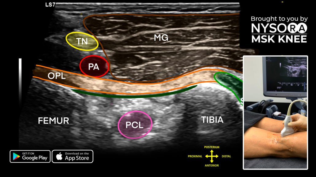
The OPL runs from the lateral femoral condyle, where it blends with the plantaris muscle, to the medial tibial plateau, where it is attached together with the semimembranosus insertion. It forms a Y-shaped structure with the semimembranosus muscle and functions to support the knee joint.
Scanning steps:
- With the patient in the prone position, place the transducer longitudinal, bridging the lateral femoral condyle and medial border of the tibial plateau.
- Identify the OPL running between the femur and tibia.
- The following structures can also be identified on ultrasound:
- Medial gastrocnemius muscle: Superficial
- Popliteal artery
- Tibial nerve: Located superficial to the popliteal artery
- Semimembranosus muscle: Located in line with the OPL
- Joint capsule: Located deep to the OPL
- Posterior cruciate ligament: Hypoechoic structure located in between the femur and tibia
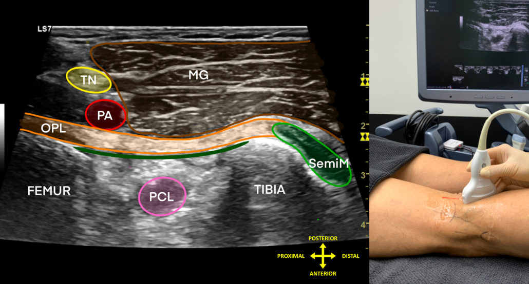
Sonoanatomy of the oblique popliteal ligament. OPL, oblique popliteal ligament; TN, tibial nerve; PA, popliteal artery; MG, medial gastrocnemius muscle; PCL, posterior cruciate ligament; SemiM, semimembranosus muscle.
Download the MSK App and start your free 7-day trial to read more about OPL and access the most practical and applicable techniques in musculoskeletal ultrasound anatomy and regenerative therapy of the knee.