
Identifying and Classifying the Severity of Hydronephrosis
Hydronephrosis is characterized by obstructive uropathy and thus enlargement of one or both kidneys due to the build-up of urine. Potential causes include kidney or bladder stones, benign prostatic hyperplasia (BPH), various cancers (bladder, renal, uterine, colon, cervical), and dysfunctional bladder (urinary retention).
Let’s explore how ultrasound helps to identify hydronephrosis:
In the absence of obstruction, the central sinus fatty tissue looks homogenous and collapsed with dark small pockets of urine within the sinus.
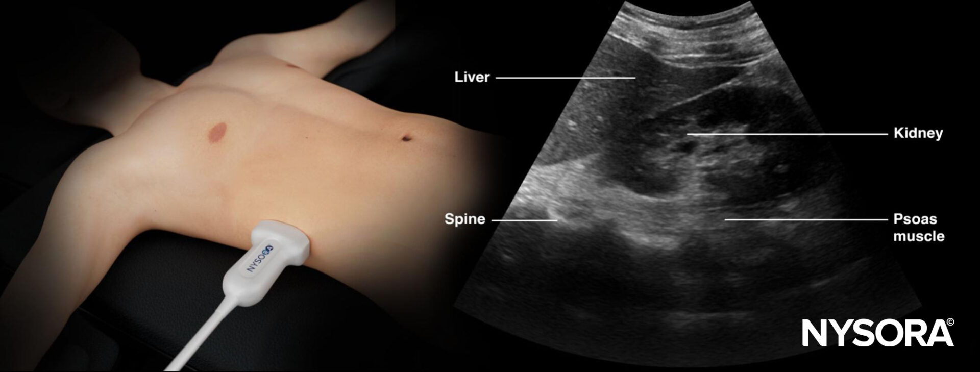
Sonoanatomy of a normal kidney.
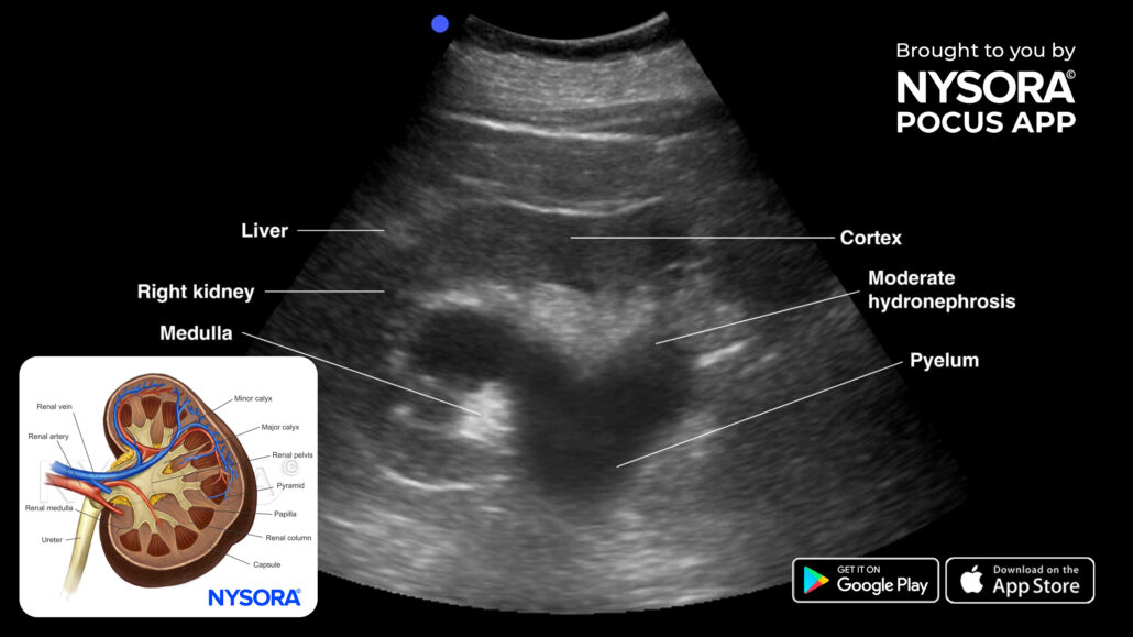
In the presence of hydronephrosis, a central collection of hypoechoic fluid or urine will be present in the center of the kidney.
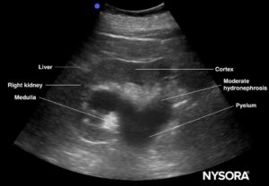
Sonoanatomy of a kidney with moderate hydronephrosis.
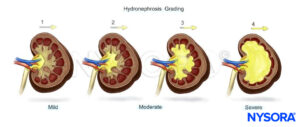
The size of the collection will determine the degree of hydronephrosis:
- Mild: Enlargement of calices with preservation of papilla (and renal anatomy)
- Moderate: Rounding of calices with obliteration of papilla and blunting of the pyramids (bear claw)
- Severe: Calciceal ballooning with cortical filling and grossly dilated renal pelvis
NOTE: Always combine a hydronephrosis scan with a bladder ultrasound.
Unleash the potential of POCUS with NYSORA’s POCUS App and elevate your practice, expand your capabilities, and deliver exceptional patient care. Download HERE.


