Case study: Carpal tunnel syndrome – Injection
A 56-year-old man, a cook by occupation, presented with 2 years history of right wrist pain along with severe pain of the radial 3 and a half fingers, increasing at night. Despite the use of splints, the pain persisted, and the patient was further evaluated.
Physical examination
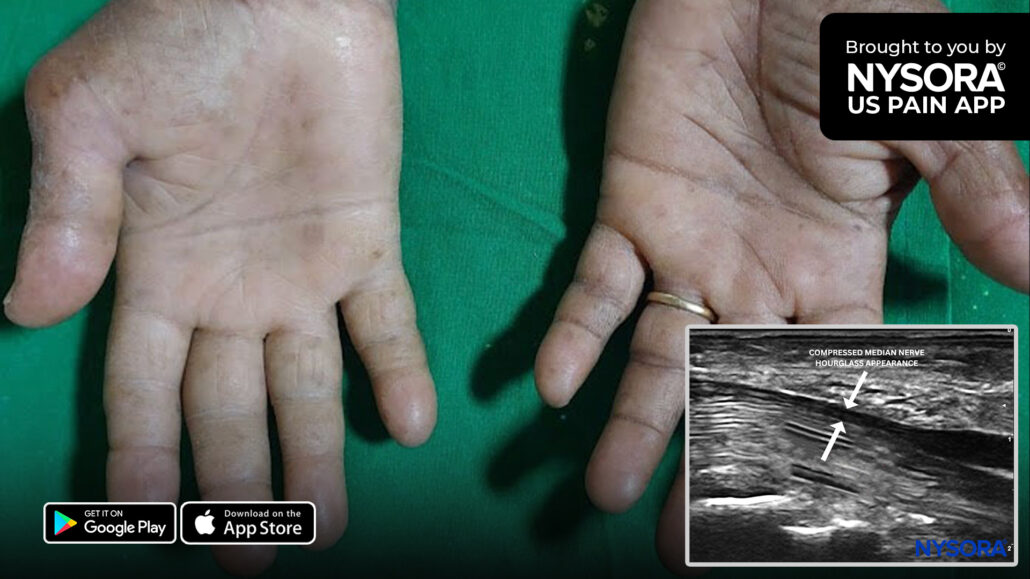
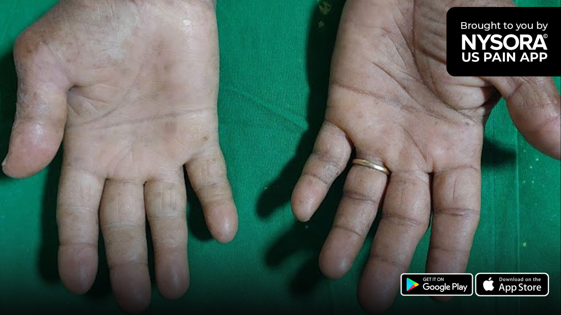
On clinical examination, the right hand and wrist looked shiny, and the classical “ape hand” deformity was present due to atrophy of the thenar eminence.
The following tests were performed:
- Phalen’s test: Positive
- Reverse Phalen’s test: Positive
- Tinel’s sign: Positive
- Durkan’s test: Positive
Ultrasound findings
- Bifid median nerve
- Persistent medial artery
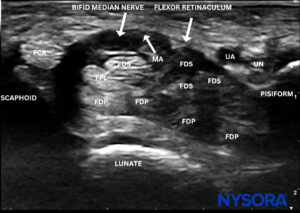
Sonoanatomy in a patient with carpal tunnel syndrome with a bifid median nerve and persistent medial artery. FCR, flexor carpi radialis tendon; FDS, flexor digitorum superficialis tendons; FDP, flexor digitorum profundus tendons; FPL, flexor pollicis longus tendon; MA, medial artery; UA, ulnar artery; UN, ulnar nerve.
- Compressed median nerve (“hourglass sign”)
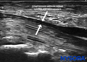
Long-axis view of the median nerve showing compression of the nerve.
- Median nerve cross-sectional area: 24.25 mm2
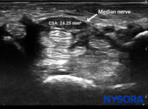
Cross-sectional area (CSA) of the median nerve.
Diagnosis
The patient was diagnosed with carpal tunnel syndrome, which is caused by compression of the median nerve, where it passes through the carpal tunnel. Symptoms include numbness, tingling, pain, and weakness in the region supplied by the median nerve (thumb, index and middle fingers, and lateral half of the ring finger).
To read more about the treatment, patient outcome, and other case studies, download the most practical app on ultrasound-guided chronic pain blocks for management of the chronic pain HERE.