Nerve Blocks App
 Pain Medicine Assistant App
 POCUS App
 IV Access App
 MSK Knee App
 VetRA App
 Nerve Block Manual
 Regional Anesthesia Updates
 Anesthesiology Manual
 Anesthesiology Review
 Anesthesia Updates 2025
 Anesthesia Updates 2026
 Pediatric Anesthesia Updates
 Airway Management Updates
 US Interventional Pain Manual
 Pain Medicine Updates
 Mastering Difficult IV Access
 PACU Nursing Manual
 RA Veterinary Manual
 About
About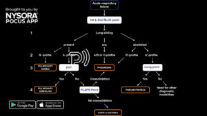
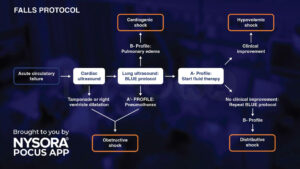
The BLUE (Bed Lung Ultrasound in Emergency) protocol, with an accuracy of more than 90% for diagnosing the cause of acute respiratory failure, can be used to
- Troubleshoot the etiology of acute respiratory failure
- Allow differentiation between pneumothorax, pneumonia, pulmonary embolism, pulmonary edema, and COPD or asthma
Here’s how we apply it in practice.
- Scan the 2 BLUE points on the left and right thorax
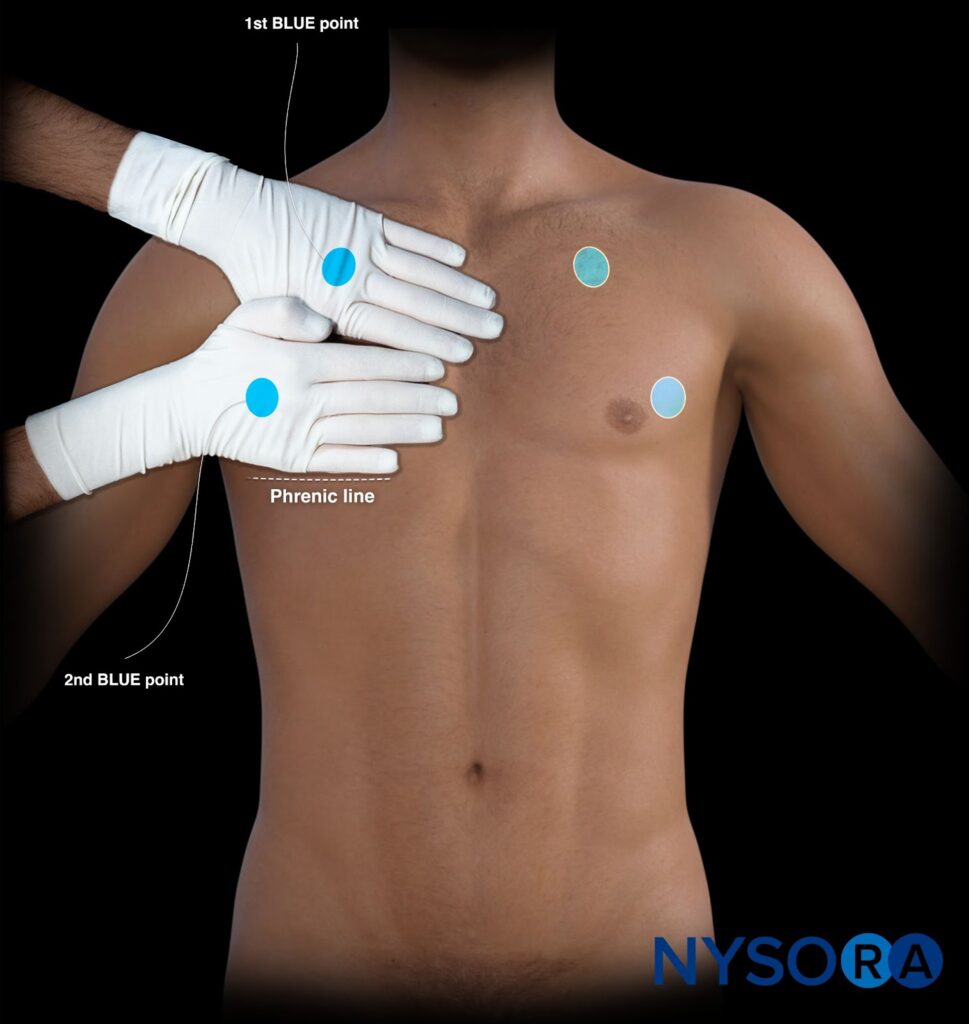
- Evaluate the presence of lung sliding (the absence of lung sliding will be indicated with a ‘).
- Check for lung artifacts (A-lines, B-lines, C-lines).
- Determine the profile based on the findings in all 4 BLUE points.
- A-profile: A-lines in all 4 BLUE points.
- B-profile: 3 or more B-lines in all 4 BLUE points.
- C-profile: A consolidation (C-line) present in one of the BLUE points.
- A/B profile: Various findings of A-lines and 3 or more B-lines in the 4 BLUE points.
- An A-profile or A’-profile requires further scanning.
- In case of an A-profile, thrombosed veins need to be excluded using the DVT protocol. When negative, the PLAPS point should be assessed to rule out consolidation.
- In case of an A’-profile, the chest wall should be scanned to rule in a lung point.
Unleash the potential of POCUS with NYSORA’s POCUS App and elevate your practice, expand your capabilities, and deliver exceptional patient care. Download HERE