Piriformis syndrome occurs when the piriformis muscle irritates the sciatic nerve, which comes into the gluteal region beneath the muscle, causing pain in the buttocks and referred pain along the sciatic nerve (i.e., sciatica). It will often improve with a conservative regimen of physical therapy and analgesic pharmacotherapy.
Here are 4 instant tips for scanning while performing a piriformis muscle injection:
- Place the transducer transversely over the posterior superior iliac spine.
- Move the transducer laterally to visualize the ilium.
- Orient the transducer in the direction of the piriformis muscle and move caudally until the sciatic notch is identified.
- The hyperechoic shadow of the bone will disappear from the medial aspect, and two muscle layers become visible: the gluteus maximus and piriformis muscle.
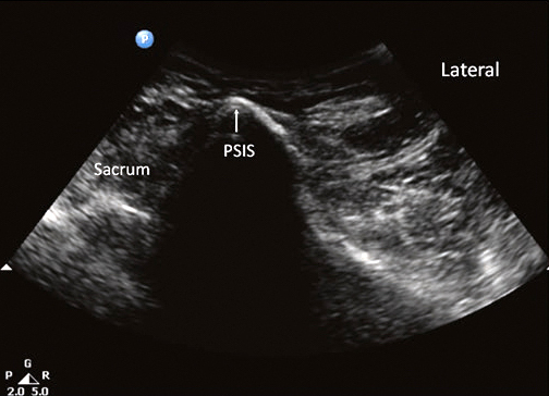
Sonoanatomy
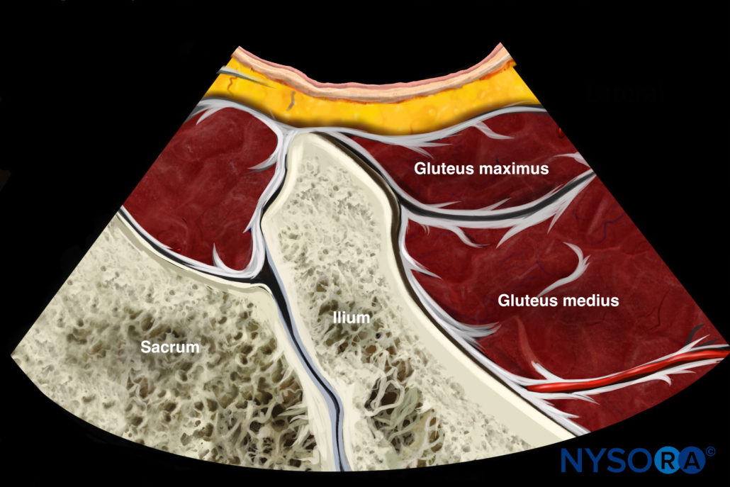
Reverse Ultrasound Anatomy
Comparison of sonoanatomy and reverse ultrasound anatomy for a piriformis muscle injection.
Download the US Pain App HERE to read other tips on managing acute and chronic pain and to access the complete guide to ultrasound-guided chronic pain blocks.


