Back to News
 2 min read

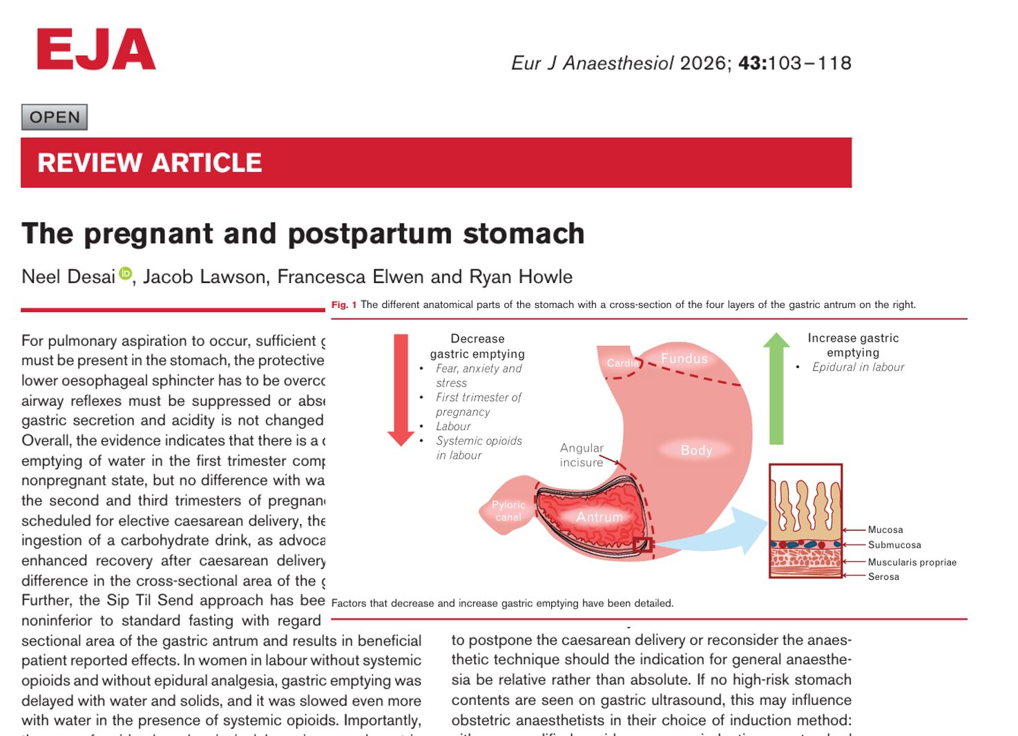
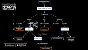
A 58-year-old male patient with acute dyspnea is assessed in the emergency department using the BLUE protocol. The scan reveals no significant lung pathology (A-profile) but prompts the consideration of deep vein thrombosis (DVT). This rapid assessment aids in detecting DVT to prevent potential complications like pulmonary embolism.
Here’s how you perform a DVT POCUS scan:
- Position the patient supine with the leg in extension and exorotation.
- Start scanning at the level of the inguinal crease with a linear transducer. For the popliteal position, the leg is flexed to allow scanning of the fossa.
- Slowly scan distally and assess compressibility every 1-2 cm.
It is impossible to compress a thrombosed vein.
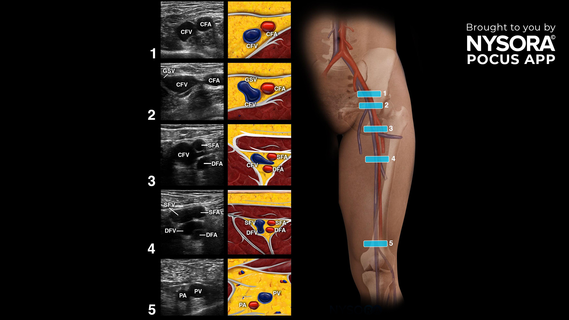
4. Pay special attention to 5 visualization key points since these are more likely for visualizing clots.
- Common femoral vein.
- Bifurcation of the common femoral vein and the saphenous vein.
- Bifurcation of the common femoral vein and the lateral perforator vein.
- Bifurcation of the superficial femoral vein and the deep femoral vein.
- Popliteal vein.
5 key scanning points for DVT visualization. CFV, common femoral vein; GSV, great saphenous vein; SFA, superficial femoral artery; DFA, deep femoral artery; SFV, superficial femoral vein; DFV, deep femoral vein; PA, popliteal artery; PV, popliteal vein.
Transform your practice with the power of POCUS using NYSORA’s POCUS App. Enhance your skills, broaden your diagnostic capabilities, and provide outstanding patient care. Experience the difference today – Download the app HERE.