 Nerve Blocks App
 Anesthesia Assistant App
 Ultrasound Pain App
 Pain Medicine Assistant App
 POCUS App
 IV Access App
 MSK Knee App
 VetRA App
 Nerve Block Manual
 Regional Anesthesia Updates
 Anesthesiology Manual
 Anesthesiology Review
 Anesthesia Updates 2025
 Anesthesia Updates 2026
 Pediatric Anesthesia Updates
 Airway Management Updates
 US Interventional Pain Manual
 Pain Medicine Updates
 Mastering Difficult IV Access
 PACU Nursing Manual
 RA Veterinary Manual
 Books
Books About
About
 Back to News

 1 min read
Baker’s cysts – communication between the posterior joint capsule and the gastrocnemius-semimembranosus bursa – are fluid-filled distended synovial-lined lesions arising in the popliteal fossa between the medial gastrocnemius and semimembranosus tendons. They are usually located at or below the joint line.
Here are 3 tips to scan Baker’s cyst (transverse scan)
- Position the patient prone.
- Place the transducer transverse over the intersection of the medial gastrocnemius and semimembranosus muscles.
- Identify the following structures:
-
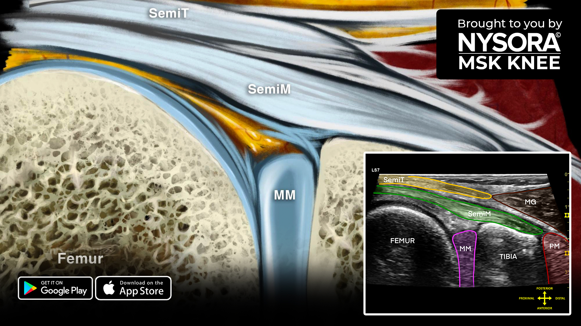
- Semimembranosus muscle: Located superficial to the femur.
- Semitendinosus muscle: Located on top of the semimembranosus.
- Sartorius muscle and tendon: Located medial to the semimembranosus.
- Gracilis muscle: Located on top of the semimembranosus.
- Medial gastrocnemius muscle: Located lateral to the semimembranosus and semitendinosus.
- Popliteal artery.
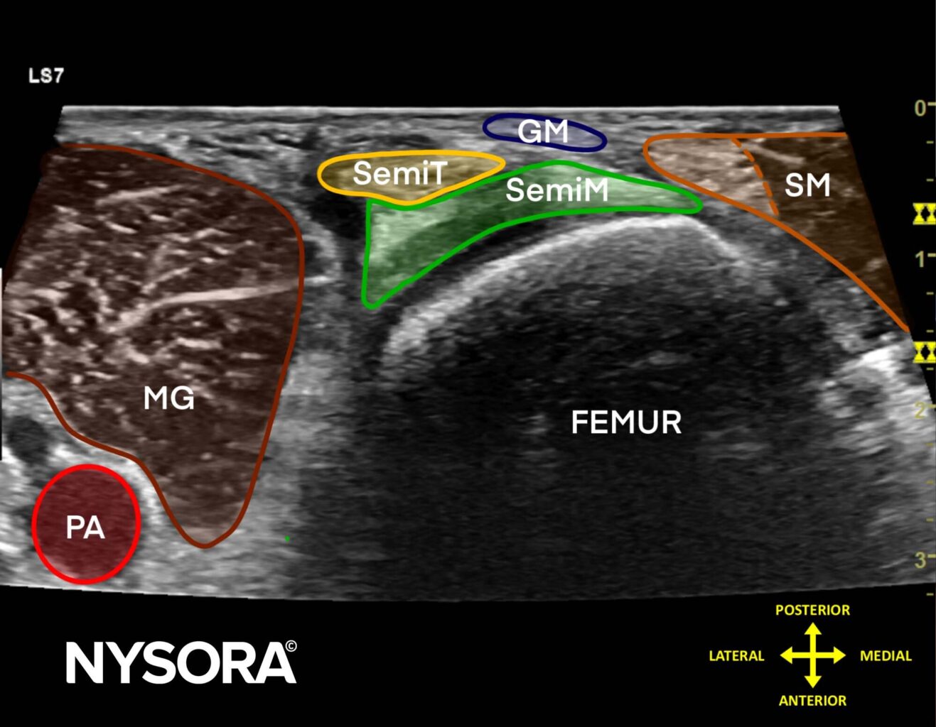
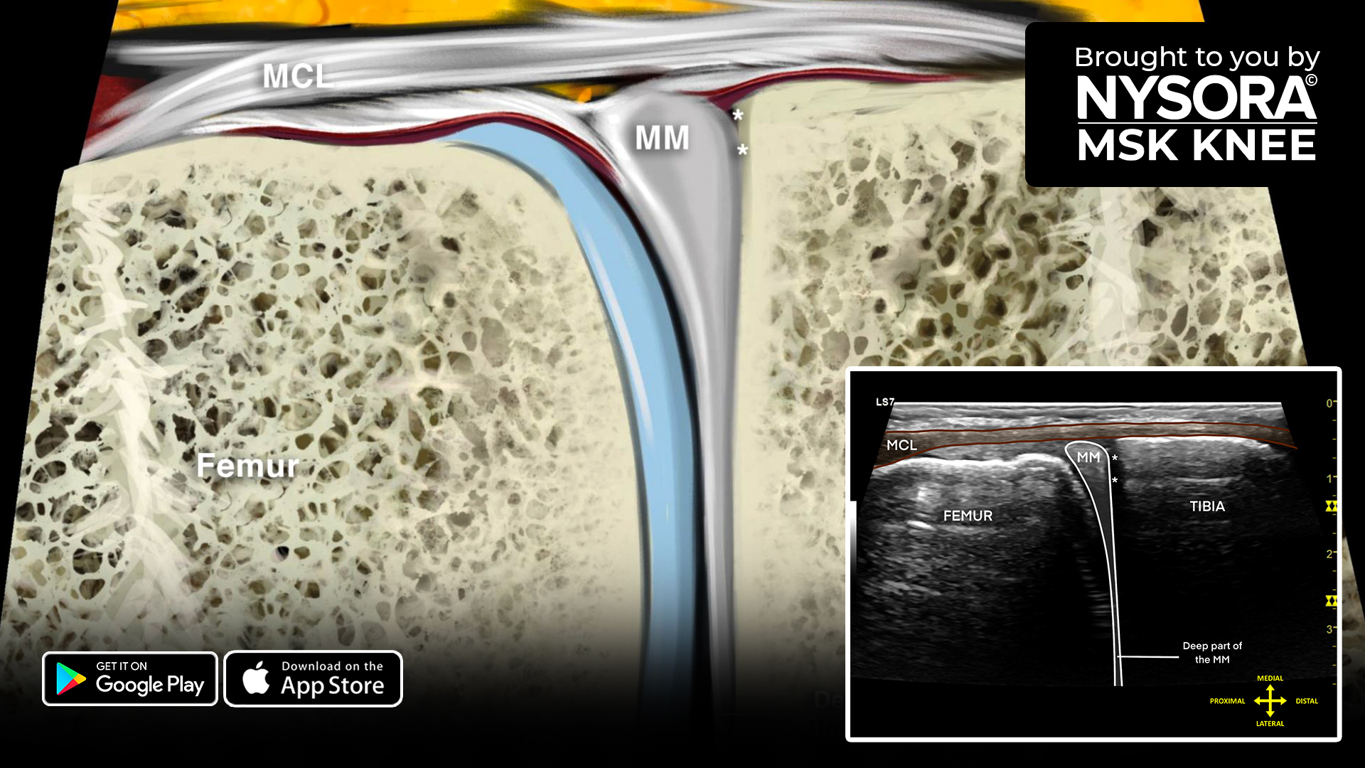
Sonoanatomy
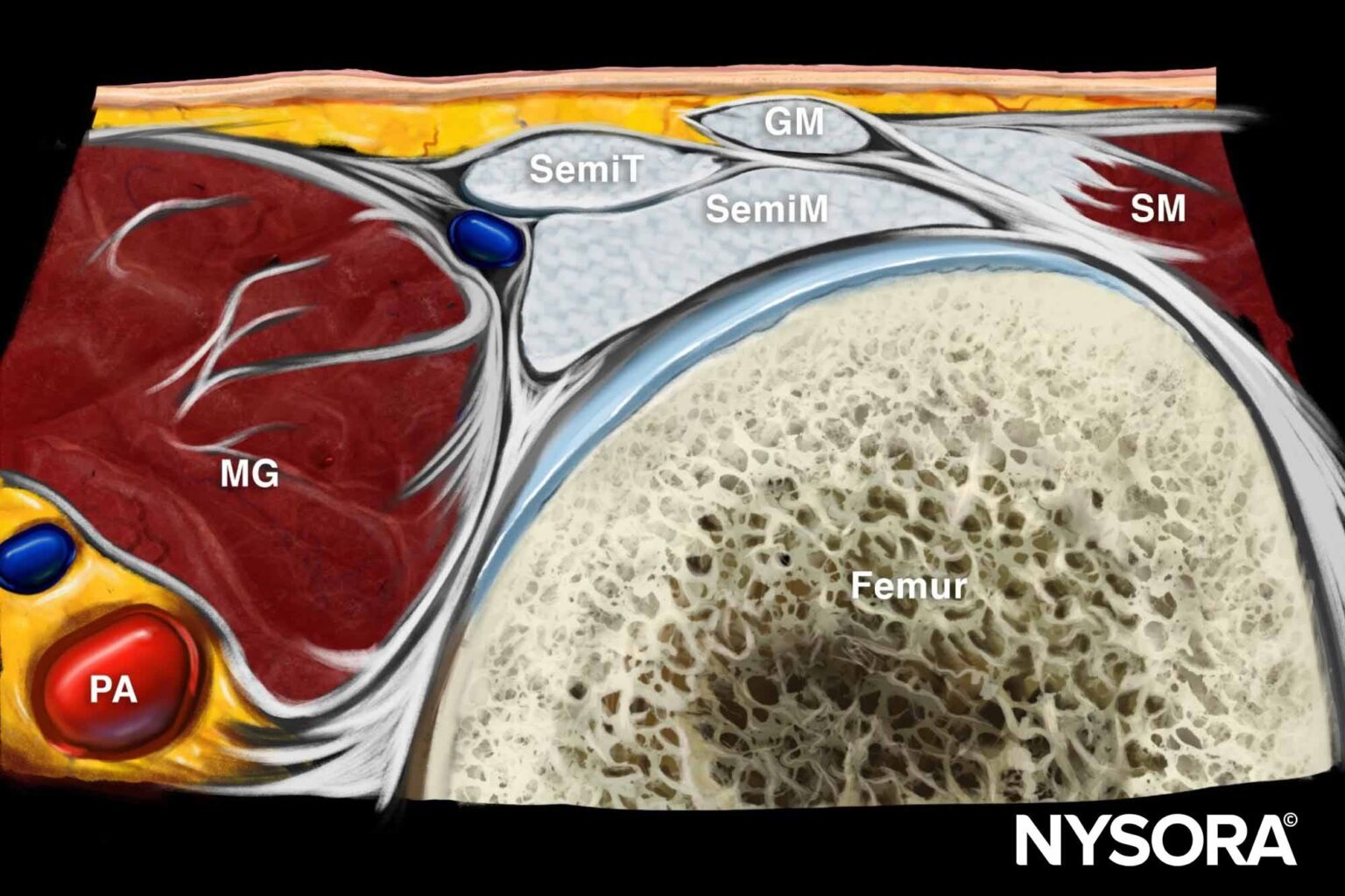
Reverse Ultrasound Anatomy
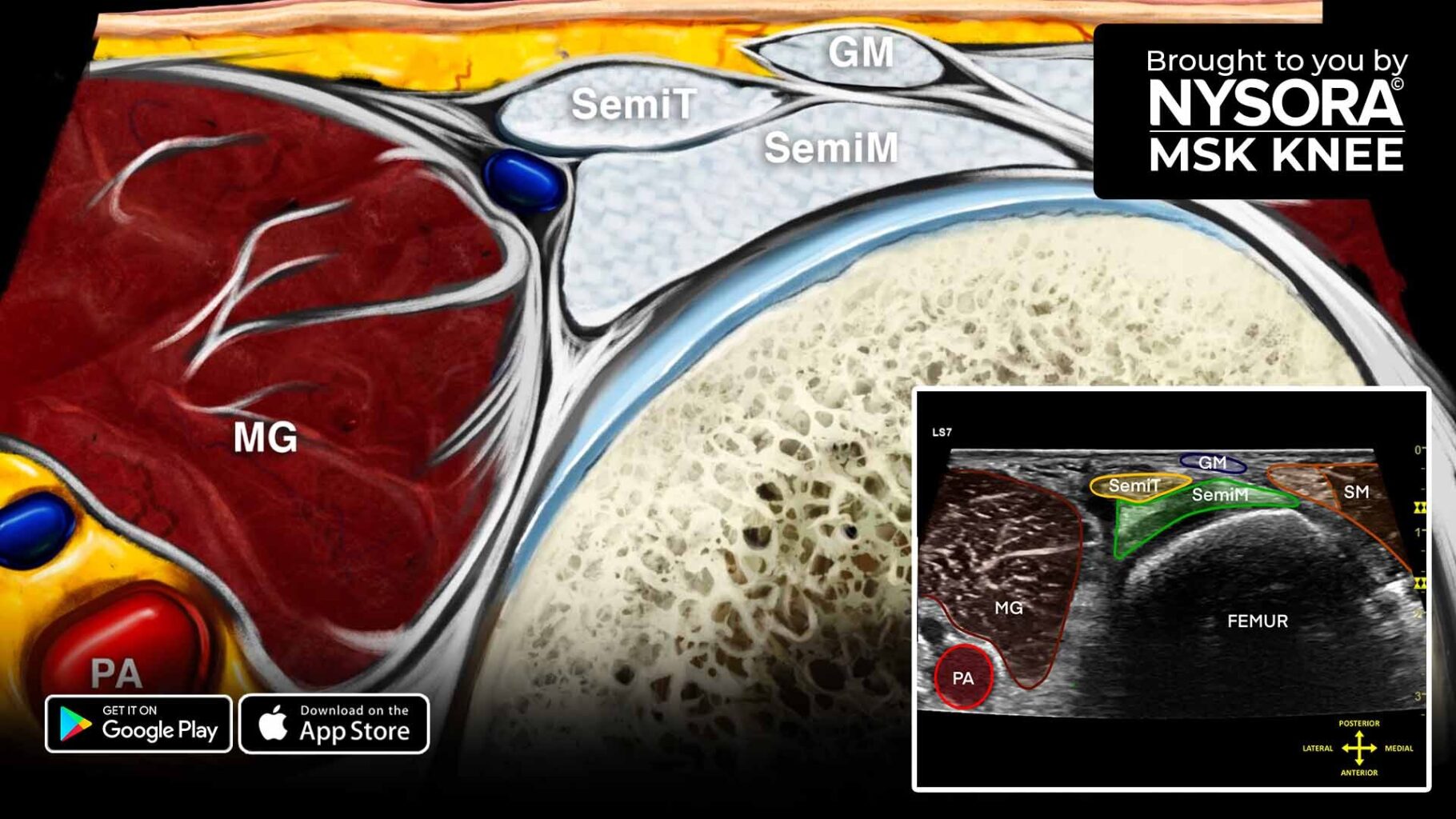
Comparison of sonoanatomy and reverse ultrasound anatomy of Baker’s cyst (transverse scan).
Download the MSK App for more tips and the most practical and applicable techniques in musculoskeletal ultrasound anatomy and regenerative therapy of the knee.