Nerve Blocks App
 Pain Medicine Assistant App
 POCUS App
 IV Access App
 MSK Knee App
 VetRA App
 Nerve Block Manual
 Regional Anesthesia Updates
 Anesthesiology Manual
 Anesthesiology Review
 Anesthesia Updates 2025
 Anesthesia Updates 2026
 Pediatric Anesthesia Updates
 Airway Management Updates
 US Interventional Pain Manual
 Pain Medicine Updates
 Mastering Difficult IV Access
 PACU Nursing Manual
 RA Veterinary Manual
 About
About
Learning objectives
- Describe and classify mitral regurgitation
- Describe the causes and mechanisms of mitral regurgitation
- Manage patients with mitral regurgitation
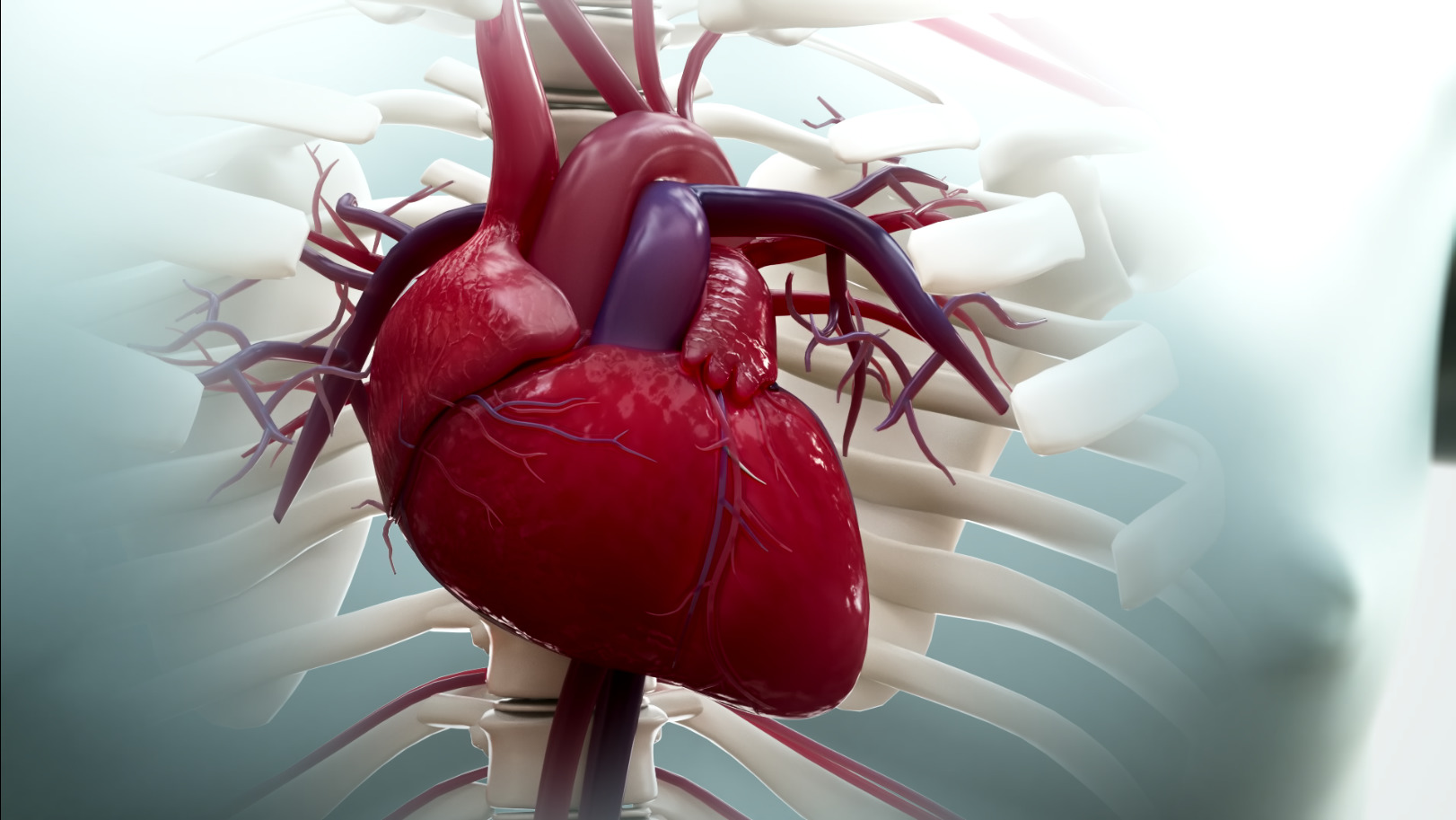
Definition & mechanisms
- Mitral regurgitation (MR) can be acute or chronic in onset, and primary or secondary in nature
- Primary MR is due to pathology of the valve preventing normal closure
- Secondary MR is caused by left ventricle dysfunction that affects the closing of the mitral valve
- The left atrium dilates as blood is ejected back into it
- Atrial fibrillation is common
- Overload of the pulmonary circulation causes dyspnea
- The LV is volume overloaded in, with dilation of the ventricle further worsening MR
- Acute MR:
- Can be caused by any disruption to the normal mechanism of the valve (growth of vegetations on the leaflets in infective endocarditis, chordae rupture in patients with pre-existing degenerative disease, or papillary muscle rupture due to an ST-elevation)
- The left atrium is unable to compensate acutely for the increased pressure caused by blood refluxing back into it
- Patients can present with sudden-onset dyspnea and require rapid stabilization and treatment
- Chronic MR:
- Primary: Abnormality of the leaflets that prevents them from closing normally
- Secondary: Anatomy of the valve is normal, but its function is impaired due to left ventricle pathology
Classification
Transoesophageal echocardiography remains the gold standard for defining the severity of the MR:
| Mild | Moderate | Severe | |
|---|---|---|---|
| Regurgitant fraction | <30% | 30–49 | ≥50% |
| Regurgitant orifice area | <0.20 cm2 | 0.2–0.39 cm2 | ≥0.40 cm2 |
| Regurgitant volume | <30 mL/beat | 30–59 mL/beat | ≥60 mL/beat |
Treatment
- Medical
- Acute MR: Filling pressure reduction with nitrates or diuretics and afterload reduction with vasodilators or an intra-aortic balloon pump as bridging to definitive treatment
- Chronic MR: Treatment is in line with standard heart failure management including β-blockers, angiotensin-converting enzyme inhibitors, and aldosterone antagonists, with diuretics where heart failure is present
- Surgical
- Primary MR:
- Surgery is indicated if the MR is severe and acute in nature and if the MR is chronic and causing symptoms, with no contraindications to surgery
- Valve repair, rather than replacement, is preferred
- Secondary MR:
- In symptomatic patients with severe left ventricle failure, the benefits of surgery are controversial unless the underlying condition can be reversed
- Primary MR:
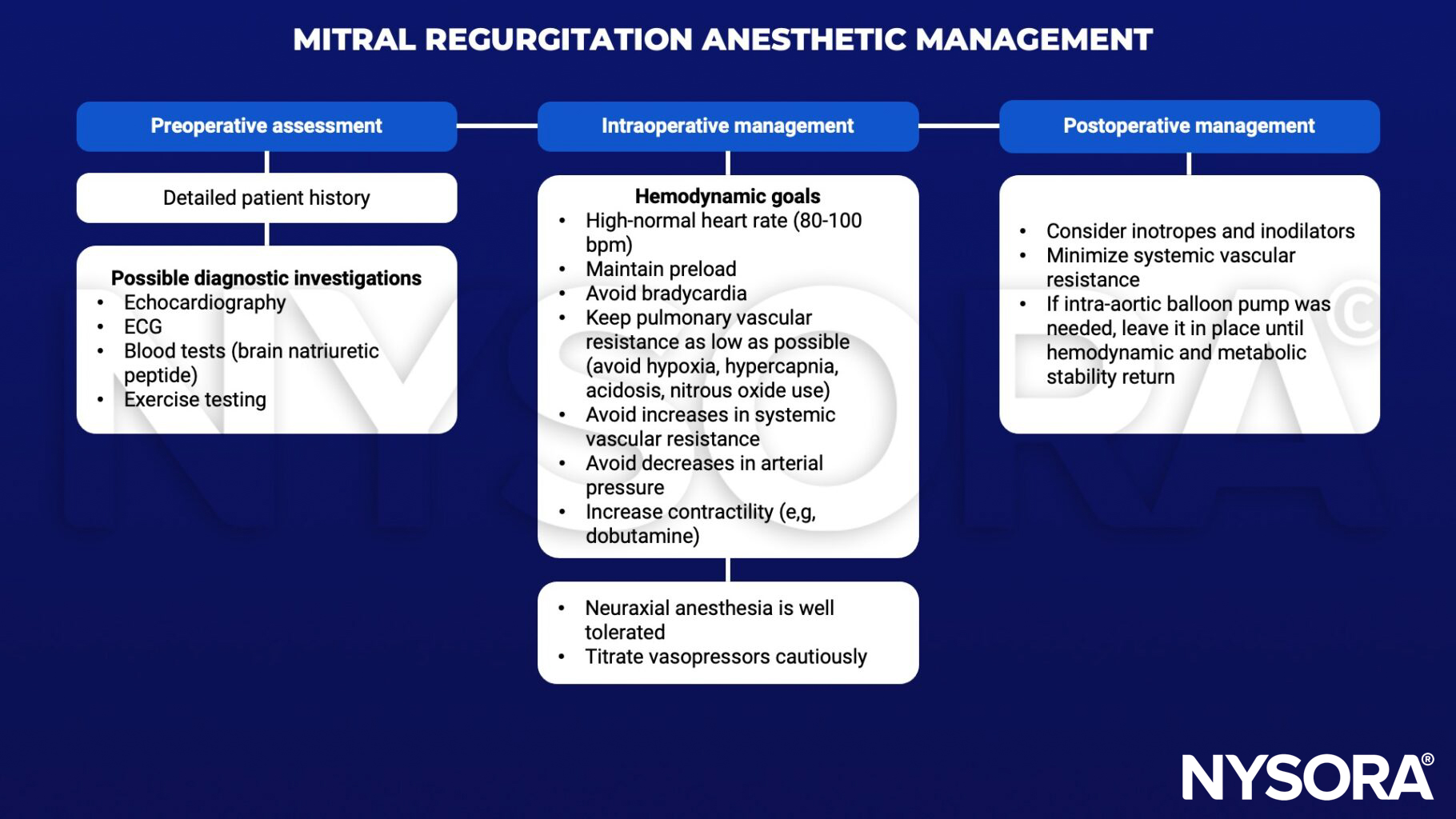
Anesthetic management
Suggested reading
- Holmes K, Gibbison B, Vohra HA. Mitral valve and mitral valve disease. BJA Education. 2017;17(1):1-9.