
Unexpected diagnosis of endocarditis
A 68-year-old woman presented at the emergency department with acute respiratory failure and fever. Lung ultrasound showed a B-profile in all four BLUE points, suggesting pulmonary edema. This prompted us to do a cardiac ultrasound.
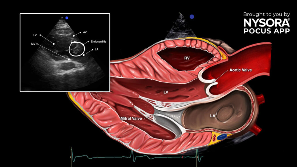
Consecutively, a focused cardiac ultrasound was performed. The parasternal long-axis view showed a lesion suggestive of endocarditis which was then confirmed by an official cardiologist ultrasound.
Endocarditis is a serious medical condition that affects the inner lining of the heart and the valves. The causal pathogen may be bacterial but occasionally fungal or viral. Endocarditis is often diagnosed by the formation of vegetations on the heart valves or other endocardial surfaces. These vegetations can interfere with the normal function of the heart, leading to complications such as severe regurgitation, cardiac failure, stroke, or systemic infections if bacteria from the heart enter the bloodstream. Endocarditis requires prompt diagnosis and treatment with antibiotics or, in severe cases, surgery to repair or replace damaged valves.
Point-of-care ultrasound (POCUS) is a valuable tool in assessing endocarditis, offering real-time imaging capabilities that aid in detecting vegetations, abscesses, and valvular abnormalities.
When evaluating endocarditis through ultrasound, distinctive pathology characteristics to observe include:
- Mobile lesion
- Hyperechoic density
- Endocarditis is often accompanied by regurgitation
Parasternal long-axis view revealing endocarditis. LV, left ventricle; AV, aortic valve; LA, left atrium; MV, mitral valve.
Transform your practice with the power of POCUS using NYSORA’s POCUS App. Enhance your skills, broaden your diagnostic capabilities, and provide outstanding patient care. Experience the difference today – Download the app HERE.


