
Tips for scanning Baker’s Cyst in a longitudinal orientation
February 23, 2023
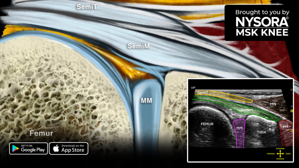
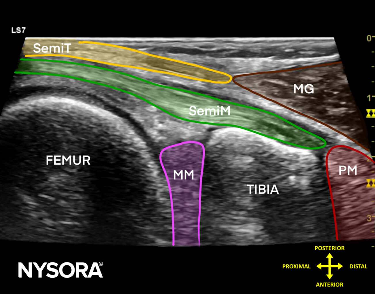
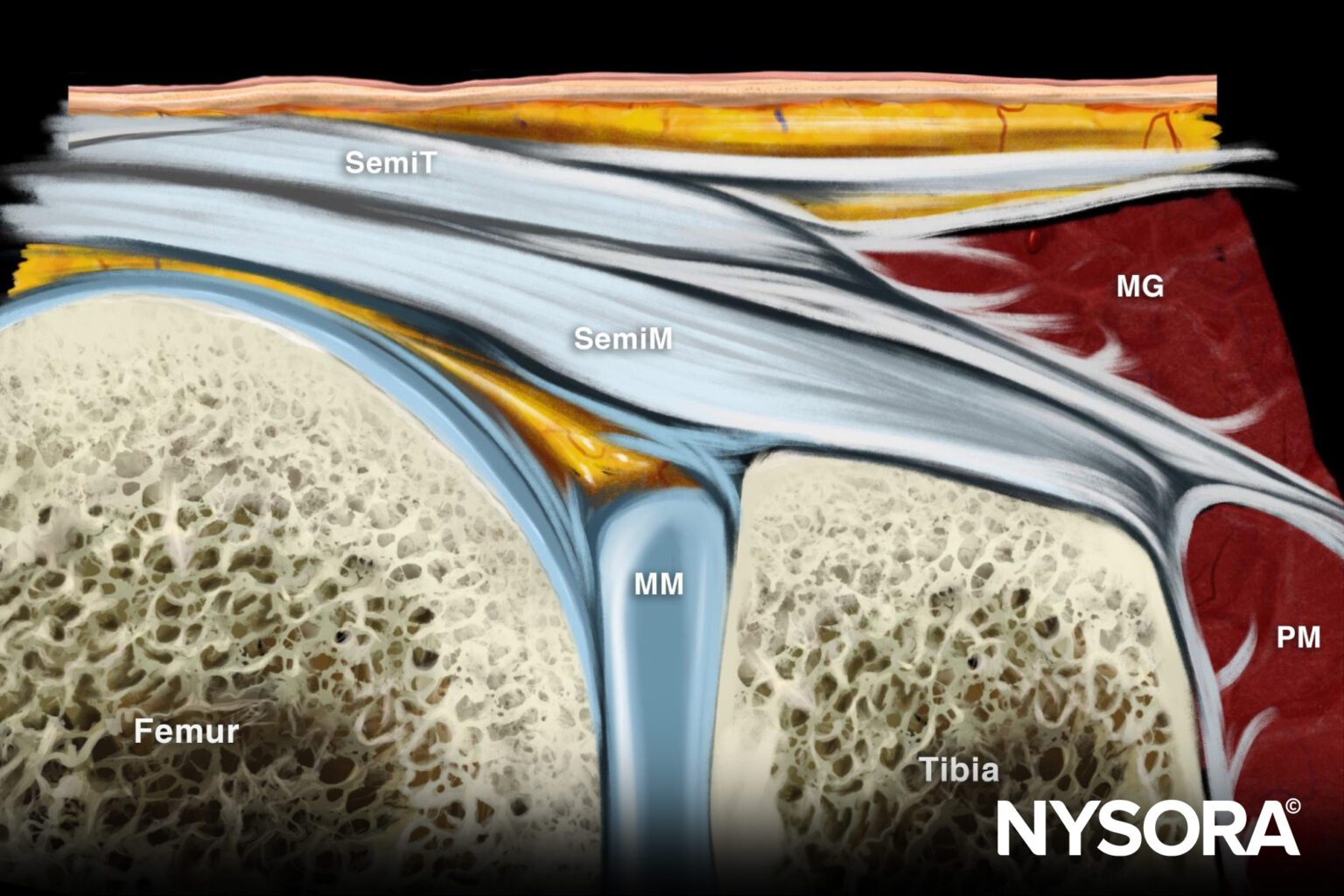
The intersection of the medial gastrocnemius and semimembranosus muscles is called the Baker’s cyst neck position.
Check out these 3 tips to scan Baker’s cyst (longitudinal scan)
- Position the patient prone.
- Place the transducer longitudinal over the intersection of the medial gastrocnemius and semimembranosus muscles.
- Identify the following structures:
- Semimembranosus muscle: Inserting onto the tibial plateau.
- Popliteus muscle: Located immediately distal to the semimembranosus.
- Semitendinosus muscle: Located on top of the semimembranosus.
- Posterior horn of the medial meniscus: Located between the tibia and femur.
- Medial gastrocnemius muscle: Overlapping the distal part of the semimembranosus and semitendinosus muscles.
Sonoanatomy
Reverse Ultrasound Anatomy

Comparison of sonoanatomy and reverse ultrasound anatomy of Baker’s cyst (longitudinal scan).
Download the MSK App for more tips and the most practical and applicable techniques in musculoskeletal ultrasound anatomy and regenerative therapy of the knee.


