Case study: Carpal tunnel release
Case presentation
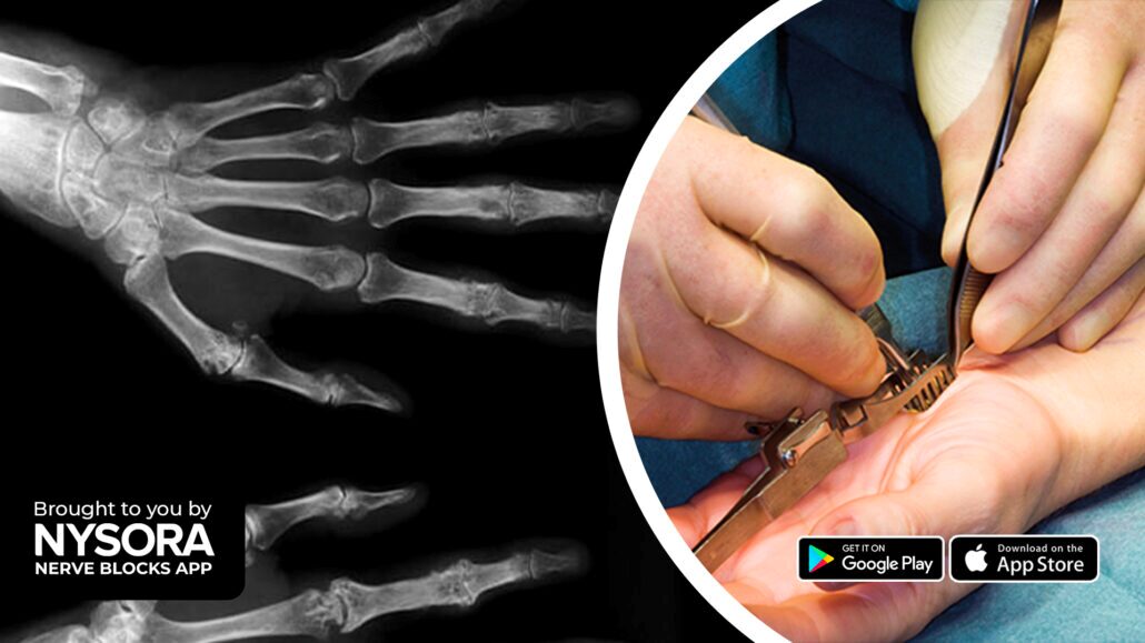
A 45-year-old female patient with a confirmed diagnosis of carpal tunnel syndrome in the right hand was scheduled for elective carpal tunnel release surgery. The patient experienced significant pain, numbness, and tingling in the affected hand, affecting daily activities and quality of life. As part of the comprehensive pain management plan, an ultrasound-guided wrist block was proposed to provide localized anesthesia and minimize postoperative pain.
Nerve block technique
The wrist block technique consists of a mid-forearm level block of the median and ulnar nerves, followed by a subcutaneous infiltration of the local anesthetic at the wrist crease for any remaining cutaneous branches of the musculocutaneous, radial, or ulnar nerves.
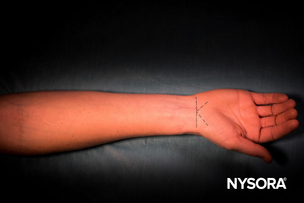
Subcutaneous infiltration.
Under ultrasound guidance, the median and ulnar nerves were identified between the superficial and deep flexors of the wrist and fingers.
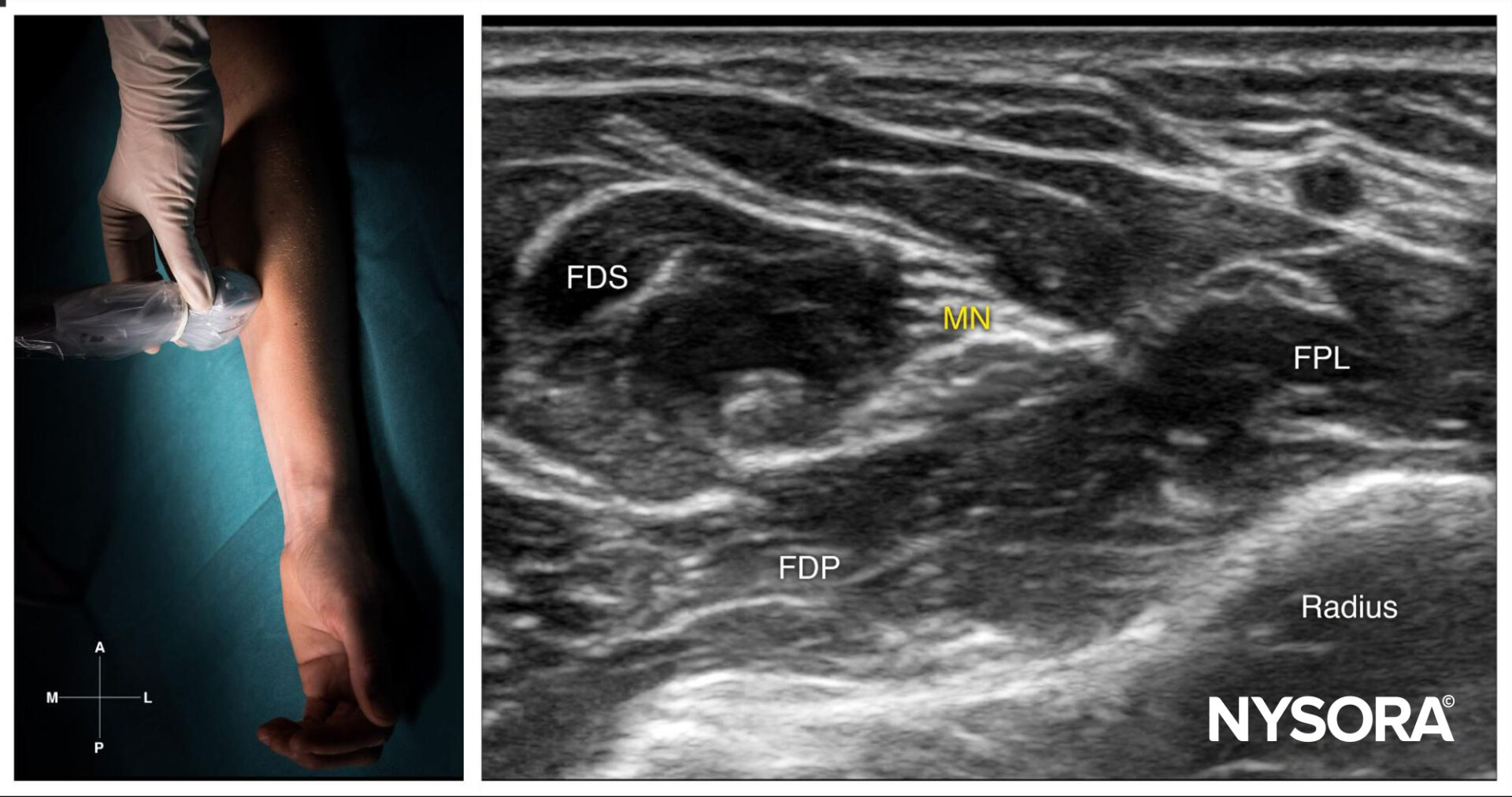
Median nerve block at the level of the wrist; transducer position and sonoanatomy. MN, median nerve; FPL, flexor pollicis longus muscle; FDS, flexor digitorum superficialis muscle; FDP, flexor digitorum profundus muscle.
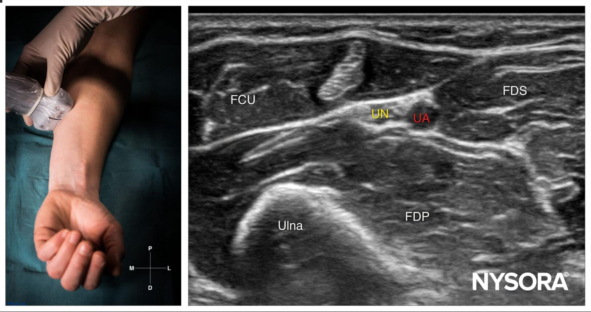
Ulnar nerve block at the level of the wrist; transducer position and sonoanatomy. UN, ulnar nerve; UA, ulnar artery; FCU, flexor carpi ulnaris; FDP, flexor digitorum profundus muscle; FDS, flexor digitorum superficialis muscle.
A 25-gauge needle was then inserted in-plane or out-of-plane and 4 mL of lidocaine 2% was injected into the fascia containing the nerves. The onset of anesthesia was observed within 10 minutes.
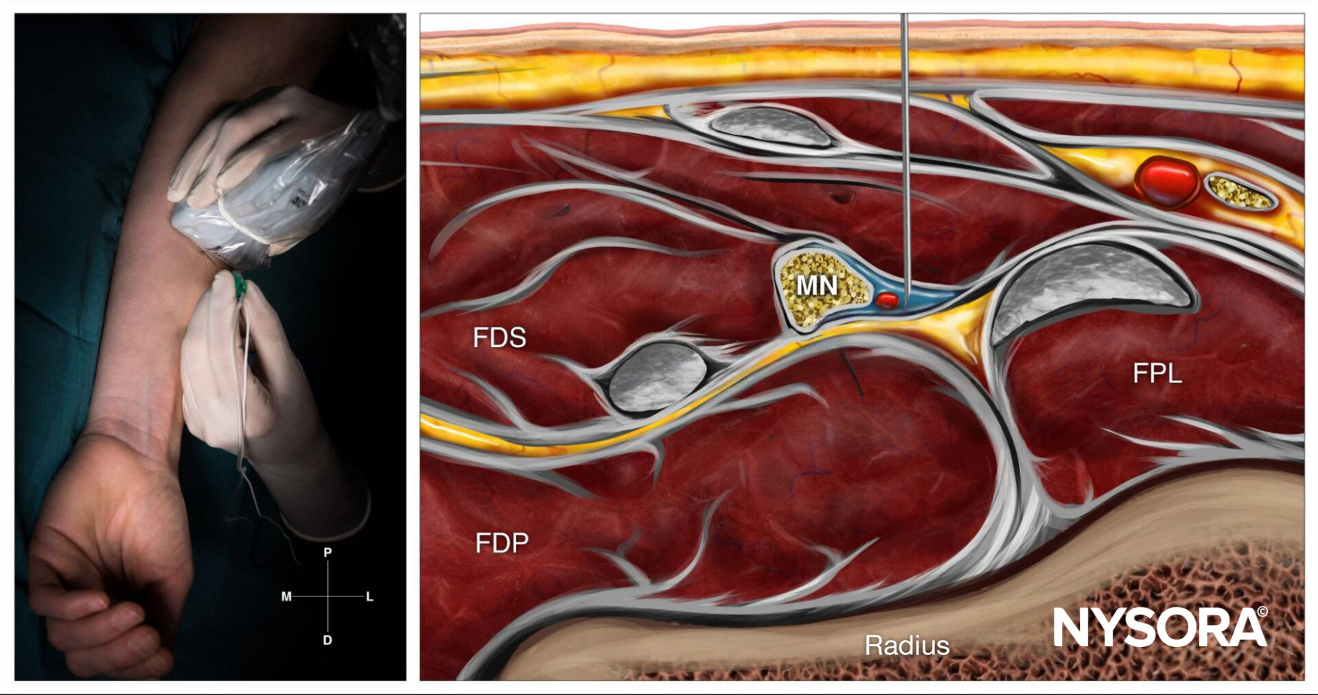
Median nerve block at the level of the wrist; Reverse Ultrasound Anatomy with needle insertion out-of-plane and local anesthetic spread (blue). MN, median nerve; FPL, flexor pollicis longus muscle; FDS, flexor digitorum superficialis muscle; FDP, flexor digitorum profundus muscle.
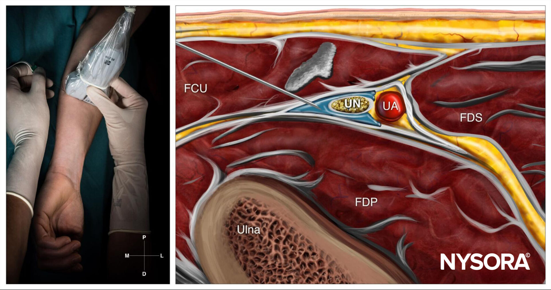
Ulnar nerve block at the level of the wrist; Reverse Ultrasound Anatomy with needle insertion in-plane and local anesthetic spread (blue). UN, ulnar nerve; UA, ulnar artery; FCU, flexor carpi ulnaris; FDP, flexor digitorum profundus muscle; FDS, flexor digitorum superficialis muscle.
Patient outcome
Following the ultrasound-guided wrist block, the patient reported immediate relief of pain and paresthesia in the affected hand. After surgery, the patient’s hand function improved, allowing for early mobilization and participation in rehabilitation exercises. The need for systemic opioids was minimized, thereby reducing the risk of associated side effects. The patient did not experience any complications or adverse events related to the wrist block.
For more case studies like these and the complete guide to the 60 most frequently used nerve blocks, download the Nerve Blocks App HERE. Don’t miss the chance to get the bestselling NYSORA Nerve Blocks App also in book format – the perfect study companion with the Nerve Blocks app!