Case study: Free flap surgery in a high-risk patient
Case presentation
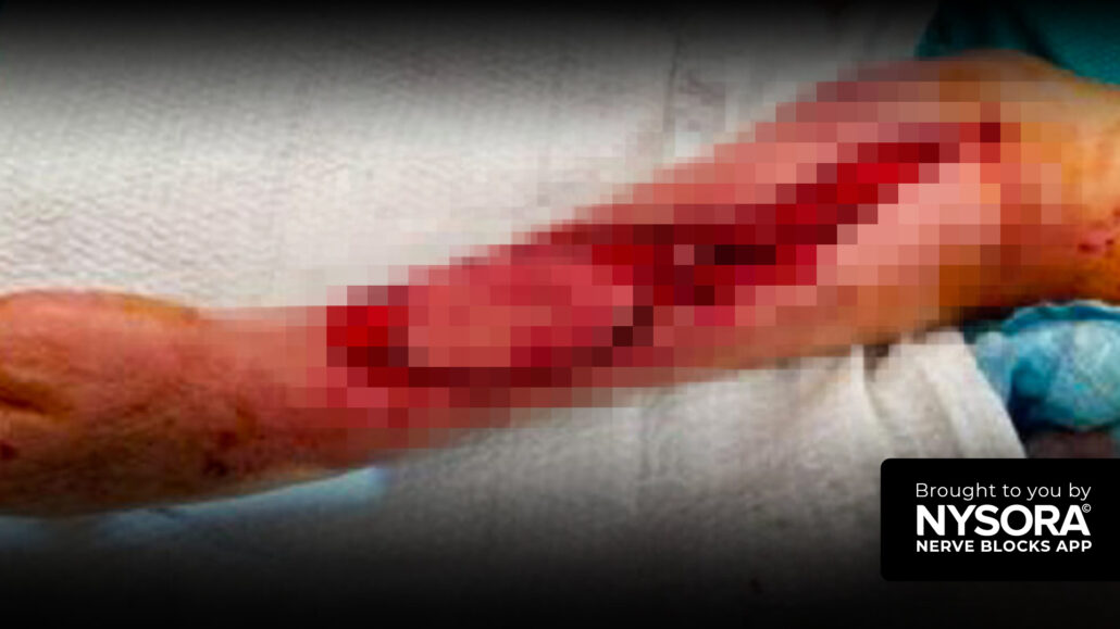
An 80-year-old patient with a history of critical aortic stenosis and a documented DNR order was presented to the operating room. The patient had a history of recurring episodes of congestive heart failure and was described as “walking on a thin line.” The primary surgical intervention was the repair of an open wound over the elbow using a free flap from the ipsilateral forearm. Additionally, a skin graft was harvested from the anterolateral thigh.
Nerve block techniques
- Costoclavicular brachial plexus block: Under ultrasound guidance, the brachial plexus was visualized in the costoclavicular space. 15 mL of 0.5% ropivacaine was injected after confirming the correct needle position. A catheter was placed for continuous infusion to ensure sustained analgesia and better perfusion longevity of the graft in the postoperative period.
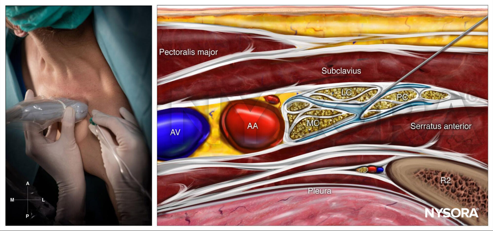
Costoclavicular brachial plexus block; Reverse Ultrasound Anatomy with needle insertion in-plane and local anesthetic spread (blue). AA, axillary artery; AV, axillary vein; LC, lateral cord; MC, medial cord; PC, posterior cord; R2, second rib.
- Lateral femoral cutaneous nerve and femoral nerve blocks: The blocks were performed to provide anesthesia to the anterolateral thigh, where the skin graft was harvested. Using ultrasound guidance, both the lateral femoral cutaneous nerve and femoral nerve were identified, and 5 mL of 0.25% bupivacaine was injected around each nerve.
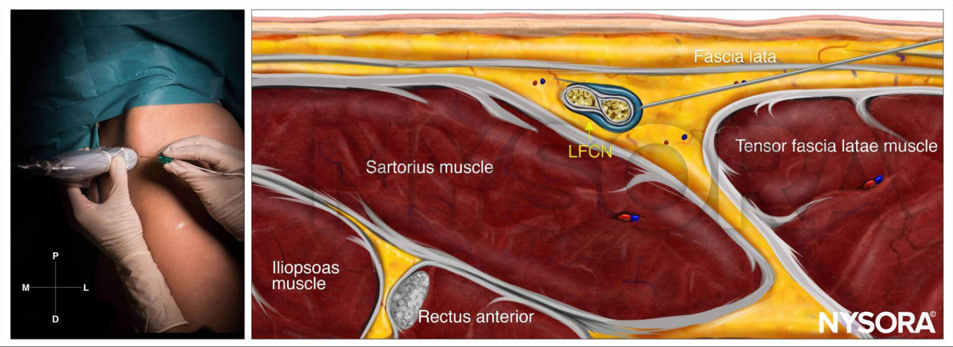
Lateral femoral cutaneous nerve block; Reverse Ultrasound Anatomy with needle insertion in-plane and local anesthetic spread (blue). LFCN, lateral femoral cutaneous nerve.
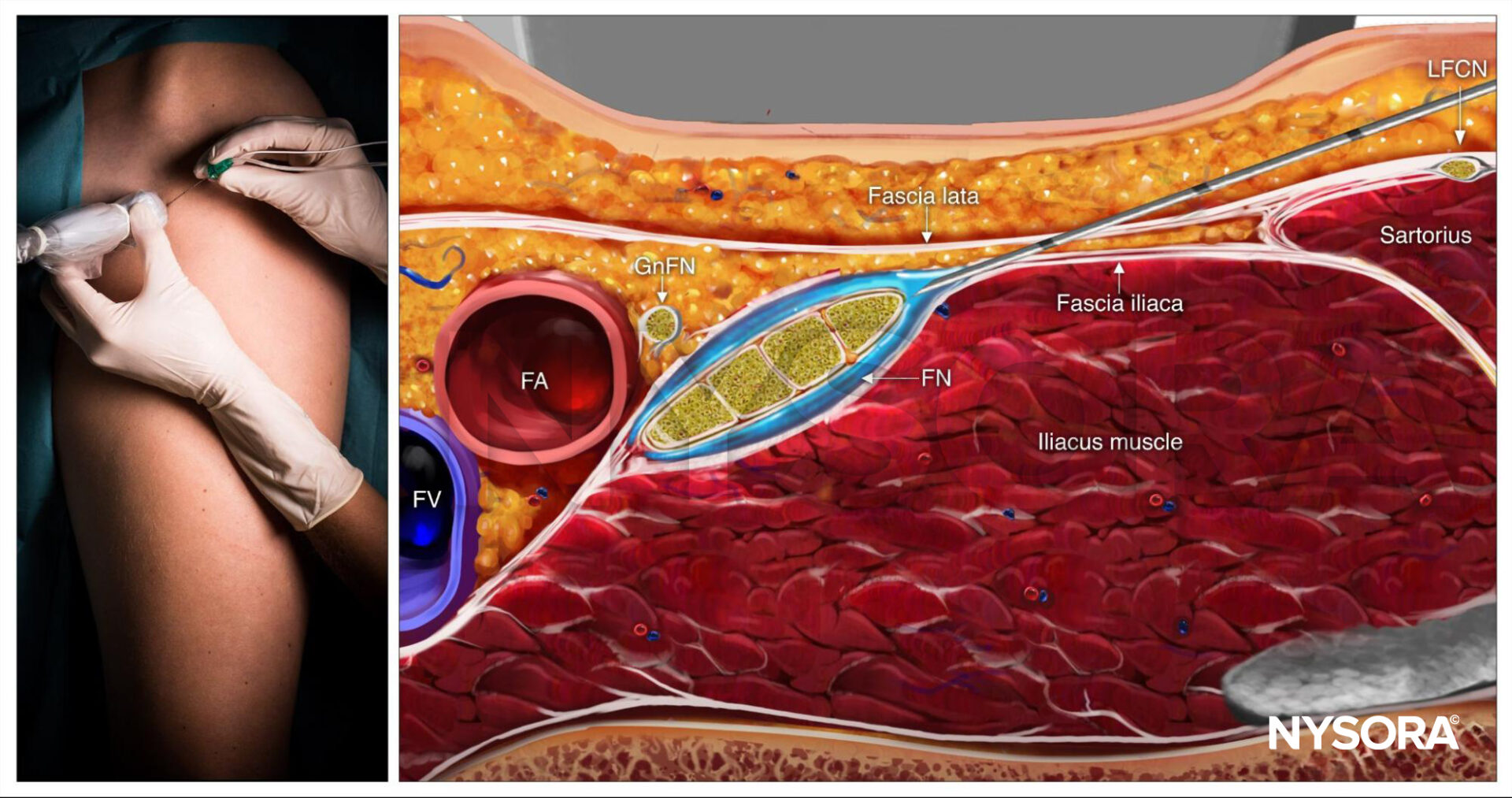
Femoral nerve block; Reverse Ultrasound Anatomy with needle insertion in-plane and local anesthetic spread (blue). FA, femoral artery; FV, femoral vein; FN, femoral nerve; GnFN, genitofemoral nerve; LFCN, lateral femoral cutaneous nerve.
Patient outcome
The surgery proceeded without complications or incidents. The blocks provided adequate anesthesia for the procedure, eliminating the need for additional anesthetic agents. The grafting of the wound site was successful. The patient had effective pain control during and after the surgery due to the nerve blocks and the continuous infusion through the catheter.
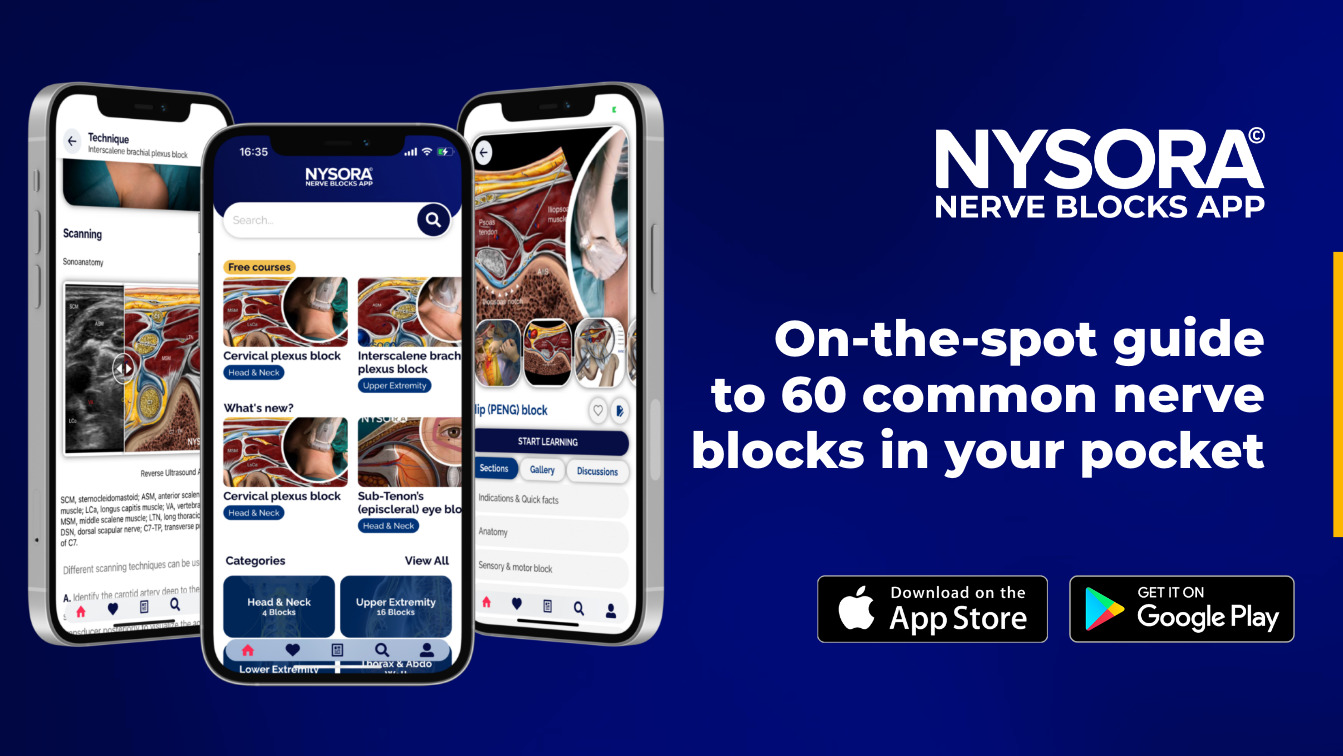
For more case studies like these and the complete guide to the 60 most frequently used nerve blocks, download the Nerve Blocks App HERE. Don’t miss the chance to get the bestselling NYSORA Nerve Blocks App also in book format – the perfect study companion with the Nerve Blocks app!