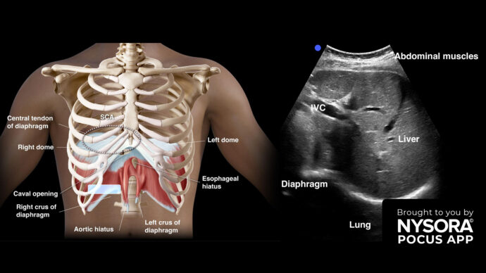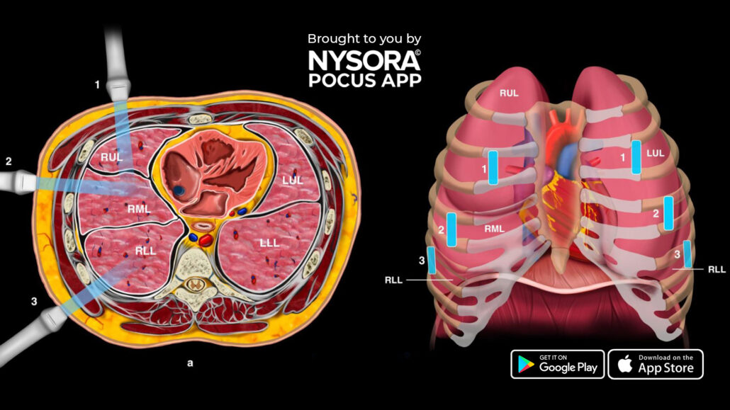
Ultrasound Characteristics of Lung Consolidation: Shred Sign and Hepatization
Lung consolidation refers to a condition where the air-filled spaces in the lung’s alveoli are replaced by fluid, pus, blood, or other substances. This results in the affected lung tissue becoming more solid and less able to exchange oxygen, leading to impaired respiratory function. It can be visualized using ultrasound, with distinct patterns such as the shred sign and hepatization helping to identify the consolidation type.
There are two types of lung consolidation:
1. Non-translobar consolidation (shred sign or C-line): This type displays an irregular boundary, often resembling a fractal line, which separates the consolidated lung from the aerated lung underneath. This pattern is commonly observed in non-translobar consolidations, most frequently associated with pneumonia.
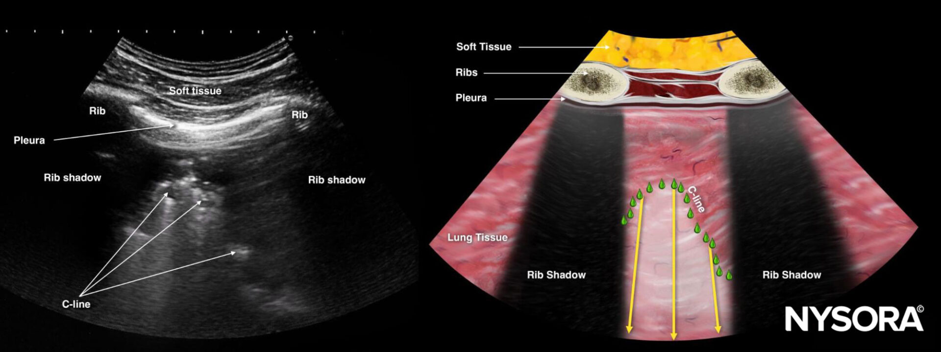
Shred sign or C-line.
2. Translobar consolidation (hepatization): In this case, the ultrasound image bears a resemblance to ultrasound imaging of the liver. The “hepatized” lung is lung tissue that is visible due to the absence of air, creating a texture similar to organ tissue. Translobar consolidations are often linked to pneumonia or atelectasis.
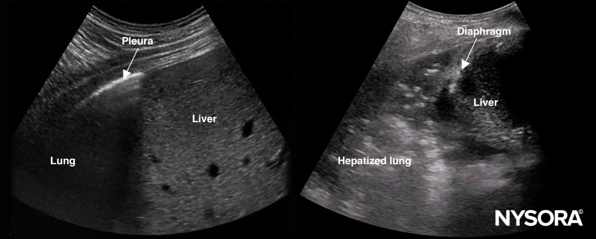
Normal lung on the left and hepatized lung on the right. Normal lung tissue cannot be visualized with ultrasound due to reflections of the air while hepatized lung tissue resembles organ tissue.
Unleash the potential of POCUS with NYSORA’s POCUS App and elevate your practice, expand your capabilities, and deliver exceptional patient care. Download HERE.