Tips for scanning the medial meniscus in a longitudinal orientation
The medial meniscus (internal semilunar fibrocartilage) is a fibrocartilage semicircular band that spans the knee joint medially, located between the medial condyle of the femur and the medial condyle of the tibia. It is a common site of injury, especially if the knee is twisted.
Here are 3 top tips to scan the medial meniscus (longitudinal scan)
- Place the patient in a supine position with the knee flexed 90°.
- Position the transducer longitudinal over the medial joint line, bridging the medial femoral condyle and tibia.
Identify the medial meniscus wedged between the medial femoral condyle and tibia. Tip: Increase the depth until both the outer rim and deep part of the medial meniscus are visualized.
Points of interest (during scanning):
- Gapping between the tibia and medial meniscus.
- Excessive hyperechoic signals in the deep part or white zone and the center of the medial meniscus. These signals correspond to mucoid degeneration of the medial meniscus.
![]()
Sonoanatomy
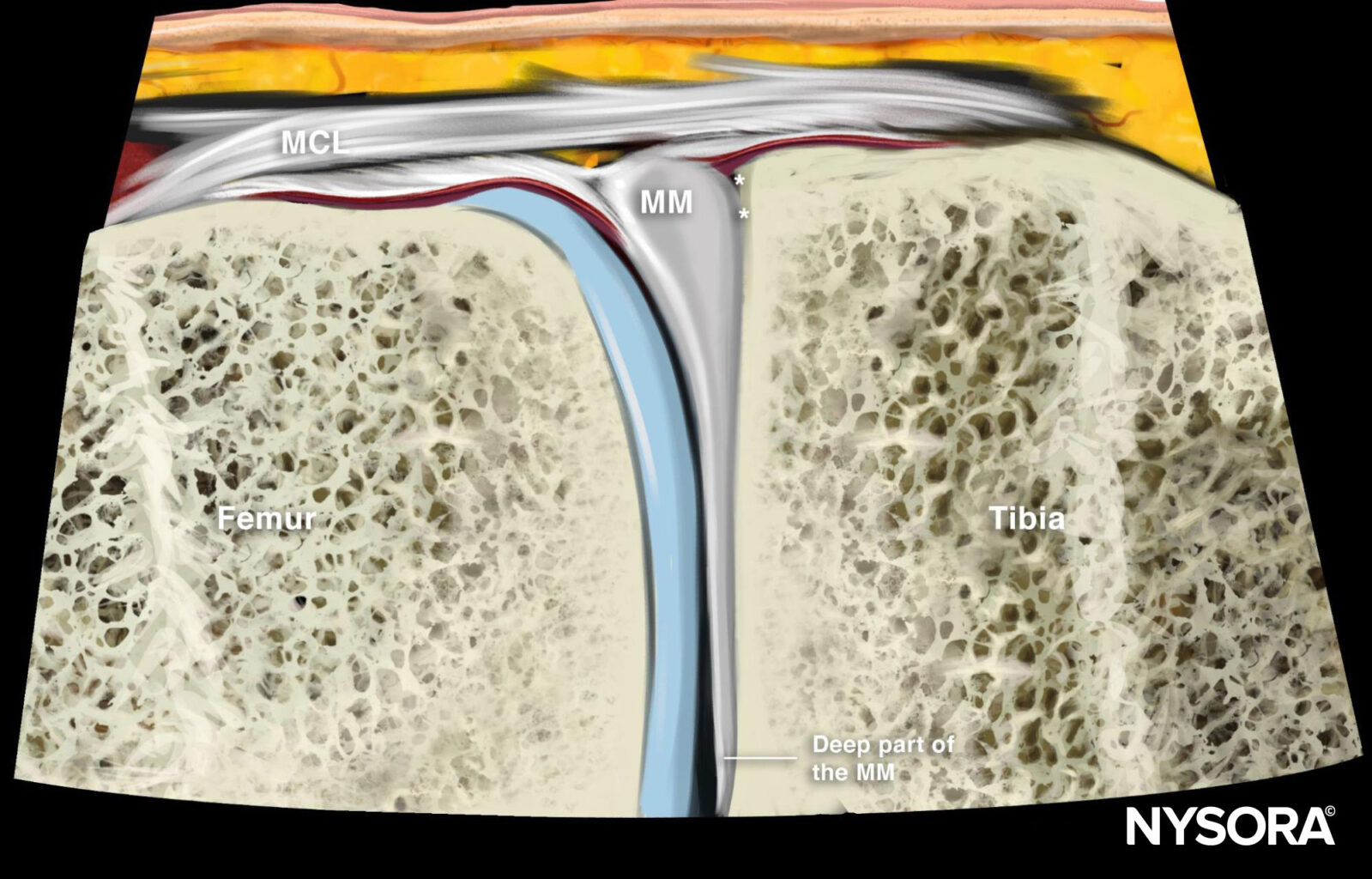
Reverse Ultrasound Anatomy
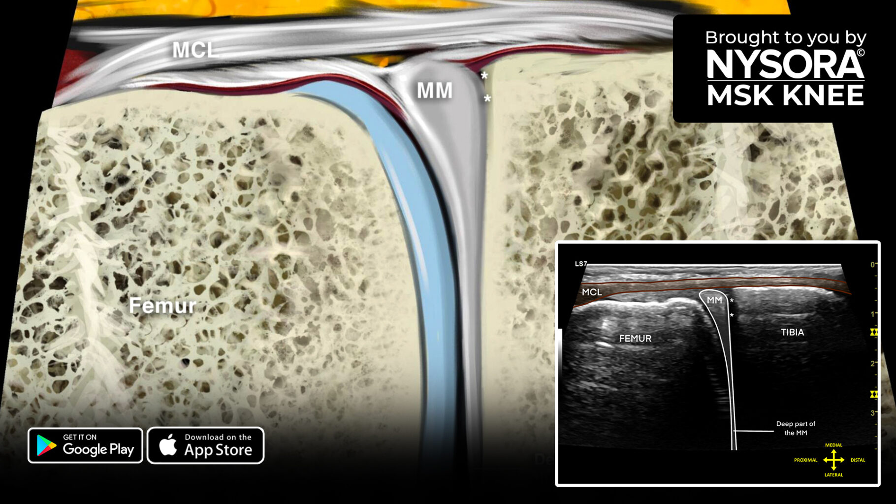
Comparison of sonoanatomy and reverse ultrasound anatomy of the medial meniscus (longitudinal scan).
Download the MSK App for more tips and the most practical and applicable techniques in musculoskeletal ultrasound anatomy and regenerative therapy of the knee.