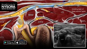
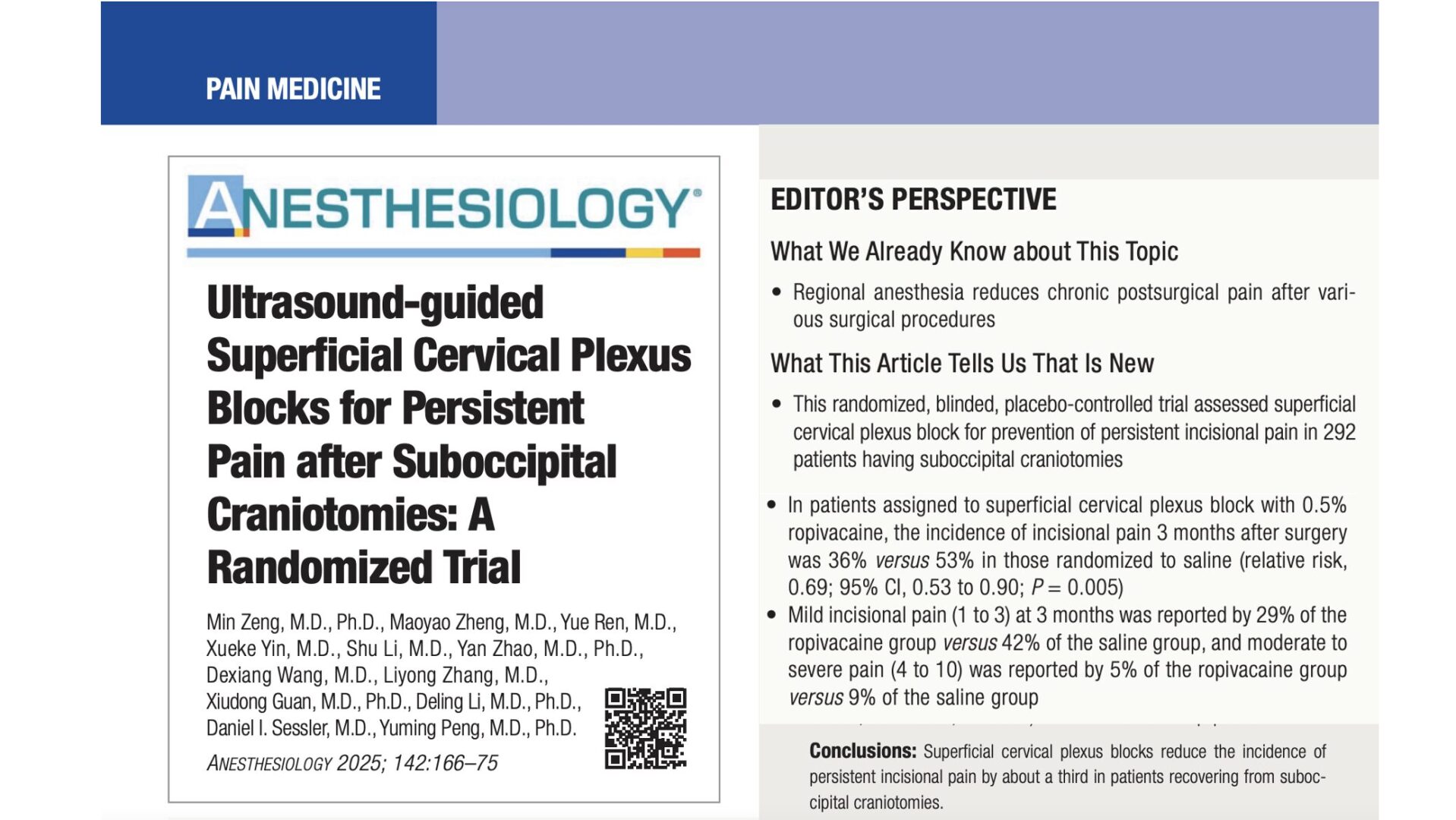
Superficial Cervical Plexus Block
Overview
- Indications: Carotid endarterectomy, neck surgery
- Landmarks:
- Mastoid process
- Sternocleidomastoid muscle; posterior border of the clavicular head
- Transverse process of C6
- Equipment: 1½" 25-gauge needle
- Local Anesthetic: 15-20 mL
- Complexity level: Basic
Regional anesthesia anatomy
The superficial cervical plexus supplies innervation to the skin of the anterolateral neck
through anterior primary rami of C2 through C4. The individual nerves emerge as four distinct
nerves from the posterior border of the sternocleidomastoid muscle. The lesser occipital nerve
usually is a direct branch from the main stem of the second cervical nerve. The larger remaining
part of this stem then unites with a part of the third cervical nerve to form a trunk that arises
as the greater auricular and the transverse cervical nerves. Another part of the third cervical
nerve runs downward to unite with a major part of the fourth to form a supraclavicular trunk, which
then divides into the three groups of supraclavicular nerves.
Distribution of anesthesia
|
The superficial cervical plexus innervates the skin of the anterolateral neck.
|
|
Patient positioning
|
The patient is in the same position as in the deep cervical plexus block.
The patient's head should be facing away from the side to be blocked.
|
|
Equipment
|
A standard regional anesthesia tray is prepared with the following equipment:
- Sterile towels and 4"x4" gauze packs
- 20-mL syringe with local anesthetic
- Sterile gloves, marking pen, and surface electrode
- A 1½" 25-gauge needle for block infiltration
|
|
Landmarks
Surface landmarks
The following surface anatomy landmarks are helpful for estimating the location of the
posterior border of the sternocleidomastoid muscle and for estimating the site of needle
injection:
- Mastoid process
- Chassaignac's tubercle of C6
The fingers of the palpating hand should be stretched to outline the posterior border of
the clavicular head of the sternocleidomas-toid muscle and to help visualize the line
connecting the mastoid process with the C6 transverse process.
Anatomic landmarks
A line extending from the mastoid to C6 is drawn. The site of needle insertion is marked
at the midpoint of the line connecting the mastoid process with Chassaignac's tubercle of
C6 transverse process. This is the location of the branches of the superficial cervical
plexus as they emerge behind the posterior border of the sternocleidomastoid muscle.
|
Technique
After skin cleansing with an antiseptic solution, a skin wheel is raised at the site of
needle insertion using a 25-gauge needle. Next, using a "fan" technique with superior-inferior
needle redirections, the local anesthetic is injected alongside the posterior border of the
sternoclei-domastoid muscle 2-3 cm below and above the needle insertion site. This injection
technique should be adequate to achieve blockade of all four major branches of the superficial
cervical plexus.
Goal
The goal of the injection is to infiltrate the local anesthetic subcutaneously and behind the
sternocleido-mastoid muscle. Attention should be paid to avoid deep needle insertion (e.g., less
than 1-2 cm).
TIPS:
- The transverse process is never sought with the superficial cervical plexus technique.
- Paresthesia is occasionally elicited during needle insertion. However, paresthesia is nonspecific
and should not be routinely sought.
Choice of local anesthetic
A superficial cervical plexus block requires 10-15 mL of local anesthetic (3-5 mL per each
redirection/ injection). Most patients benefit from the use of a long-acting local anesthetic.
Since motor block is not sought with this technique, some anesthesiologists suggest using a
low-concentration of local anesthetic (e.g., 0.2-0.5% ropivacaine or 0.25% bupivacaine).
Although a low concentration may suffice when the needle is ideally placed in the vicinity of
the cervical plexus nerves, this is often not the case and the higher concentration results in
both a higher success rate and a longer duration of blockade.
|
Onset (min) |
Anesthesia (hrs) |
Analgesia (hrs) |
| 1.5% Mepivacaine (+ HCO3; + epinephrine |
10-15 |
2.0-2.5 |
3-6 |
| 2% Lidocaine (+ HCO3; + epinephrine |
10-15 |
2-3 |
3-6 |
| 0.5% Ropivacaine |
10-20 |
3-4 |
4-10 |
| 0.25% Bupivacaine (+ epinephrine) |
10-20 |
3-4 |
4-10 |
Block Dynamics and Perioperative Management
This block is associated with minor patient discomfort. Small doses of midazolam 1-2 mg
for sedation and alfentanil 250 to 500 µg for analgesia just before needle insertion
should produce a comfortable and cooperative patient during nerve localization. Similarly
to the deep cervical plexus blockade, the sensory coverage of the neck is complex and a
degree of cross-coverage from the cervical plexus branches from the opposite side of the
neck should be expected. The onset time for this block is 10-15 minutes and the first sign
of the blockade is the decreased sensation in the area of the distribution of the respective
components of the cervical plexus. Excessive sedation should be avoided before and during head
and neck procedures because many procedures require a fully conscious, oriented, and cooperative
patient during the entire surgical procedure. In addition, airway management, when it becomes
necessary, may prove difficult because of the shared access to the head and neck with the
surgeon.
TIP: A subcutaneous midline injection of the local anesthetic extending from the
thyroid cartilage distally to the suprasternal notch will block the branches crossing from
the opposite side. This injection can be considered as a "field" block. It is very useful
for preventing pain from surgical skin retractors on the medial aspect of the neck.
Complications and How to Avoid Them
| Infection |
- Low risk
- A strict aseptic technique is used
|
| Hematoma |
- Avoid multiple needle injections, particularly in anticoagulated patients
- Keep a 5 minute steady pressure on the site when the cartoid artery is inadvertenly punctured
|
| Phrenic nerve blockade |
- Phrenic nerve blockade (diaphragmic paresis) invariably occurs with a deep cervical plexus block
- A deep cervical block should be carefully considered in patients with significant respiratory disease
- Bilateral deep cervical block in such patients may be considered contraindicated
- Blockade of the phrenic nerve does not occur after superficial cervical plexus block
|
| Local anesthetic toxicity |
- Central nervous system toxicity is the most serious consequence of the cervical plexus block. This complication occurs because of the rich vascularity of the neck, including vertebral and carotid artery vessels; it is usually caused by an inadvertent intravascular injection of local anesthetic rather then absorption
- Careful and frequent aspiration should be performed during the operation
|
| Nerve injury |
- Local anesthetic should never be injected against resistance or when the patient complains of severe pain on injection |
| Spinal anesthesia |
- This complication may occur with injection of larger volume of local anesthetic inside the dural sleeve that accompanies the nerves of the cervical plexus
- It should be noted that a negative aspiration test for CSF does not rule out the possibility of intrathecal spread of local anesthetic
- Avoidance of high volume and pressure during injection are the best measures to avoid this complication
|
Bibliography
- Aunac S, Carlier M, Singelyn F, De Kock M: The analgesic efficacy of bilateral combined superficial and deep cervical plexus block administered before thyroid surgery under general anesthesia. Anesth Analg 2002; 95:746-50.
- Diedone N, Gomola A, Bonnichon P, Ozier YM: Prevention of postoperative pain after thyroid surgery: A double-blinded randomized study of bilateral superficial cervical plexus blocks. Anesth Analg 2001; 92:1538.
- Jankovic D, Wells C: Regional Nerve Blocks. 2nd edition. Wissenchafts-Verlag Berlin, Blackwell Science, 2001.
- Masters RD, Castresana EJ, Castresana MR: Superficial and deep cervical plexus block: technical considerations. AANA J 1995; 63:235-43.
- Mulroy MF: Regional Anesthesia: An Illustrated Procedural Guide. 3rd edition. Philadelphia, Lippincot, 2002.
- Murphy TM: Somatic Blockade of Head and Neck. In Cousins, M.J., and Bridenbaugh PO(eds): Neuronal Blockade in Clinical Anesthesia and Management of Pain. Philadelphia, J.B. Lippincott-Raven Publishers, 1988, pp 489-514.
- Pandit JJ, Bree S, Dillon P, Elcock D, McLaren ID, Crider B: A comparison of superficial versus combined (superficial and deep) cervical plexus block for carotid endarterectomy: a prospective, randomized study. Anesth Analg 2000; 91:781-6.
- Stoneham MD, Doyle AR, Knighton JD, Dorje P, Stanley JC: Prospective, randomized comparison of deep or superficial cervical plexus block for carotid endarterectomy surgery. Anesthesiology 1998; 89:907-12.
- Winnie AP, Ramamurthy s, Durrani Z,Radonjic R: Interscalene cervical plexus block: A single-injection technique. Anesth Analg 1975; 54:370-5.
GO TO TOP
|