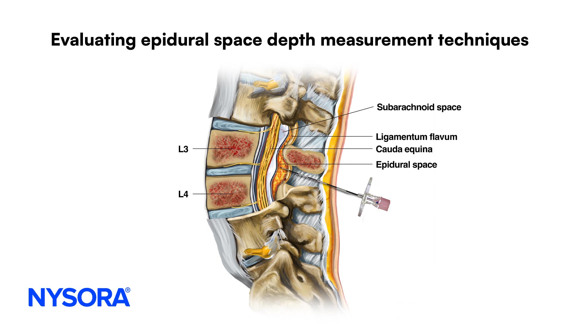
Continuous Infraclavicular Brachial Plexus Block

Overview
  • Indications: Elbow, forearm, hand surgery
  • Landmarks: Medial clavicular head, coracoid process
  • Nerve stimulation: Hand twitch at 0.2-0.3 mA current
  • Local anesthetic: 30-45mL
  • Complexity level: Advanced
Image
General considerations

A continuous infraclavicular block is an advanced regional anesthesia technique and considerable experience with the single-shot technique is necessary for its safe and successful implementation. The use of a catheter significantly increases the utility of infraclavicular block. The infraclavicular technique is especially suitable for insertion of catheters because the technique and course of the catheter are straightforward. The brachial plexus is encountered at a relatively deep location, which decreases the chance of inadvertent catheter dislodgement. Additionally, the catheter insertion site is easily approachable for maintenance and inspection. Typically, the initial dose of local anesthetic is injected through the needle, followed by infusion of a more dilute local anesthetic at a desired rate. This technique may be utilized for prolonged surgery on the hand, wrist, elbow, or distal arm and surgery at the same anatomic locations requiring pro-longed postoperative pain management or sympathetic block.

Regional anesthesia anatomy
Image

The boundaries of the infraclavicular fossa are the pectoralis minor and major muscles anteriorly, ribs medially, clavicle and the coracoid process superiorly, and humerus laterally. At this location, the brachial plexus is composed of cords. The sheath surrounding the plexus is delicate. It contains the subclavian/axillary artery and vein. Axillary and musculocutanous nerves leave the sheath at or before the coracoid process in 50% of patients. Consequently, the deltoid and biceps twitches should not be accepted as reliable signs of brachial plexus identification.

Distribution of anesthesia
Image

A typical distribution of anesthesia after an infraclavicular brachial plexus block includes the hand, wrist, forearm, elbow, and distal arm. The skin of the axilla and proximal medial arm (unshaded areas) is not anesthetized (intercosobrachial and medium cutaneous brachii nerves).

Patient positioning
Image

The patient is in the same position as in the single-shot technique. However, it is imperative that the anesthesiologist be in the ergonomic position to allow for maneuvering during catheter insertion. It is also very important that all equipment as well as the catheter should be immediately available and prepared in advance, because small movements of the needle while trying to prepare the catheter may result in needle dislodgement from its position in the brachial plexus sheath.

Equipment
Image A standard regional anesthesia tray is prepared with the following equipment:
  • Sterile towels and 4x4" gauze packs
  • 20-mL syringe with local anesthetic
  • Sterile gloves, marking pen, and surface electrode
  • One 1½" 25-gauge needle for skin infiltration
  • A 5-cm long, insulated stimulating needle (Tuohy style or Quincke tip)
  • Catheter
  • Peripheral nerve stimulator
Landmarks

The landmarks are the same as for the single-shot technique:

  1. Medial clavicular head
  2. Coracoid process
  3. Midpoint of line connecting 1 and 2

The point of needle insertion is labeled 3 cm caudal to the midpoint between the medial clavicular head and the coracoid process.

Image Image

TIP: Attention should be paid to avoid a too medial insertion of the needle as well as a too steep angle of the needle, which carries a risk of pneumothorax.

Technique
Image

The subcutanoues tissue at the projected site of needle insertion is anesthetized with local anethetic. The block needle is then attached to the nerve stimulator (1.5 mA, 2 Hz, 100 µsec) and to a syringe with local anethetic. The fingers of the left hand should be firmly positioned on the pectoralis muscle and the needle is inserted at a 45 degree angle to the horizontal plane and approximately parallel to the medial clavicular head-coracoid process line. The needle bevel should be facing up. As the needle is advanced beyond the subcutaneous tissue, direct stimulation of the pectoralis muscle is obtained as the needle enters the body of the muscle. Stimulation of the brachial plexus is encountered soon after the pectoralis twitches cease. The sought response is that of the medianus nerve, indicated by rhythmic flexion of the wrist and fingers.

Image

Once the medianus response is obtained at a 0.5 mA current level or less, the initial dose of local anesthetic is injected (30-45 mL). The catheter then is inserted while making sure that the needle remains firmly seated in the position in which the stimulation of brachial plexus was obtained. The catheter tip should be advanced some 5 cm beyond the needle tip. The needle is then carefully withdrawn while simultaneously advancing the catheter to prevent its dislodgment. The most important aspect of this technique is stabilization of the needle for catheter insertion after the brachial plexus is localized. In patients in whom the needle encounters the brachial plexus at very shallow location, sometimes it best to have an assistant advance the catheter to assure that the needle does not move out of its original position.

TIPS:
  • This block should be avoided in patients with abnormal coagulation because the combination of the large needle diameter and inability to apply pressure in case of accidental puncture of the infraclavicular vessels (subclavian or axillary artery and vein) carries a risk of hematoma.
  • The stimulation characteristics of the current, Tuohy-style needles appear to be somewhat different from that of the single-shot needles. This likely is the effect of the larger needle diameter and the coating of the needle tip. A slight needle rotation or angle change can make a significant difference in the ability to stimulate.
  • Slight caudal orientation of the needle may be required to facilitate the catheter insertion.
  • Some needles currently on the market have particularly blunt tips and tend to get hung up on the skin advancement. For this reason, sometimes it is best to create a small skin "nick" with a scalpel or side of a sharp tipped, 18-gauge needle before attempting to insert such a needle through the skin.
Image

The catheter is checked for inadvertent intravascular placement and secured to the chest. A number of techniques to secure the catheter to the skin have been proposed. We find that a benzoin prep followed by application of a clear dressing is the simplest and the most efficacious. The infusion port should be clearly marked as: "continuous infraclavicular block".

Management of a continuous infusion catheter

The catheter is activated by injecting a bolus of local anesthetic (10-15 mL) followed by an infusion at 8 mL/hr. A PCA (patient-controlled analgesia) dose of 3-5 mL every 30 minutes can be added for breakthrough pain, in which case the basal infusion should be decreased to 5 ml/min. For continuous infusion, we typically use 0.2% ropivacaine.

Patients should be seen and instructed on the use of PCA at least twice a day. During each visit, the insertion site should be checked for erythema and swelling. The extent of motor and sensory blockade should be documented. The infusion and PCA dose are adjusted accordingly. When the patient complains of breakthrough pain, the extent of blockade should be checked first. A bolus of dilute local anesthetic (e.g., 10-15 mL of 0.5% ropivacaine) should be injected to reactivate the catheter. Increasing the infusion alone will never result in improvement of analgesia. When the bolus failes to result in blockade after 30 minutes, the catheter should be labeled as migrated and should be removed. Every patient receiving a continuous nerve block infusion should be prescribed an immediately available alternative pain management protocol because incomplete analgesia and catheter dislodgment can occur. For inpatients, this is best done using a back-up IV PCA.

TIPS:
  • Breakthrough pain in patients on continuous infusion is always managed by administering a bolus of local anesthetic. Increasing the rate of infusion alone is never adequate.
  • With patients on the ward, a higher concentration of a shorter acting and epinephrine-containing local anesthetic (e.g., 1% mepivacaine or lidocaine with 1:300,000 epinephrine) is useful and safe to both manage the pain and test the position of the catheter.
Image
Disposition of radio opaque dye in the brachial plexus sheath after injection of 2ml through the infraclavicular catheter
Goal

The goal is to achieve a hand twitch (preferably medianus) using a current of 0.2-0.3mA.

TIPS:
  • Twitches from the biceps or deltoid muscles should not be accepted, since the musculocutaneous and axillary nerve, respectively, may depart the brachial sheath before the caracoid process.
  • Hand stabilization and precision is crucial with this block as the sheath of the brachial plexus is very thin at this location and small movements of the needle may result in injection of local anesthetic outside the sheath. This in turn, results in a weak block with a slow onset.
  • A twitch of the pectoralis muscle is observed first and indicates a too shallow placement of the needle. As contractions of the pectoralis muscle cease, the needle is slowly advanced until the twitches of the brachial plexus are elicited. This usually occurs at a depth of 5-8 cm.
  • After the twitches of the pectoralis muscle cease, the stimulating current is lowered to below 1.0 mA to decrease patient discomfort. The needle is then slowly advanced or withdrawn until hand twitches are obtained at 0.2-0.3 mA.
  • The success rate with this block decreases when local anesthetic is injected after obtaining stimulation with a current intensity above 0.3 mA.
  • In the absence of the medianus response, stimulation of the radialis or ulnar nerve can also be accepted, as long as the twitch of the hand is clearly visible.
  • The twitch of the biceps (musculocutaneous nerve) or deltoid (axillary nerve) muscles should not be accepted as these nerves often leave the brachial plexus sheath proximal to the coracoid process.

Failure to obtain nerve stimulation on the first needle pass

When insertion of the needle does not result in brachial plexus stimulation, the following maneuvers should be undertaken:

  1. Keep the palpating hand in the same position, with the palpating finger firmly seated in the pectoralis and the skin between the fingers stretched.
  2. Withdraw the needle to the skin, redirect 10º cephalad, and repeat the procedure.
  3. Withdraw the needle from the skin, redirect 10º caudal, and repeat the procedure.
Image

TIPS:

  • When these maneuvers fail to result in motor response, withdraw the needle and assess the landmarks.
  • Check that the nerve stimulator is properly connected and delivering the set stimulus.
  • Consider inserting the needle 2-cm laterally and repeating the above steps.

Interpreting Responses to Nerve Stimulation

Some common responses to nerve stimulation and the course of action to obtain the proper response

Stimulation Motor response Explanation Corrective Action
Pectoralis muscle - direct muscle stimulation Arm adduction Too shallow placement of the needle Continue advancing the needle
Latissimus dorsi Arm adduction Too deep placement of the needle Withdraw the needle to skin level and reinsert in another direction (superior/inferior)
Axillary nerve Deltoid muscle Needle placed too inferiorly Withdraw the needle to skin level and reinsert with a superior orientation
Musculocutaneous nerve Biceps twitch Needle placed too superiorly Withdraw the needle to skin level and reinsert with a light caudal orientation
Choice of local anesthetic

The infraclavicular brachial plexus requires a relatively large volume of local anesthetic to achieve anesthesia of the entire plexus. The choice of the type and concentration of local anesthetic should be based on whether the block is planned for surgical anesthesia or pain management. Due to the high vascular content of the area and potential for inadvertent intravascular injection, the local anesthetic solution should be injected slowly with frequent aspiration.

Onset
(min)
Anesthesia (hrs) Analgesia (hrs)
2%-Chloroprocaine (plus HCO3 + epi) 5-10 1.5 2.0
1.5% Mepivacaine (plus HCO3 + epi) 5-15 2.5-4 3-6
2% Lidocaine (plus HCO3 + epi) 5-15 3-6 5-8
0.5% Ropivacaine 15-20 6-8 8-12

TIP: Always assess the risk-benefit ratio of using large volumes and concentrations of long-acting local anesthetic for a lumbar plexus block.

Block Dynamics and Perioperative Management

Adequate sedation and analgesia are crucially important for this block to ensure patient comfort and to facilitate interpretation of responses to nerve stimulation. For instance, a midazolam 2-6 mg IV can be used to achieve sedation. A short-acting narcotic (e.g., alfentanil 250-750µg) is added just before needle insertion. A typical onset time for this block is 5-15 minutes, depending on the local anesthetic chosen. Waiting beyond 20 minutes will not result in further enhancement of the blockade. The first sign of the impending successful blockade is loss of muscle coordination within minutes after the injection. This loss can be tested easily by asking the patient to touch his nose, while paying attention that the patient does not miss the nose and injure his/her eye. The loss of motor coordination typically occurs before sensory blockade can be documented. In case of inadequate skin anesthesia despite the apparent timely onset of the blockade, local infiltration at the site of the incision by the surgeon is often all that is needed to allow the surgery to proceed. Before and after the surgery, both the patient and the surgeons should be informed about the expected duration of the blockade.

Complications and How to Avoid Them
Hematoma - Avoid multiple needle insertions through the pectoralis muscle
- Apply firm pressure over the site of needle insertion after needle withdrawal
- Carefully review indications for the single shot and avoid continuous infraclavicular block in patients with abnormal coagulation
Systemic toxicity - Limit the volume/dose of long-acting local anesthetic
- Carefully review risks and benefits of using long-acting local anesthetics for each and every patient/operation
- Inject local anesthetic with frequent aspiration to rule out intravascular injection, carefully assesing the patient for signs of local anesthetic toxicity
- Inject local anesthetic SLOWLY to avoid "channeling" of local anesthetic to smaller veins/lymphatic channels that may have been punctured during needle advancement
Nerve injury - Use the nerve stimulator to confirm the needle position! This technique requires deep needle insertion and the use of paresthesia is not acceptable
- Make sure that the nerve stimulator is fully functional and connected properly
- Advance the needle slowly when the twitches of the pectoralis muscle cease
- Orient the bevel of the needle down to facilitate nerve stimulation and avoid contact of the plexus complements (and vascular walls) with the advancing tip of the needle
- Do not inject against high pressures! In this scenario, withdraw the needle, check its patency by flushing it and repeat the procedure
- Stop injecting immediately when patients complain of pain on injection!
Pneumothorax - This is an often feared by exceedingly rare complication
- The needle direction is actually away from the chest cavity (as opposed to interscalene or supraclavicular blocks)
- Attention should be paid to the site and angle of needle insertion to ensure that the needle assumes a plane away from the chest wall
Bibliography
  • Chelly JE, Casati A, Fanelli G: Continuous peripheral nerve block techniques. An illustrated guide. London, Mosby International Limited, 2001.
  • Greher M, Retzl G, Niel P, Kamolz L, Marhofer P, Kapral S: Ultrasonographic assessment of topographic anatomy in volunteers suggests a modification of the infraclavicular vertical brachial plexus block. Br J Anaesth 2002; 88:632-6.
  • Ilfeld BM, Morey TE, Enneking FK: Continuous infraclavicular brachial plexus block for postoperative pain control at home: a randomized, double-blinded, placebo-controlled study. Anesthesiology 2002; 96:1297-304.
  • Koscielniak-Nielsen ZJ, Rotboll Nielsen P, Risby Mortensen C: A comparison of coracoid and axillary approaches to the brachial plexus. Acta Anaesthesiol Scand 2000; 44:274-9.
  • Klaastad O, Lilleas FG, Rotnes JS, Breivik H, Fosse E: Magnetic resonance imaging demonstrates lack of precision in needle placement by the infraclavicular brachial plexus block described by Raj et al. Anesth Analg 1999; 88:593-8.
  • Rodriguez J, Barcena M, Rodriguez V, Aneiros F, Alvarez J: Infraclavicular brachial plexus block effects on respiratory function and extent of the block. Reg Anesth Pain Med 1998; 23:564-8.
  • Raj PP: Infraclavicular approaches to brachial plexus Anesthesia. Techniques in Reg Anesth and Pain Mangagement 1997; 1:169-77.
  • Raj PP, Pai U, Rawal N: Techniques of regional anesthesia in adults. In Clinical Practice of Regional Anesthesia Edited by Raj. New York, Churchill Livingstone, 1991, pp 276-300.
  • Whiffler K: Coracoid block--a safe and easy technique. Br J Anaesth 1981; 53:845-8.
  • Wilson JL, Brown DL, Wong GY, Ehman RL, Cahill DR: Infraclavicular brachial plexus block: parasagittal anatomy important to the coracoid technique. Anesth Analg. 1998; 87:870-3.

GO TO TOP


Site map

Acknowledgments


Valid XHTML 1.0!


COPYRIGHT © 1996, 2006 NYSORA.COM
No part of this web page may be reproduced without the permission of the authors.

DISCLAIMER: The material presented on this Web page has not been peer-reviewed. The indications, techniques and dosages on this Web page have been recommended in the medical literature and/or conform to OUR clinical practice. The medications and equipment have not necessarily been approved by the Food and Drug Administration (FDA) for use in the techniques and dosages for which they are recommended. The package insert for each drug and/or equipment should be consulted for use and dosage as recommended by the FDA. Because standards, practices and recommendations change, it is advisable to keep abreast of revised recommendations, particularly those concerning new drugs and techniques. While the techniques and dosages described are successfully used in our practice, they should be followed with a discretion since their complications may be dependent on the operator, patient and/or other accompanying clinical circumstances. The development and maintenance of this web page has not been supported by any pharmaceutical or medical manufacturing industry. The medications and/or equipment discussed in the web page is shown solely for teaching purposes. Similar equipment or medications from other manufacturers may produce similar clinical results to ours.