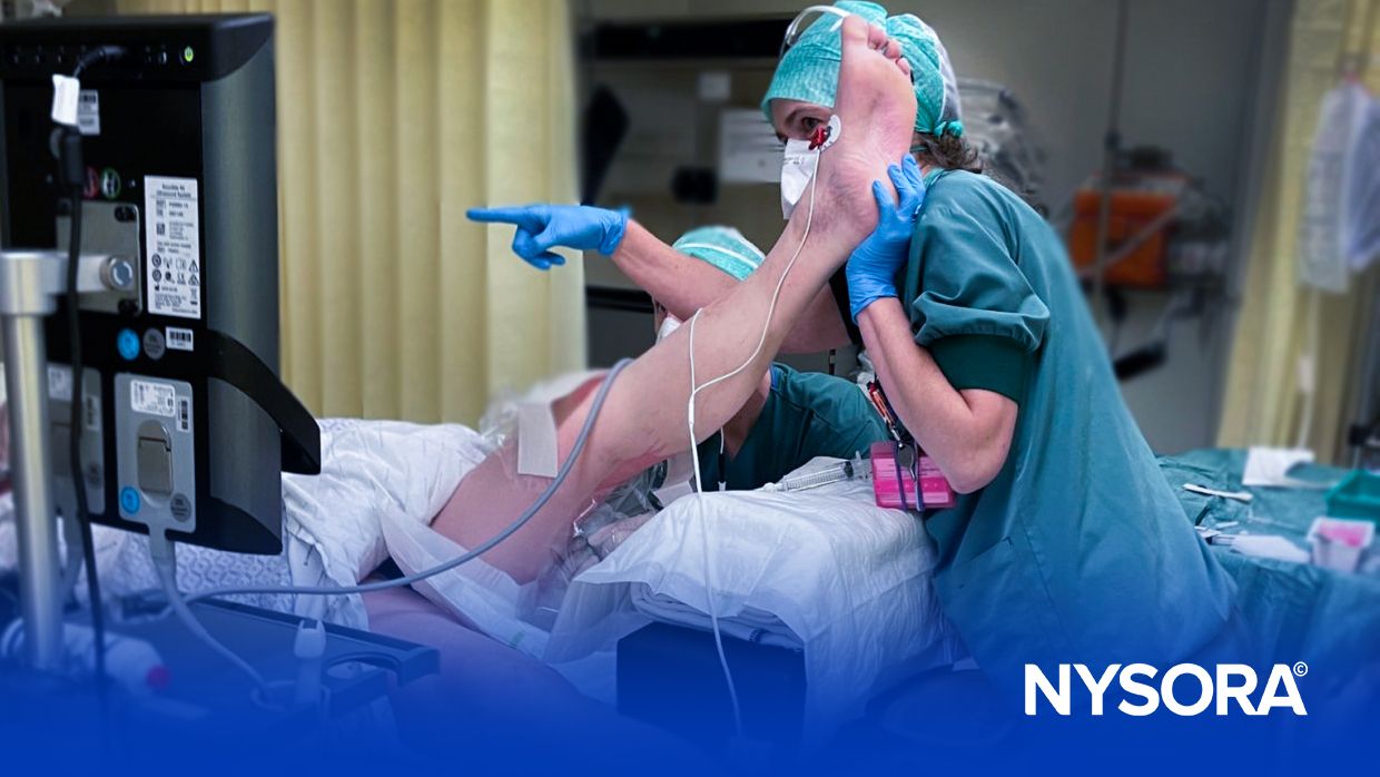
Home | Nerve Blocks | Nerve Stimulator Techniques | Popliteal Block: Lithotomy Approach

Popliteal Block: Lithotomy Approach

image


Description of the Technique

Image
Figure 1.
Image
Sciatic nerve block in the popliteal fossa (also called popliteal block) is a well-suited anesthetic technique for operations below the knee. However, difficulty in positioning patients in the prone position for the block often precludes its use in patients who are potentially the greatest beneficiaries of peripheral nerve blockade. Here we describe an alternative approach to popliteal block that can be reliably performed with a patient in the supine position, while relying on commonly used anatomic landmarks.

Image
Figure 2.
1. Popliteal fossa crease; 2. Biceps femoris muscle; 3. Semitendinosus; 4. Insertion point according to the classical, posterior approach

With a patient in the supine position, the leg is flexed at both the hip and knee joints, and supported by an assistant (Figure 1). The anatomical landmarks (see video) of the popliteal fossa (popliteal crease inferior, the semimembranosus and semitendinosus muscles medial, and the long head of the biceps femoris muscle lateral) are identified in this position (Figure 2). An insulated needle, attached to a peripheral nerve stimulator is inserted 7cm above the popliteal crease, 1 cm lateral to the midline and directed 45° cephalad (Figure 3; Video 2). Upon obtaining either dorsal or plantar flexion of the foot (Video 3), using the output current of 0.5 mAmp or less, 30ml to 40ml of solution of local anesthetic is injected while intermittently aspirating for blood.

Image
Figure 3.

Alternatively (read: preferentially), the block can be performed using the landmarks used in the "anatomical" approach. Instead of relying on frequently obscure boundaries of the popliteal fossa "triangle", a 50mm insulated needle is inserted at the midpoint between the tendons of the biceps femoris and semitendinosus muscles, 7cm above the popliteal fossa crease, and advanced cephalad at a 60° angle relative to the horizontal plane. The needle is slowly advanced while seeking a plantar or dorsi-flexion of the foot or toes. When the nerve is not localized on the first needle insertion, the needle is slowly withdrawn to the skin and reinserted through the same skin puncture. The first reinsertion should be at an angle of 5°, and the second at 10° lateral to the initial insertion plane. Failure to obtain stimulation of the sciatic nerve should prompt removal of the needle. The needle is then reinserted through a new puncture site 5 mm lateral to the initial insertion site and the maneuver is repeated. This technique is repeated through new insertion sites in 5mm incremental lateral insertions, until the desired response is obtained.


Case 1

A 32 yr. old, 72 kg, 170-cm man presented for emergency open reduction and internal fixation (ORIF) of an open fracture of his left tibial malleolus. The patient was injured in a high speed automobile accident 48 h before the surgery. Pre-anesthetic evaluation revealed an alert and oriented, mentally retarded male. He answered all questions appropriately, but with difficulty due to the pain from the extensive soft tissue trauma on the right side of his face. His airway was judged to be Mallampati - 4, and the cervical spine was stabilized with a hard collar. Examination of the heart and lungs were normal. The severe pain caused by body movements limited examination of the back. The x-ray survey revealed multiple pelvic injuries including a comminuted fracture of the right iliac wing with extension into the acetabulum, fracture of both pubic rami on the right, and fracture of the right ischium with involvement of the right hip joint. Computed tomography (CT) revealed a large pelvic hematoma, that considerably decreased intrapelvic volume, as well as an extensive hematoma extending from the left iliac crest into the region of the gluteal muscles and both thighs. Computed tomography of the cervical spine showed multiple fractures of cervical vertebrae C6 and C7, and subluxation of C6 on C7. The orthopedic surgeons were concerned that the pelvic injuries might be extended, and the right hip joint disrupted, during the transfer and positioning of the patient on the operating room table.

The unstable cervical spine injury and potentially difficult airway raised concerns about the use of general anesthesia. Inability to place the patient in the supine or lateral position and extensive hematoma on his back precluded the use of neuraxial anesthesia. Sciatic nerve block was considered, but the extensive trauma to the buttocks and pelvis contraindicated the approaches at the level of the pelvis. Inability to position the patient in the prone position precluded the use of the classical approach to popliteal block. Thus, while minimizing rotation of the right thigh, the alternative approach to popliteal block in the supine position was employed. After obtaining rhythmic dorsiflexion of the foot with a stimulating current of 0.5mAmp, 40ml bupivacaine 0.375% was injected. Then, a left femoral nerve block was performed using 15ml of bupi-vacaine 0.375% [4]. Twenty five minutes after block placement, adequate anesthesia was confirmed in the femoral and sciatic nerve distributions, and the surgeons proceeded with the ORIF of the left medial malleolar fracture. Supplemental sedation was achieved with intravenous midazolam 1 mg and fentanyl 150µg. A tourniquet was not used. The intraoperative and the postoperative course were uneventful.


Case 2

A 33 yr old, 88 kg, 168-cm woman in her 28th week of pregnancy was scheduled for open reduction and internal fixation of an open fracture of her right ankle. Her vital signs were: BP 90/50mmHg, HR 80bpm, RR 16. Cardiopulmonary and laboratory examinations were normal. The x-ray of the right ankle showed a trimalleolar fracture. The patient refused general anesthesia and expressed extreme reluctance in receiving either spinal, or epidural anesthesia. She readily consented to peripheral nerve block anesthesia. The patient was positioned in the supine position with a wedge under her right hip and the supine approach to popliteal block was used. Using an output current of 0.5mA, rhythmic dorsiflexion of the foot was obtained and 40ml mepivacaine 1.5% were injected. Additionally, 5ml mepivacaine 1.5% were injected subcutaneously in the medial aspect of the leg at the level of the tibial tuberosity to block the saphenous nerve. Thirty minutes later, anesthesia was confirmed and the surgery proceeded uneventfully. A tourniquet was applied below the knee. There was no need for supplemental intravenous analgesia or sedation during the surgery. The postoperative course was uncomplicated.


Case 3

A 62yr old, 60kg, 175cm man with gangrene of his right foot was scheduled for emergency amputation of the affected leg below the knee. The patient had a history of poorly controlled diabetes mellitus and severe ischaemic cardiomyopathy requiring frequent hospitalizations for congestive heart failure. Eleven months before this admission, he had suffered a myocardial infarction. After an above-knee amputation of his left leg under general anesthesia during the current admission, his postoperative course was complicated by myocardial infarction and pulmonary edema.

During the pre-anaesthetic evaluation, the patient appeared to be in distress due to the severe pain in his leg. Blood pressure was 150/90mmHg, HR 75, RR 20. Airway evaluation revealed Mallampati 2 and multiple loose teeth. The extension of the neck was severely limited. Bilateral rales were present at the lung bases. His heart rate was regular with audible S3/S4 gallops. His entire skin, including lower back, was covered with extensive psoriatic, scaling lesions. Large decubiti were present on his sacrum and buttocks. An echocardiography report was indicative of multi-segmental wall motion abnormalities, severe mitral regurgitation, and an estimated ejection fraction of 20%. His medications included insulin, metoprolol 50mg bid, captopril 25mg tid, digoxin 0.12mg qd, isosorbide dinitrate 10mg qid, furosemide 40mg qd.

Because of his fragile cardiovascular status, we decided to perform the amputation of the involved leg under regional anesthesia. His skin condition and extensive decubiti contraindicated both neuroaxial anesthesia and a sciatic nerve block at the level of the pelvis. Since the patient was unable to assume the prone position due to weakness and extreme pain in his right foot and the stump of his left leg, the supine approach to popliteal block was used. After the right popliteal nerve was identified using the output current of 0.5mA, 40ml mepivacaine 1.5% were injected. Additionally, the saphenous nerve was blocked using 5ml mepivacaine 1.5%. Twenty five minutes later the surgeon proceeded with the operation. A tourniquet was not used during the operation. His intraoperative and postoperative course were uneventful.


Discussion

Sciatic nerve block has been successfully used for surgical anesthesia and postoperative pain control for various operations below the knee. The common techniques of sciatic nerve blockade include approaches at the pelvis level: (anterior [5], lateral [6], posterior [7] and supine position [8] approaches) and the classical approach at the knee level with the patient in the prone position [9]. In this report we presented three cases where the sciatic nerve block in the popliteal fossa was judged to be the most suitable anaesthetic technique, but the inability to position patients in an optimal position for the performance of the block precluded the use of the classical approach. In each case, the popliteal block was successfully performed in the supine position using the described technique. We routinely use a peripheral nerve stimulator when performing the popliteal block, seeking either dorsal or plantar flexion of the foot at the output current of 0. mAmp or less. Upon obtaining desired response, 30ml to 40ml of solution of local anaesthetic is injected while intermittently aspirating for blood.

Volume of 30 to 45ml of local anesthetic solution is used in popliteal nerve blockade by many authors [1, 9]. We also prefer larger volumes of local anaesthetics for this block, because the large content of fat in the popliteal fossa and a thick epineurium of the sciatic nerve [10] may decrease the amount of LA (local anesthetic) that reaches the nerve and thus adversely affect the success rates of popliteal nerve block [11]. Additionally, larger volumes of local anaesthetic may also extend the duration of action of the block and play a role in optimizing post-operative pain relief. When anesthesia of the medial foot and leg is required, the popliteal block is supplemented with either the saphenous, femoral nerve block [12], depending on the planned surgical procedure.

This approach to popliteal block proved to be convenient and reliable, and has become a routine technique in our clinical practice.


References

  1. Rorie DK, Byer DE, Nelson DO, et al.: Assessment of block of the sciatic nerve in the popliteal fossa. Anesth Analg 1980;59:371-6.
  2. Gouverneur JM.: Sciatic nerve block in the popliteal fossa with atraumatic needles and nerve stimulation. Acta Anaesth Belgica 1985;4:391-9.
  3. Vloka JD, Hadzic A, Lesser J, Mulcare R, Kitain E.: Anaesthesia for short saphenous vein stripping: A comparison of popliteal nerve block with posterior cutaneous nerve of the thigh block versus spinal anaesthesia. Reg Anesth 1995;20(2S):104.
  4. Brown DL. Femoral nerve block. In: Brown DL, ed. Atlas of regional anaesthesia. Philadelphia: W.B. Saunders, 1992:89-95.
  5. Beck GP. :Anterior approach to sciatic nerve block. Anesthesiology 1963;24:222-4.
  6. Ichiyanagi K.: Sciatic nerve block: lateral approach with the patient supine. Anesthesiology 1959;20:601-4.
  7. Moore DC. :Block of sciatic and femoral nerves. In: Moore DC, ed. Regional Block. Illinois: C.C. Thomas, 1965:275-288.
  8. Raj PP, Parks RI, Watson TD, Jenkins MT.: A new single position supine approach to sciatic-femoral nerve block. Anesth Analg 1975;54:489-93.
  9. Brown DL., ed. : Atlas of regional anesthesia. Philadelphia: W.B. Saunders, 1992:109-13.
  10. Sunderland S.:The sciatic nerve and its tibial and common peroneal divisions. Anatomical features. Nerves and Nerve Injuries. Edinburgh and London: E. & S. Livingstone LTD., 1968:1012-95.
  11. Kilpatrick AWA, Coventy DM, Todd JG.: A comparison of two approaches to sciatic nerve block. Anaesthesia 1992;47:155-7.
  12. Winnie AP, Ramamurthy S, Durrani Z.: The inguinal paravascular technic of lumbar plexus anesthesia: the "3-in-1 block". Anesth Analg 1973;52:989-996.
  • email Email to a friend
  • print Print version
  • Plain text Plain text
Image gallery
3.88