
Explore NYSORA knowledge base for free:
Explore NYSORA knowledge base for free:
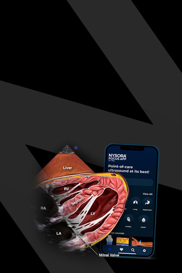
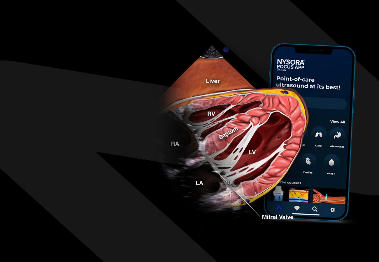

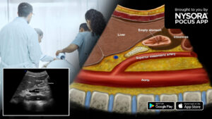
Master your emergency diagnostics skills on the go!
Convenience meets excellence with immersive walkthroughs to the most commonly used POCUS techniques, accompanied by proprietary NYSORA learning aids and clinical pearls.
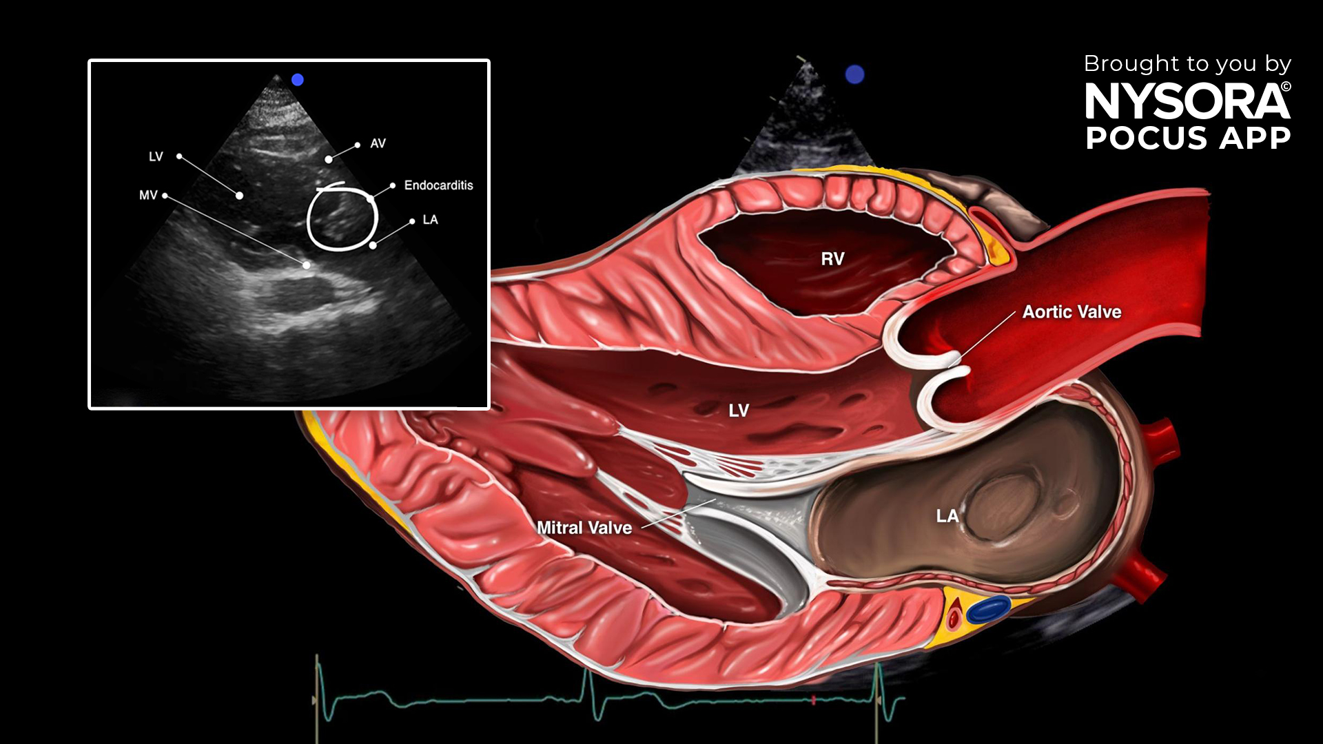
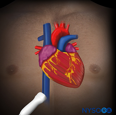
A 68-year-old woman presented at the emergency department with acute respiratory failure and fever. Lung ultrasound showed a B-profile in all four BLUE points, suggesting pulmonary edema. This prompted us to do a cardiac ultrasound. Consecutively, a focused cardiac ultrasound was performed. The parasternal long-axis view showed a lesion suggestive of endocarditis which was then confirmed by an official cardiologist ultrasound. Endocarditis is a serious medical condition that affects the inner lining of the heart and the valves. The causal pathogen may be bacterial but occasionally fungal or viral. Endocarditis is often diagnosed by the formation of vegetations on the heart valves or other endocardial surfaces. These vegetations can interfere with the normal function of the heart, leading to complications such as severe regurgitation, cardiac failure, stroke, or systemic infections if bacteria from the heart enter the bloodstream. Endocarditis requires prompt diagnosis and treatment with antibiotics or, in severe cases, surgery to repair or replace damaged valves. Point-of-care ultrasound (POCUS) is a valuable tool in assessing endocarditis, offering real-time imaging capabilities that aid in detecting vegetations, abscesses, and valvular abnormalities. When evaluating endocarditis through ultrasound, distinctive pathology characteristics to observe include: Mobile lesion Hyperechoic density Endocarditis is often accompanied by regurgitation Parasternal long-axis view revealing endocarditis. LV, left ventricle; AV, aortic valve; LA, left atrium; MV, mitral valve. Transform your practice with the power of POCUS using NYSORA’s POCUS App. Enhance your skills, broaden your diagnostic capabilities, and provide outstanding patient care. Experience the difference today – Download the app HERE.
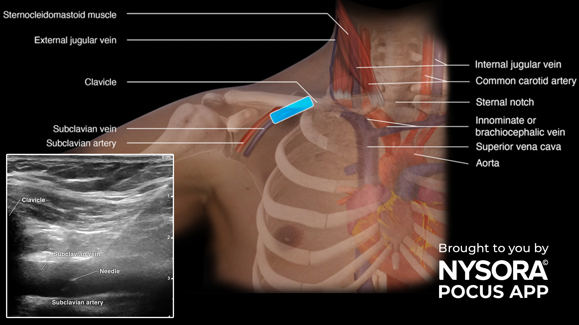
Subclavian vein cannulation is an essential medical procedure used to access central veins for various clinical purposes. The incorporation of point-of-care ultrasound (POCUS) has significantly enhanced the precision and safety of this technique. POCUS allows healthcare providers to visualize and navigate the subclavian vein with accuracy, reducing complications and improving the overall success of the procedure. When it comes to subclavian vein cannulation, these expert tips can make a significant difference: Always identify the vein, artery, pleura and ribs before starting the procedure. Subclavian vein cannulation carries the least risk for catheter-related infections, while femoral vein cannulation has a higher risk. In cases involving small or flat veins in intubated patients, techniques like the Valsalva maneuver or positive end-expiratory pressure (PEEP) can enhance vein distention, simplifying the cannulation procedure. Always augment your ultrasound-guided subclavian vein cannulation with a lung ultrasound together with a quick cardiac ultrasound to check for the rapid atrial swirl sign (RASS). This will allow you to rule out pneumothorax and confirm the position of the catheter insertion. Transform your practice with the power of POCUS using NYSORA’s POCUS App. Enhance your skills, broaden your diagnostic capabilities, and provide outstanding patient care. Experience the difference today – Download the app HERE.

In our last post we taught you how to identify fluid content inside the stomach. Fluid content however may be the result of endo- or exogenous factors. Let’s quickly recap our case from the last post: A 40-year-old man with a wrist fracture presented after having a drink 3 hours prior to his admission. You performed a gastric ultrasound since you are dealing with emergency surgery and you visualized fluid content: Ultrasound and Reverse Ultrasound Anatomy of a stomach with fluid content Endoluminal gastric content can be caused by numerous factors such as delayed gastric emptying by stress or opioids, non compliance, gastric secretions, etc. The following tutorial will teach you how to differentiate a low from a high risk stomach. For fluid content, the cross-sectional area (CSA) of the antrum should be measured in the lateral decubitus position to assess the risk of aspiration. This is done by tracing the outer layer of the antrum or the serosa. Cross-sectional area of the antrum. By using the CSA and the age of the patient, the volume can be estimated using this formula: Gastric volume = 27.0 + (14.6) x (CSA of antrum in right lateral decubitus position) – 1.28 x age If the fluid content is >1.5 mL/kg, the stomach is considered full and thus high risk. Fluid content <1.5 mL/kg is compatible with a fasting state and thus low risk. Transform your practice with the power of POCUS using NYSORA’s POCUS App. Enhance your skills, broaden your diagnostic capabilities, and provide outstanding patient care. Experience the difference today – Download the app HERE.
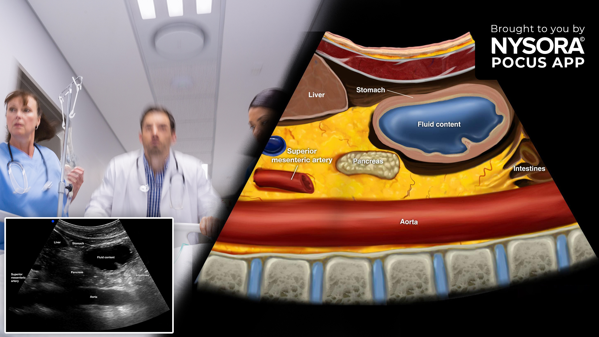
Emergency surgery presents a unique challenge as patients may not be fasted. Assessing gastric content, whether full or empty, becomes pivotal, influencing anesthesia choices and perioperative strategy to mitigate aspiration risks during intubation. By employing ultrasound to assess gastric content, medical professionals can swiftly and confidently make decisions regarding anesthesia and airway management. This non-invasive technique allows for the visualization of gastric contents, helping differentiate between clear, empty stomachs and those with residual contents. Imagine the following case: A 40-year-old patient broke his wrist after falling from the stairs. 3 hours before the accident, he had a couple of glasses at an after-work celebration. Your surgeon is eager to operate on him. What’s your strategy? In these cases, gastric ultrasound can be of immense value. Imagine you use your POCUS knowledge and scan the stomach. You see the following ultrasound image: Ultrasound and Reverse Ultrasound Anatomy of a stomach with fluid content. Here, the stomach is filled with hypoechogenic intraluminal content. Fluid in the stomach leads to distinctive visual indicators. This includes a noticeable rounding and distension of the antrum, along with thinning of the stomach walls. When assessed sonographically, a clear distinction can be made between two types of fluids: clear fluids and non-clear fluids (such as suspensions or milk). Clear fluids are anechoic. Non-clear fluids appear hyperechoic. However, it is important to understand that the stomach itself also produces fluid. Gastric ultrasound can help to differentiate between endo- and exogenous fluid content. In our next post, we will teach you how much fluid is considered too much. Stay tuned. Clinical decision pathway for anesthesia in emergency surgery based on gastric ultrasound. Transform your practice with the power of POCUS using NYSORA’s POCUS App. Enhance your skills, broaden your diagnostic capabilities, and provide outstanding patient care. Experience the difference […]
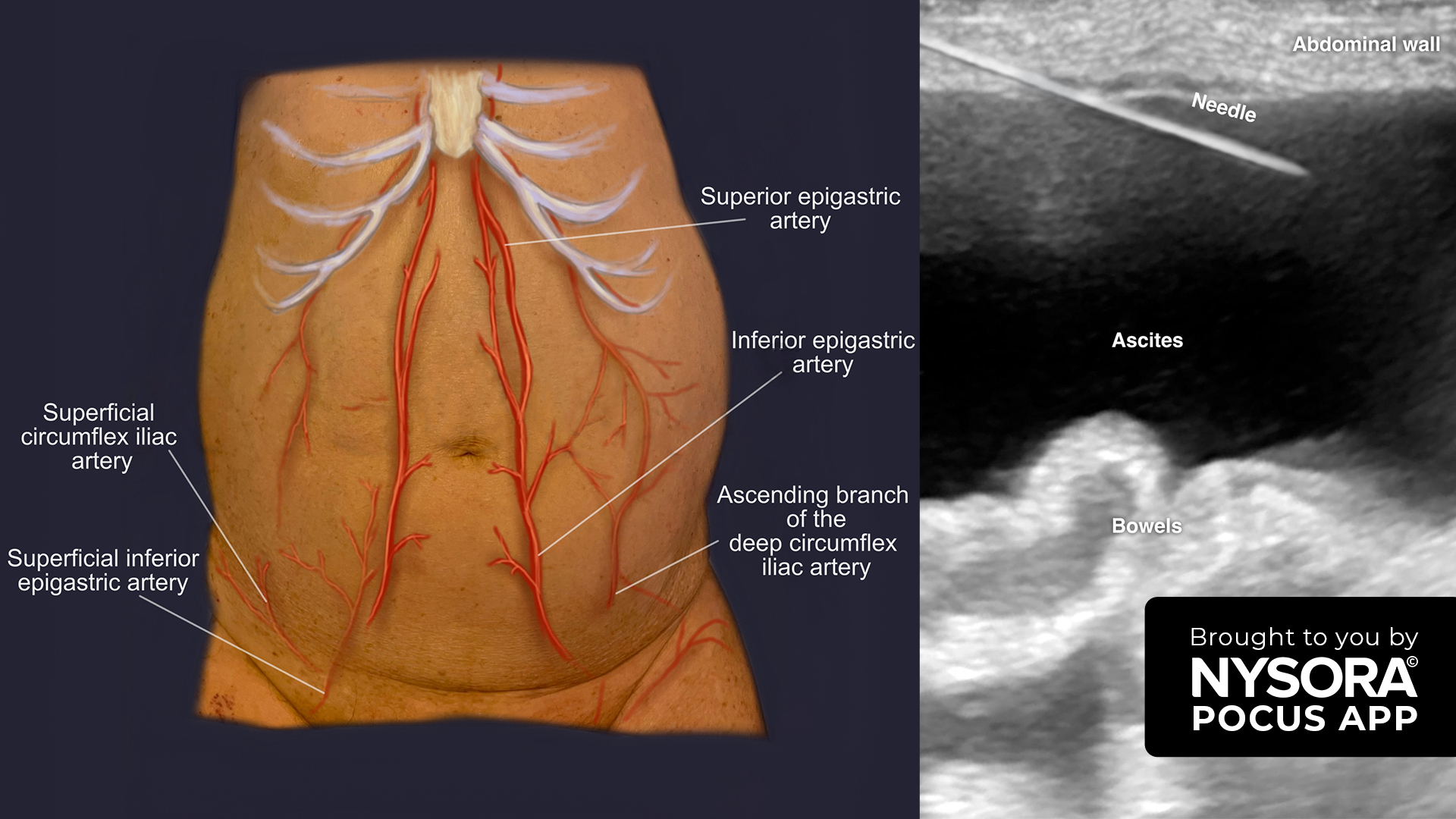
Paracentesis, a procedure to puncture and access free intraperitoneal fluid or ascites, is used for both diagnostic and therapeutic purposes. Causes of ascites include liver disease, heart disease, malignancy, kidney disease, chronic inflammation, or hypoalbuminemia. Ultrasound guidance is crucial in this procedure for site determination and needle guidance, reducing risks like vessel or bowel injury. Here are some key tips for a successful ultrasound-guided paracentesis: Use a curvilinear transducer to detect free fluid presence and a linear transducer for the ultrasound-guided puncture. This method minimizes bleeding risks at the insertion site, puncture site infections, and abdominal wall hematomas. It’s essential to identify and avoid puncturing dilated veins (caput Medusa) in ascites patients. Locate and steer clear of the inferior epigastric artery, typically 5-6 cm lateral from the midline, using color Doppler. Avoid needle insertion through the suprapubic area due to the vicinity of the urinary bladder. Remember that visceral structures like bowels move autonomously and tend to float due to their air content. Monitor these floating structures closely during needle insertion to reduce bowel puncture risks and needle contamination. Transform your practice with the power of POCUS using NYSORA’s POCUS App. Enhance your skills, broaden your diagnostic capabilities, and provide outstanding patient care. Experience the difference today – Download the app HERE.
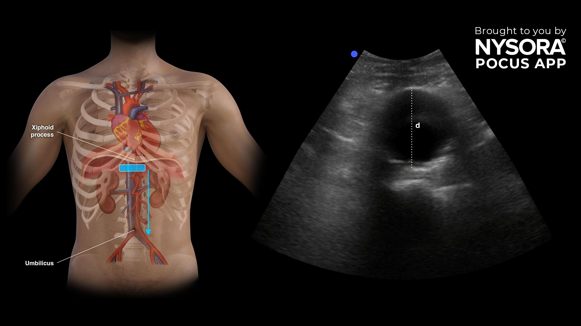
We are thrilled to announce the launch of a brand new course in the ‘Vascular’ category in NYSORA’s POCUS app: Abdominal Aortic Aneurysm (AAA). This comprehensive course is designed for healthcare professionals who wish to expand their expertise in ultrasonography for assessing AAAs. What you’ll learn: Sonoanatomy of the abdominal aorta: Discover the detailed anatomy of the abdominal aorta through high-quality ultrasound images and illustrations. Scanning technique: Learn how to properly scan the abdominal aorta to look for signs of AAA. Identification and assessment of AAA: Gain crucial skills in recognizing and evaluating AAAs, enhancing your diagnostic abilities. Correlate ultrasound findings with interventions: Enhance your decision-making with practical tips and clear algorithms. Transform your practice with the power of POCUS using NYSORA’s POCUS App. Enhance your skills, broaden your diagnostic capabilities, and provide outstanding patient care. Experience the difference today – Download the app HERE.
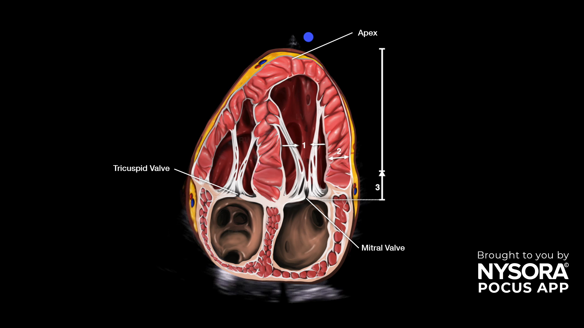
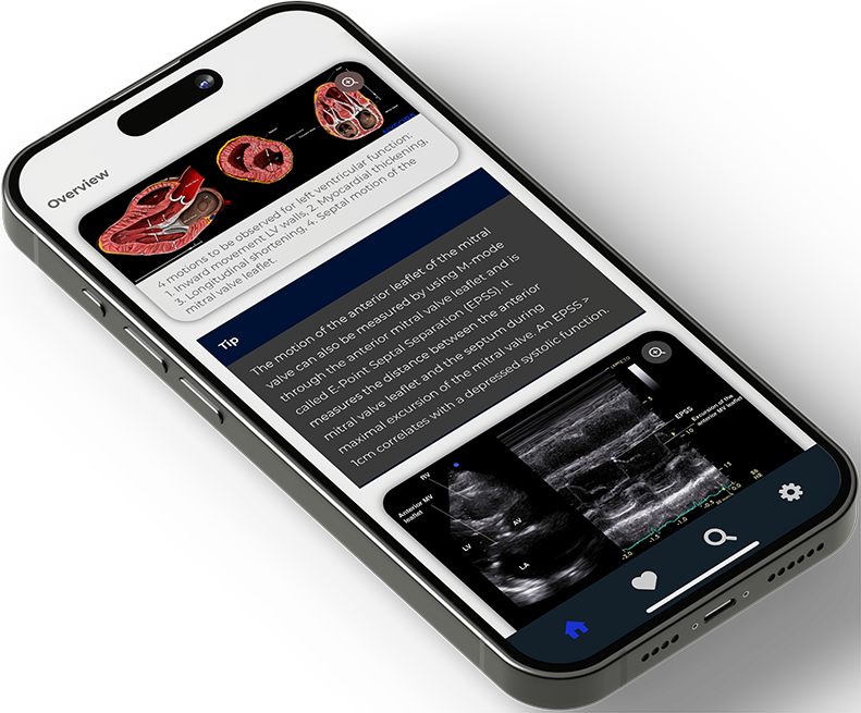
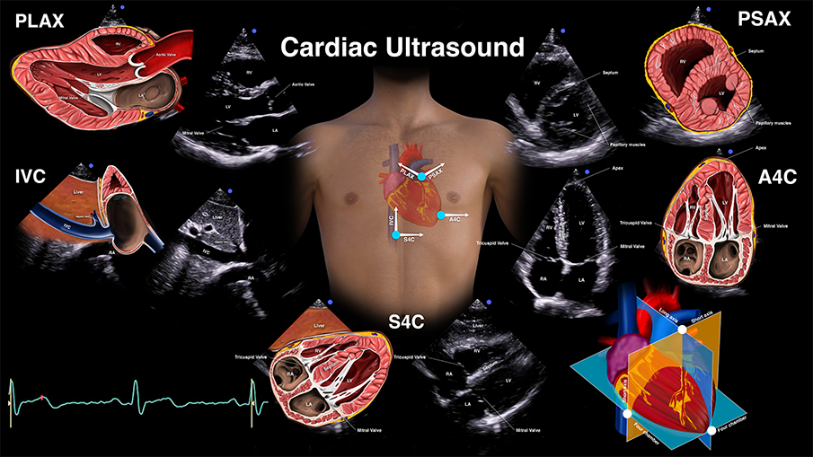
A 71-year-old male with a medical history of coronary artery disease, chronic hypertension, and type 2 diabetes presents with chest pain. Upon arrival, standard protocol led to the execution of an ECG, chest X-ray, and blood tests. While awaiting these results and during the clinical examination, a point-of-care ultrasound scan focusing on the heart was conducted, comprising five crucial ultrasound views. Each view can be used to evaluate the left ventricular function, and, in this case, the apical four-chamber view was used. The technique, also known as eyeballing, evaluates the inward motion of the endocardium, the thickening of the left ventricular wall during systole, and the longitudinal shortening or movement of the mitral valve apparatus toward the heart’s apex. Closely assessing these 3 signs in the apical four-chamber view can distinguish a normal from a decreased left ventricular function. Unleash the potential of POCUS with NYSORA’s POCUS App and elevate your practice, expand your capabilities, and deliver exceptional patient care. Download HERE.
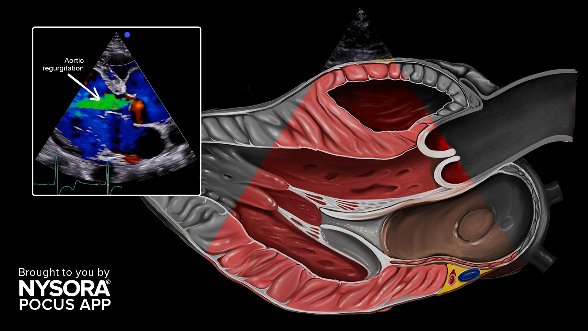
Point-of-Care Ultrasound (POCUS) is excellent for cardiac assessment. Major valvular diseases of the tricuspid, mitral, and aortic valves are also easy to identify. Aortic regurgitation, also known by aortic insufficiency, is a significant valvular disease characterized by the backflow of blood from the aorta. This condition occurs when the aortic valve, which separates the left ventricle from the aorta, does not close properly. As a result, the heart needs to work harder to pump the volume overload. Aortic regurgitation can have various underlying causes, with degenerative valve conditions, congenital defects, or other heart-related issues being the most common culprits. Upon diagnosis, it is essential to prioritize a formal transthoracic echocardiography, preferably from a certified healthcare professional. When evaluating aortic regurgitation through ultrasound, distinctive pathology characteristics to observe include: Aliasing with large jets, seen as multiple color blood flow on color Doppler. The regurgitation is severe if the jet occupies the majority (65%) of the left ventricle outflow tract. Jets that are not seen in the center of the valve tend to be underestimated and have to be considered severe until proven otherwise. Aortic regurgitation. Unleash the potential of POCUS with NYSORA’s POCUS App and elevate your practice, expand your capabilities, and deliver exceptional patient care. Download HERE.
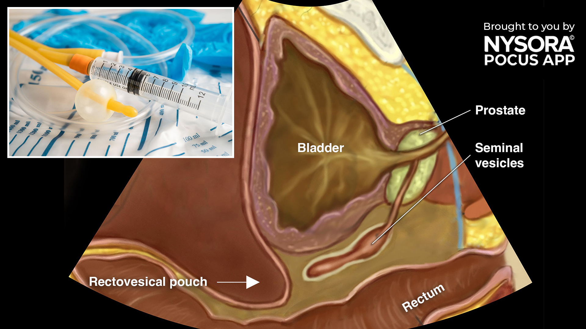
A Foley catheter is a flexible, sterile tube inserted into the bladder to facilitate urine drainage. It features an inflatable balloon that ensures it stays anchored securely in the bladder. It is frequently used in patients who cannot empty their bladder naturally, those undergoing specific surgeries, or for continuous urine output monitoring in hospitalized individuals. Ultrasound can be utilized to confirm the catheter’s correct position. An ideally positioned catheter should show either an empty or non-distended bladder with an intraluminal balloon. If the balloon appears to be surrounded with hypoechogenic fluid, the foley may be obstructed. Endoluminal balloon in an obstructed foley. Reverse Ultrasound Anatomy of the male pelvis. Unleash the potential of POCUS with NYSORA’s POCUS App and elevate your practice, expand your capabilities, and deliver exceptional patient care. Download HERE.
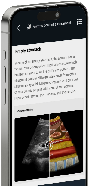
Gastric content assessment via ultrasound can provide valuable information when determining whether a patient’s stomach is full or empty, particularly in the context of preparing anesthesia for emergency surgery. Emergency surgery is common and there are no fasting guidelines for emergency surgery. When the stomach is empty, the antrum typically presents as either a round or elliptical shape, commonly known as the “bull’s eye pattern.” This distinct pattern is characterized by a thick, hypoechogenic wall built out of muscularis propria with central and external hyperechoic layers, the mucosa, and the serosa. Unleash the potential of POCUS with NYSORA’s POCUS App and elevate your practice, expand your capabilities, and deliver exceptional patient care. Download HERE.

Lung consolidation refers to a condition where the air-filled spaces in the lung’s alveoli are replaced by fluid, pus, blood, or other substances. This results in the affected lung tissue becoming more solid and less able to exchange oxygen, leading to impaired respiratory function. It can be visualized using ultrasound, with distinct patterns such as the shred sign and hepatization helping to identify the consolidation type. There are two types of lung consolidation: 1. Non-translobar consolidation (shred sign or C-line): This type displays an irregular boundary, often resembling a fractal line, which separates the consolidated lung from the aerated lung underneath. This pattern is commonly observed in non-translobar consolidations, most frequently associated with pneumonia. Shred sign or C-line. 2. Translobar consolidation (hepatization): In this case, the ultrasound image bears a resemblance to ultrasound imaging of the liver. The “hepatized” lung is lung tissue that is visible due to the absence of air, creating a texture similar to organ tissue. Translobar consolidations are often linked to pneumonia or atelectasis. Normal lung on the left and hepatized lung on the right. Normal lung tissue cannot be visualized with ultrasound due to reflections of the air while hepatized lung tissue resembles organ tissue. Unleash the potential of POCUS with NYSORA’s POCUS App and elevate your practice, expand your capabilities, and deliver exceptional patient care. Download HERE.

When conducting peripheral vascular access procedures, such as radial artery cannulation, executing the technique smoothly is crucial to reduce risks and enhance patient comfort. Proper visualization of the needle tip is essential when performing an out-of-plane technique. Follow these tips to optimize needle visualization in an out-of-plane technique: Utilizing the creep technique enables improved tracking of the needle tip during out-of-plane procedures, thereby enhancing needle tip visualization and consequently increasing safety. 2. Optimizing the angle and tilting of the ultrasound transducer is essential for achieving optimal beam reflections, which in turn enhances needle visualization. When the needle is not visible, this phenomenon is referred to as anisotropy. Anisotropy occurs when it is impossible to visualize the desired structure of interest due to the loss of sound waves, resulting from decreased reflection back toward the transducer. Employing these strategies can effectively mitigate risks, counteract anisotropy, and substantially elevate the level of patient safety and comfort throughout the procedure. Unleash the potential of POCUS with NYSORA’s POCUS App and elevate your practice, expand your capabilities, and deliver exceptional patient care. Download HERE.


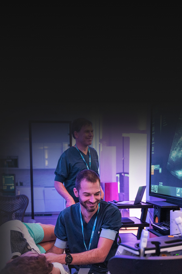
POCUS is becoming the most reliable decision-making tool for diagnostics in emergency medicine and critical care. The POCUS app helps master it on your terms.


We recently partnered with Dr. Ray on POCUS. He is an anesthesiologist and critical care physician and he explains that the transition from regional anesthesia into POCUS is a natural step that considerably changes your practice. Therefore, we designed an app together to empower healthcare professionals with advanced guidance on POCUS wherever they go. We sat down with him to discuss POCUS, its history, and NYSORA’s role in the app publication.
Point-of-care ultrasound (POCUS) refers to the use of (portable) ultrasound devices at the bedside or point of care to provide real-time diagnostic imaging. Unlike traditional ultrasound, which is performed in dedicated imaging departments, POCUS allows healthcare providers to quickly assess patients and guide clinical decision-making directly at the patient’s bedside.
While both ultrasound and point-of-care ultrasound (POCUS) utilize the same imaging technology, they differ in their application and setting. Traditional ultrasound typically involves scheduled appointments in specialized imaging departments, whereas POCUS is performed by healthcare providers directly at the patient’s bedside or point of care to provide immediate diagnostic information and guide treatment decisions in real time.
The objective of point-of-care ultrasound (POCUS) is to facilitate rapid clinical decision-making by providing real-time diagnostic information directly at the patient’s bedside. It allows healthcare providers to quickly assess patients, guide interventions, monitor treatment responses, and expedite patient care, particularly in critical or emergency situations.
The four main types of ultrasound scanning techniques are:
- B-mode ultrasound: Produces two-dimensional grayscale images to visualize anatomical structures.
- Doppler ultrasound: Assesses blood flow by detecting changes in the frequency of sound waves reflected by moving blood cells.
- Color Doppler ultrasound: Combines B-mode imaging with Doppler technology to visualize blood flow direction and velocity, typically represented in color.
- Power Doppler ultrasound: Is more sensitive in detecting blood flow than color Doppler, but does not provide information on direction and speed of blood flow.
- Spectral Doppler ultrasound: A way to visualize the Doppler principle by means of graphical peaks.
- M-mode ultrasound: Displays motion over time, often used to assess cardiac function and fetal heart rate.
Point-of-care ultrasound (POCUS) can be performed by various healthcare providers, including physicians, nurse practitioners, physician assistants, paramedics, and other trained personnel with appropriate certification or training in ultrasound imaging. Proper education and training are essential to ensure proficiency and safety when performing POCUS.
Point-of-care ultrasound (POCUS) is utilized across various medical specialties to aid in diagnosis, treatment, and patient management. Some specialties that commonly use POCUS include emergency medicine, critical care, internal medicine, anesthesia, obstetrics and gynecology, surgery, cardiology, and primary care. POCUS is also increasingly integrated into pre-hospital and point-of-injury care by paramedics and emergency medical technicians.