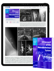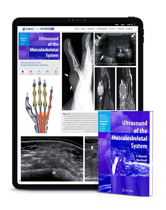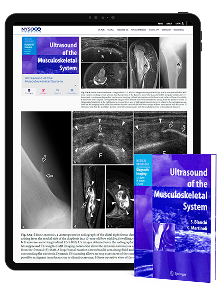
Ultrasound of the Musculoskeletal System, Review
Introduction of Ultrasound of the Musculoskeletal System Ultrasound of the Musculoskeletal System is an invaluable text comprising 19 chapters and approximately one thousand pages and figures. Over the last 15 years, musculoskeletal ultrasonography has become an important imaging modality used in sports medicine, joint disorders, and rheumatology. With the rapid development and sophistication of this modality, essential information for a better understanding of the pathophysiologic assessment of many disorders has been established. This, in turn, has aided both in making crucial decisions regarding surgical intervention and in monitoring the effects of therapy. Equally important is the ready availability, affordability, speed, and diagnostic accuracy of ultrasonography. Contents Ultrasound of the Musculoskeletal System is an invaluable text comprising 19 chapters and approximately one thousand pages and figures. The authors have designed unique schematic drawings which aid in better understanding the anatomy of the body part in terms of its sonographic characteristics discussed in each chapter. Correlations of ultrasonography with CT and MRI findings are applied throughout the text, demonstrating not only the exact indications for its use, but also highlighting its limitations. Development Technical advances continue to improve the utility of ultrasonography as a diagnostic technique in musculoskeletal imaging. Drs. Bianchi and Martinoli have successfully capitalized on the collaboration between radiologists, orthopedists, and rheumatologists as exemplified by their representative images and correlative discussions. Many of the techniques described in the text have been pioneered or improved by Dr. Bianchi and Dr. Martinoli. This text should become a key library reference source for radiologists, orthopedists, and rheumatologists. It is extremely readable and its illustrations help in the clarification of points made in the text. Ultrasound of the Musculoskeletal System is the most comprehensive work of its kind to date. It establishes a higher standard in musculoskeletal imaging and should remain a classic for

